www.fm.viamedica.pl O R I G I N A L A R T I C L E
Address for correspondence: Prof. Jerzy St. Gielecki, Department of Anatomy, Silesian Medical University, ul. Medyków 18, 40–752 Katowice Ligota, Poland, tel: +48 32 252 64 87, fax: +48 32 252 64 87, e-mail: [email protected]
Digital-image analysis of the aortic arch’s
development and its variations
Jerzy St. Gielecki, Renata Wilk, Bożena Syc, Magdalena Musiał-Kopiejka,
Aneta Piwowarczyk-Nowak
Department of Anatomy, Silesian Medical University, Katowice, Poland
[Received 18 June 2004; Revised 20 August 2004; Accepted 1 September 2004]
The study was performed on 103 human foetuses (58 female, 45 male) sponta-neously aborted at between 14 and 30 weeks of gestation. The arteries were filled with latex, preserved in formalin and then dissected under the microscope and digitalised using a camera system. The following measurements were taken with the use of special computer software: the external diameter, the length and the volume of the arch of the aorta. The increase in diameter and length in relation to age corresponded to a linear function with values ranging from 1.77 mm to 4.09 mm for the diameter and from 4.94 mm to 13.31 mm for the length. The increase in volume corresponded to a square root function with values ranging from 13.42 mm3 to 173.96 mm3. Analysis of arch of the aorta variations revealed 11 cases of a common trunk for the brachiocephalic trunk and the left common carotid artery and 7 cases with the left vertebral artery arising directly from the arch of the aorta. In 2 cases the brachiocephalic trunk was absent, the right subclavian artery branching directly from the arch of the aorta at the level of the left subclavian artery or from the descending aorta just below the arterial duct ostium.
Key words: human foetus, morphometric measurements, variability, aortic arch
INTRODUCTION
The primary arterial system undergoes many com-plicated changes during development. Between the first 6 and 8 weeks of foetal life primary arterial arch-es arise from the arterial sac to evolve into the final pattern of great arterial vessels. The left 4th
arch builds the aortic arch, which grows intensively and joins the dorsal aorta [15].
The aortic arch is short and its limits are diffi-cult to define. As the upper limit we assumed the level of the 2nd right sternocostal joint and as the
lower the narrowing (often termed the isthmus) lying above the arterial duct ostium to the de-scending aorta. Arterial variations of the aortic
MATERIAL AND METHODS
The study was performed on 103 human foetus-es obtained from spontaneous abortions (58 female, 45 male) aged from 14 to 30 weeks of gestation. The foetal age (in weeks) was established by mea-suring the length of their humeral and femoral bones using USG equipment (Table 1). The foetal arteries were filled with latex and preserved in 4% formalin and then dissected under a surgical microscope. The dissected arteries were digitalised in 24 bit BMP. For approximation and scaling of the microscopic pic-tures (2272 × 1704 pix) the Olympus AnalySIS com-puter program was used. In order to make measure-ments of the vessels under examination, a special computer program, MORPHO04, was developed. The program’s operation is based on B-splines modelled with Bezier’s Curves generated over textured graphic (pictures) of the vessels examined (Fig. 1). Moreover, on the assumption that the vessel cross-section is cir-cular, it was possible to calculate its volume [8].
The measurements were taken as follows: the ex-ternal diameter, the length, and the volume of the aortic arch. The results were correlated to foetal age so as to establish the growth dynamics of the arteries. To make the statistical analysis, the foetuses were split into 5 groups as follows: 14–16 weeks, 17–20 weeks, 21–24 weeks, 25–28 weeks and 29–30 weeks. These groups correspond to the 4th, 5th, 6th, 7th and
8th
months of gestation.
The results were analysed using statistical tests (ANOVA, NIR, and RIR) for equal and unequal amounts. The Pearson test, based on the method of the smallest squares, was used to establish the lin-ear correlation for each parameter against gestation-al age. The results obtained were presented in ta-bles and figures.
The aortic arch variations were analysed and dif-ferent types of abnormalities in the origin and posi-tion of the aortic arch branches were distinguished based on Niżankowski’s classification [13].
RESULTS
RESULTS
RESULTS
RESULTS
RESULTS
Variation analysisUsually 3 main arterial trunks emerge from the aortic arch (Fig. 2A). The distances between their origins are the same, although the arteries can change their position, arising from a single arterial trunk or very close together. This usual pattern of 3 branches was found in 83 specimens (80.6%). In the remaining specimens (28 specimens — 27.2%) it was possible to distinguish aortic arches where the left common carotid artery branched very close to the brachiocephalic trunk (Fig. 2B). In 11 cases (10.7%), only a single arterial trunk was found for the brachiocephalic trunk and the left common ca-rotid artery (Fig. 2C, 3). In 7 cases (6.8%) the left vertebral artery sprang directly from the aortic arch between the left common carotid and left subcla-vian arteries (Fig. 2D, 4). In one case the right sub-clavian artery originated from the posterior surface of the aorta at the level of the left subclavian tery, while in another case the right subclavian
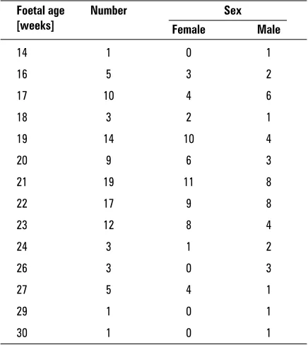
ar-Table 1. Distribution of foetal age and sex
Foetal age Number Sex
[weeks] Female Male
14 1 0 1
16 5 3 2
17 10 4 6
18 3 2 1
19 14 10 4
20 9 6 3
21 19 11 8
22 17 9 8
23 12 8 4
24 3 1 2
26 3 0 3
27 5 4 1
29 1 0 1
30 1 0 1
Figures 2A–E. Diagrams illustrating variations in the arch of the aorta. AA — arch of the aorta, BT — brachiocephalic trunk, LCC — left common carotid artery, LS — left subclavian artery, RCC — right common carotid artery, LV — left vertebral artery.
A B
C D
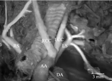
Figure 3. Single trunk for common carotid artery and brachio-cephalic trunk. AA — arch of the aorta, DA — ductus arteriosus, BT + LCC — single trunk for the common carotid artery and bra-chiocephalic trunk, LS — left subclavian artery.
tery originated from the descending aorta just be-low the arterial duct ostium (Fig. 2E, 5). In both cases the arteries were directed towards the right upper extremity, crossing the anterior surface of the vertebral column and passing posterior to the trachea and oesophagus, reaching the posterior cleft of the scalenus muscles.
Morphometric analysis
The values for all foetal age groups have been presented in Table 2.
The mean values of the aortic arch diameter ranged from 1.77 mm for the 14–16 week gesta-tional group to 4.09 mm for the 29–30 week age group. With regard to foetal age, the diameter of the arteries increased according to the linear func-tion y = a + b * x with a correlafunc-tion coefficient of 0.82. The results obtained were statistically signifi-cant (p < 0.05) (Fig. 6A) for each age group.
The length of the aortic arch ranged from 4.94 mm to 13.31 for the 14–16 and 29–30 week gesta-tional groups respectively. With regard to the aortic arch diameter, the increase in the artery length was dependent on foetal age in accordance with the lin-ear function y = a + b * x with a correlation coeffi-cient of 0.79 and the results obtained showed sta-tistical significance (p < 0.05) (Fig. 6B).
The volume of the aortic arch ranged from 13.42 mm3 to 173.96 mm3 for groups of 14–16 weeks
and 29–30 weeks of gestation respectively. The vol-ume increase runs according to the square root model y = (a + b * x)2
with a correlation coefficient of 0.86 and the results obtained were of statistical significance (p < 0.05) (Fig. 6C).
DISCUSSION
The aortic arch variations observed in the mate-rial under examination have been described earlier. Czerwiński et al. [6] reported on the basis of autop-sy specimens (118 cadavers) one case in which the left vertebral artery sprang directly from the aortic arch and one case in which the right subclavian ar-tery originated from the posterior wall of the de-scending aorta below the level of the left subclavian artery. Both the above-mentioned variations with respect to the position and number of the arteries were present in the material under examination. Czerwiński also analysed the clinical signs associat-ed with aortic arch variations. In the first case no clinical symptoms were observed in connection with the variation. In second case difficulties in swallow-ing and in breathswallow-ing were noted, especially durswallow-ing Figure 4. Left vertebral artery spreading out directly from the
aor-tic arch. AA — arch of the aorta, DA — ductus arteriosus, BT — brachiocephalic trunk, LCC — left common carotid artery, LV — left vertebral artery, LS — left subclavian artery.
upper limb abduction. Similar variations were de-scribed by Aleksandrowicz [2] and Lize [11], also on the basis of autopsy specimens. In Sora’s et al. [16] investigation a case of a single arterial trunk for the brachiocephalic trunk and left common carotid ar-tery was analysed. In Niżankowski’s work [13] the variation was described as present in 0.9% of the specimens examined, while we noted it in 7 speci-mens (8%). Roguin et al. [14] analysed the case of a 6-day-old infant with a single arterial trunk arising from the aortic arch, from which emerged the right subclavian, both carotid arteries and the left
subcla-Figure 6. Development of the average diameter [mm], length [mm] and volume (mm3) of the arch of the aorta in human
foet-uses. A. Allometric linear function: Diameter = –0.815826 + + 0.173906 * Age. Correlation coefficient 0.82. B. Allometric linear function: Length = –2.74321 + 0.497327 * Age. Correla-tion coefficient 0.79. C. Allometric square root function: Volume = = (–6.76617 + 0.658652 * Age)2. Correlation coefficient 0.86.
Table 2. The diameter, length and volume values of the arch of the aorta (SD — standard deviation)
Foetal age group [weeks] Diameter [mm] SD Length [mm] SD Volume [mm3] SD
14–16 1.77 0.42 4.94 0.73 13.42 8.08
17–20 2.52 0.40 6.62 1.15 35.05 16.32
21–24 2.91 0.38 8.09 1.33 55.52 18.61
25–28 4.07 0.27 10.19 1.22 132.61 27.33
29–30 4.09 0.26 13.31 1.10 173.96 7.88
C
A B
vian artery. The single trunk presence was associat-ed with coarctation of the aorta and patent ductus arteriosus. Białowąs et al. [4] noted 3 cases of a right-sided aortic arch which passed beyond or in front of the oesophagus and trachea. The variations in great arterial trunks in association with pathological chang-es to the heart, such as interventricular septal de-fects in human foetuses, were described by Mou-laert et al. [12]. Kozielec [10] described the case of a left subclavian artery arising from the ductus
arte-riosus. These anomalies were absent in the material
In the morphometric studies, it was found that the increase in the aortic arch external diameter was in accordance with a linear function in relation to foe-tal age. Hyett et al. [9] obtained similar results from a study of human foetuses of between 9 and 18 weeks of gestation. Ursell et al. [17] also noted linear growth of this parameter in relation to age when analysing the internal diameters of the great vessels in human foetuses from 10 to 26 weeks of gestation. Alvarez et al. [3] observed correlations between foetal weight and length and aortic arch diameter.
The results obtained from autopsy specimens have been confirmed by clinical examinations using echocar-diography or ultrasonography. According to many authors [1, 3, 5, 7] the increase in the diameter of the aortic arch has a close linear correlation to foetal age. The present studies on autopsy material have confirmed the linear growth of the diameter and length according to foetal age and, furthermore, provide data on the type of volume increase. It was found that volume growth is in accordance with a square root function.
The method used has a high degree of accuracy in view of the introduction of the main principles of vectorial computer graphics. The high sensitivity of this method allows precise evaluation of the morpho-metric parameters of the vessels. As a result, the dig-ital measurements made in accordance with Bezier’s Curves, are more precise than those solely made with a raster image visible on the computer screen.
REFERENCES
1. Achiron R, Zimand S, Hegesh J, Lipitz S, Zalel Y, Rotsh-tein Z (2000) Fetal aortic arch measurements between 14 and 38 weeks gestation: in utero ultrasonographic study. Ultrasound Obstet Gynecol, 15: 226–230. 2. Aleksandrowicz R (1967) Two cases of absence of the
brachiocephalic trunk. Folia Morphol, 26: 235–237.
3. Alvarez L, Aranega A, Saucedo R, Contreras JA, Lopez F, Aranega A (1990) Morphometric data concerning the great arterial trunks and their branches. Int J Cardiol, 29: 127–139.
4. Białowąs J, Hreczecha J, Grzybiak M (2000) Right-sid-ed aortic arch. Folia Morphol, 59: 211–216.
5. Comstock CH, Riggs T, Lee W, Kirk J (1991) Pulmo-nary-to-aorta diameter ratio in the normal and abnor-mal fetal heart. Am J Obstet Gynecol, 10: 1038–1044. 6. Czerwiński F, Mierzwa A, Krzanowski K, Michalska G (1993) Rare cases of arteries branching directly from aortic arch in the man. Folia Morphol, 53: 223–231. 7. Gembruch U, Shi C, Smrcek JM (2000) Biometry of the fetal heart between 10 and 17 weeks of gestation. Fetal Diagn Ther, 15: 20–31.
8. Gielecki JS, Cytowski J, Gacek W (1996) A new morpho-metrical method for measuring the diameter, length and volume of vessels. Folia Morphol, 55: 243–245. 9. Hyett J, Moscoso G, Nicolaides K (1995)
Morphomet-ric analysis of the great vessels in early fetal life. Hum Reprod, 10: 3045–3048.
10. Kozielec T (1977) Origin of the left subclavian artery from the arterial duct in fetal life in man. Folia Mor-phol, 36: 191–194.
11. Lize J (1970) Abnormal origin of the great vessels from the aortic arch. Folia Morphol, 29: 401–402. 12. Moulaert AJ, Bruins CC, Oppenheimer-Dekker A (1976)
Anomalies of the aortic arch and ventricular septal defects. Circulation, 53: 1011–1015.
13. Niżankowski C, Rajchel Z, Ziółkowski M (1975) Abnor-mal origin of arteries from the aortic arch in man. Fo-lia Morphol, 34: 109–116.
14. Roguin N, Sujov P, Shapir Y, Peleg H, Riss E (1982) Single arterial trunk arising from the aortic arch associated with coarctation of the aorta. Pediatr Radiol, 12: 39–40. 15. Sadler TW (1993) Langman’s Medical Embriology, Med
Tour Press International. Warszawa, pp. 207–219. 16. Sora M, Strobl B, Forstner-Streffleur S, Staykov D (2002)
Aortic arch variation analyzed by using plastination. Clin Anat, 15: 379–382.