R E S E A R C H
Open Access
Effects of MMP-9 inhibition by doxycycline on
proteome of lungs in high tidal volume
mechanical ventilation-induced acute lung injury
Adrian Doroszko
1,5, Thomas S Hurst
2, Dorota Polewicz
1, Jolanta Sawicka
1, Justyna Fert-Bober
1, David H Johnson
3,
Grzegorz Sawicki
1,4*Abstract
Background:Although mechanical ventilation (MV) is a major supportive therapy for patients with acute respiratory distress syndrome, it may result in side effects including lung injury. In this study we hypothesize that MMP-9 inhibition by doxycycline might reduce MV-related lung damage. Using a proteomic approach we identified the pulmonary proteins altered in high volume ventilation-induced lung injury (VILI). Forty Wistar rats were randomized to an orally pretreated with doxycycline group (n = 20) or to a placebo group (n = 20) each of which was followed by instrumentation prior to either low or high tidal volume mechanical ventilation. Afterwards, animals were euthanized and lungs were harvested for subsequent analyses.
Results:Mechanical function and gas exchange parameters improved following treatment with doxycycline in the high volume ventilated group as compared to the placebo group. Nine pulmonary proteins have shown significant changes between the two biochemically analysed (high volume ventilated) groups. Treatment with doxycycline resulted in a decrease of pulmonary MMP-9 activity as well as in an increase in the levels of soluble receptor for advanced glycation endproduct, apoliporotein A-I, peroxiredoxin II, four molecular forms of albumin and two unnamed proteins. Using the pharmacoproteomic approach we have shown that treatment with doxycycline leads to an increase in levels of several proteins, which could potentially be part of a defense mechanism.
Conclusion:Administration of doxycycline might be a significant supportive therapeutic strategy in prevention of VILI.
Background
Acute lung injury (ALI) and its more severe form, acute respiratory distress syndrome (ARDS), are characterized by an acute inflammation and disruption of the alveolar-capillary membranes leading to alveolar flooding with protein-rich edema fluid. Mortality rates resulting from ALI and ARDS range from 18 to 54.7% [1]. Although mechanical ventilation (MV) is an important supportive strategy for patients with ARDS, it is also known to further lung injury in certain conditions of mechanical stress, leading to ventilation-induced lung injury (VILI). The mechanisms by which conventional MV exacerbates lung injury and inflammation are of considerable clinical
significance. The potential importance of VILI in the clinical treatment of critically ill patients has been well established by recent clinical trials [2,3], where a relative risk reduction of 22% in patients ventilated with the lower tidal volume has been shown. The results from these studies indicate that mortality attributable to VILI is at least 9 to 10% in such patients. Therefore, despite the significant progress that has been recently made in emergency medicine, further research concerning the pathophysiology of ALI is needed in order to indicate new therapeutic targets preventing pulmonary damage.
Several experimental and clinical studies led to the hypothesis that the deleterious effects of MV might be mediated by local inflammation and the systemic release of inflammatory cytokines (biotrauma) [4-6]. During ALI/ARDS, various proinflammatory cytokines and che-mokines are up-regulated and contribute to the * Correspondence: [email protected]
1Department of Pharmacology, University of Saskatchewan, Saskatoon,
Saskatchewan, Canada
initiation and propagation of the inflammatory response. In clinical studies, changes in levels of biological mar-kers have been used primarily in an effort to identify VILI rather than to study disease pathogenesis. The ven-tilator-associated changes in levels of some biological markers have been correlated with patient outcomes, including duration of MV, organ failures, length of hos-pital stay, and mortality [7-9]. Despite various pharma-cologic interventions developed in the last two decades that aim at specific targets, such as cytokines and adhe-sion molecules, the therapeutic strategy for this syn-drome remains to be established. Although alveolar epithelial injury is a major determinant of outcome in patients with ALI, there is still no reliable biological marker of alveolar epithelial injury.
Recently, new mechanisms in the pathology of acute respiratory failure have shifted the focus to lung mechanics, tissue damage, remodeling, and the sys-temic effects derived from the mechanical stress imposed by the ventilator in patients with ARDS [7]. The conversion of physical signals such as contractile forces or external mechanical perturbations into che-mical signaling events is a fundamental cellular process that occurs at the cell-extracellular matrix contact, known as focal adhesions, thus modifying cell viscoe-lastic properties, which may compromise the balance of forces in the alveolar epithelium [10]. Stretch-induced cell stiffening could compromise the balance of forces at the cell-cell and cell-matrix adhesions [4].
Mascarenhas et al. [11] in an in vitro study showed
thatde novosynthesis of hyaluronate in the extracellu-lar matrix increases proinflammatory cytokines in VILI. Moreover, Taylor et al. [12] demonstrated that hyaluro-nate released after lung injury can stimulate endothelial cells to produce cytokines by activation of a Toll-like receptor 4-dependent mechanism, thus suggesting that endogenous components of the extracellular matrix can stimulate endothelial cells to trigger recognition of injury in the initial stages of the wound defense and repair response. Recent data from studies performed on pulmonary microvascular endothelial cells and iso-lated perfused rat lungs confirm the role of endothelial responses to stretch-inducing VILI [13]. High tidal volume ventilation (HVV) may induce focal adhesion formation and recruit leukocytes on the endothelial cells [14]. Haseneen and colleagues [13] demonstrated the existence of a connection between stretched endothelial cells and lung remodeling by release of matrix metalloproteinases (MMPs) activated through a membrane type-1 MMP (MT1-MMP) mechanism. The first evidence suggesting that MMPs are important in regulation of endothelial permeability was revealed by Soccal and co-workers using a lung ischemia/reperfu-sion (I/R) model [15]. Furthermore, it has been shown
that inhibition of MMP-2 and MMP-9 protects the blood-brain barrier during cerebral ischemia and that there is an increase in MMP-2 activity resulting from an increase in oxidative stress [16]. In ALI, and mainly in ARDS, increased levels of MMP-2 and MMP-9 in the bronchoalveolar lavage (BAL) have been suggested to play a role in basement membrane disruption [17]. Studies in various lung injury models show that MMPs are strongly related to the pathogenesis of lung injury [18-20] and that MMP inhibitors (MMPI) decrease the extent of lung injury [21,22]. Therefore, understanding the extracellular as well as intracellular actions of MMPs might be crucial for studying molecular mechanisms of ALI, including those related to MV. The detrimental role of increased MMP-9 activity in pathogenesis of VILI has been widely explored [21,23-25] and there is also some evidence regarding the beneficial role of doxycycline in other models of ALI [26,27]. However, it has not been clearly estab-lished whether doxycycline, by inhibition of MMP-9, might play a beneficial role in limitation of VILI.
A better understanding of pathological mechanisms of ALI should help not only to alleviate the side effects of mechanical forces, but also to develop new therapeutic strategies. Moreover, it is necessary to develop potential pharmacologic targets to modulate the molecular and cellular effects of lung stretch to minimize the iatrogenic consequences of MV.
In the present study, using the pharmacoproteomic approach, we investigated the hypothesis that increased MMP-9 activity, involved in VILI, results in changes in pulmonary proteome, which could be minimized by MMP-9 inhibition by use of doxycycline.
Examination of global changes in protein levels due to VILI should provide new insights into the mechanisms involved in lung damage. Furthermore, it should lead to the development of novel therapeutic strategies as well as in establishment of more precise and sensitive diag-nostic markers.
Results
Effect of ventilation on mechanical function of lungs and on gas exchange
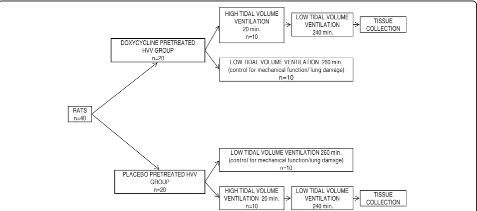
Since treatment with doxycycline had no effect on either mechanical function of lungs or on parameters of gas exchange in low tidal volume ventilated lungs which were present in high tidal volume ventilated (HVV) lungs, we decided to perform biochemical analyses of lungs from only HVV groups (Figure 1). We postulated that HVV should result in changes similar in nature but greater in magnitude as compared to those observed in lungs ventilated with low tidal volume and therefore the observed changes in lung function should be much more pronounced.
Doroszkoet al.Proteome Science2010,8:3 http://www.proteomesci.com/content/8/1/3
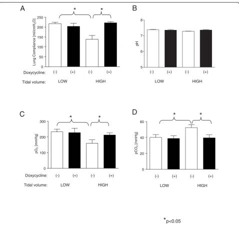
Treatment with doxycycline resulted in a significant increase in lung compliance as compared to the placebo HVV treated group (221.8 ± 5.4 ml/cm H2Ovs. 148.3 ±
5.4 ml/cm H2O in the placebo group, respectively, p =
0.028) which was comparable to levels observed in the low volume ventilation groups (Figure 2A). The values of blood pH in all subgroups remained at physiological levels which is shown in Figure 2B.
High tidal volume ventilation following placebo treatment resulted in significant worsening of gas
exchange as assessed by a decrease in PaO2 (159.6 ±
21.9 mmHg vs. 232 ± 18.1 mmHg in low ventilated
placebo group, respectively, p = 0.027) as well as an
increase in PaCO2 (52.4 ± 3.7 mmHg vs. 40.1 ± 3.6
mmHg in low ventilated placebo group, respectively, p = 0.048). An impairment in gas exchange was reduced by pretreatment with doxycycline (PaO2 = 212.6 ± 13.3
mmHg, p = 0.048 vs. PaO2 in placebo treated HVV
group and PaCO2 = 39.6 ± 3.7 mmHg vs. PaCO2 in
placebo treated HVV group, respectively) (Figure 2C and 2D).
Analysis and identification of proteins in lung homogenates separated by 2-DE
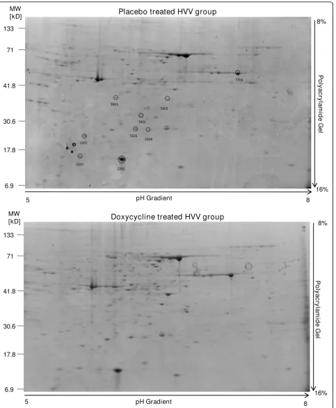
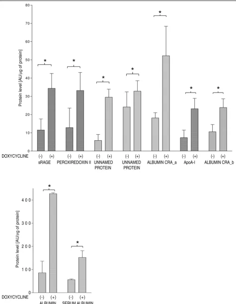
Approximately 220 protein spots were detected in each gel (Figure 3). Using an arbitrary set sensitivity thresh-old, based on minimum peak value sensitivity and a noise filter level, only nine proteins have shown signifi-cant changes between analyzed groups (Figure 4). The results of densitometric analysis of protein spots from lungs subjected to ventilation after pretreatment with doxycycline as well as from placebo treated group are
shown in Figure 4. The levels of all 9 proteins were sig-nificantly higher in the doxycycline treated HVV group as compared to the placebo treated HVV group. The results of protein identification by mass spectrometry (MS) analysis are presented in Table 1 and the struc-tural information for the peptides used for protein iden-tification is shown in Additional file 1 - Table S1. We have identified 9 proteins including: soluble receptor for advanced glycation endproduct (sRAGE), Apoliporotein A-I (ApoA-I), Peroxiredoxine II (Prx II) and four mole-cular forms of albumin. Also, two protein spots were identified as unnamed proteins.
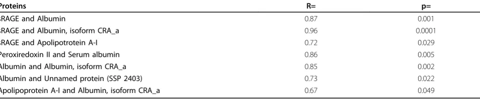
Next, the analysis of correlations between protein levels was performed in order to assess the relationships, which might reflect a common pathophysiological path-way leading to the changes in their levels. Positive, sta-tistically significant correlations between levels of identified proteins were observed and are presented in Table 2.
Mechanical ventilation and MMP-9 activity
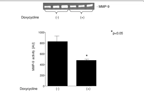
The activity of MMP-9 in lung homogenates in both groups is shown in Figure 5. Treatment with
doxycy-cline decreased MMP-9 activity (475.2 ± 28.1 AU vs.
813.8 ± 119.5 AU in placebo treated HVV group, respectively, p < 0.05).
Immunoblot analysis of identified proteins
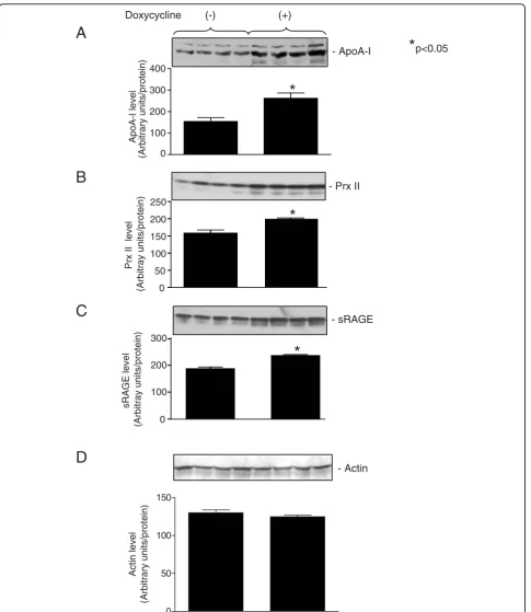
Immunoblot analysis for ApoA-I, Prx II and sRAGE has revealed significant increase in the levels of analyzed proteins in the group treated with doxycycline as com-pared to the placebo group (Figure 6). Loading control is shown in Figure 6D.
Discussion
Proteins play an important role in regulation of biologi-cal systems; therefore, they are among the most com-mon diagnostic and therapeutic targets in medicine. Hence, studying changes in the proteome should lead to the discovery of novel diagnostic markers and therapeu-tic strategies. Ventilation induced lung injury (VILI) has been shown to be associated with alteration of a broad variety of proteins within lungs (such as IL8, nuclear
factor Nrf2, surfactant associated proteins A and D as well as pro-B cell enhancing factor (PBEF)) [28-31]. In this rat model of VILI, high tidal volume ventilation (HVV) should result in changes similar in nature but greater in magnitude as compared to those observed in mechanically ventilated patients in emergency units.
We have shown that the use of doxycycline effectively reduces changes in function of lungs ventilated mechanically with high tidal volume and decreases
Figure 2Changes in lung function dependent on ventilation volume as well as on treatment with doxycycline. A. Lung compliance in four subgroups separated by volume of ventilation and by treatment with doxycycline or placebo. *p < 0.05 for indicated pairs respectively. B. pH values in four subgroups separated by volume of ventilation and by treatment with doxycycline or placebo. C. Partial pressure of O2in arterial blood at the end of mechanical ventilation in four subgroups separated by volume of ventilation and by treatment with doxycycline or placebo. *p < 0.05 for indicated pairs respectively. D. Partial pressure of CO2in arterial blood at the end of mechanical ventilation in four subgroups separated by volume of ventilation and by treatment with doxycycline or placebo. *p < 0.05 for indicated pairs respectively. Doroszkoet al.Proteome Science2010,8:3
http://www.proteomesci.com/content/8/1/3
Figure 4Quantitative analysis of proteins for which levels significantly increased after treatment with doxycycline. n = 6/group. *p < 0.05
Doroszkoet al.Proteome Science2010,8:3 http://www.proteomesci.com/content/8/1/3
activity of MMP-9 within pulmonary tissue. Moreover, as we have demonstrated, treatment with doxycycline results in up-regulation of several proteins which could potentially explain observed minimization of the sus-ceptibility of pulmonary tissue to VILI.
In this study, the proteins for which levels have signif-icantly increased have been shown to play a protective role in lung injury under various pathological condi-tions. Therefore, treatment aimed at preventing their alteration, such as hereby demonstrated with doxycy-cline, could be considered as an important therapeutic aim in limitation of VILI. We have observed significant changes in concentrations of nine proteins identified by mass spectrometry (MS) analysis (soluble receptor for advanced glycation endproduct (sRAGE), apoliporotein A-I (ApoA-I), peroxiredoxin II (Prx II), four molecular forms of albumin and two unnamed proteins). Since the antibodies for unnamed proteins identified by MS analy-sis are commercially unavailable, and albumins consti-tute a heterogeneous group of proteins with diverse antigen specificity, we decided to analyze only three recognized proteins by immunoblot (ApoA-I, sRAGE and Prx II). Detected proteins were up-regulated in dox-ycycline treated group which is thus consistent with the 2-DE results.
Our study reveals an increase in ApoA-I level in pul-monary tissue after treatment with doxycycline. Since ApoA-I overexpression has been demonstrated to play a protective role in lipopolisacharide (LPS) induced sys-temic inflammation and multiple organ damage in mice
[32], the increase in ApoA-I level, which we observed, can thus play an important role in limitation of VILI as well. ApoA-I could effectively protect against endotoxe-mia and acute lung damage. The potential mechanism might be related to inhibition of inflammatory cytokine release from macrophages.
The next altered protein which we detected in our experimental model of VILI is peroxiredoxin II (Prx II). Our results indicate that treatment with doxycycline prevents a decrease in levels of Prx II; therefore the antioxidative capacity of pulmonary tissue might be increased, which could result in lower susceptibility to ventilation induced damage. Various Prx enzymes were tested for their capacity to scavenge peroxynitrite. Some of these enzymes were reported to catalytically reduce peroxynitrite to nitrite rapidly enough to forestall the damage of cellular components [33]. Subsequently, dif-ferent Prx enzymes have been shown to be associated with peroxynitrite reductase activity [34], mammalian Prx V [35], and Prx VI [36]. The change in Prx II level in response to the treatment with doxycycline, observed in this study, could be an important defense mechanism limiting peroxynitrite-dependent lung damage during mechanical ventilation.
Furthermore, this study demonstrates an increase in the expression of soluble receptor for advanced glyca-tion endproduct (sRAGE) due to pretreatment with dox-ycycline. Recently, several carboxyl-terminal truncated isoforms of RAGE, such as soluble RAGE (sRAGE) and endogenous secretory RAGE (esRAGE) were identified Table 1 Results of identification of protein spots changed by doxycycline treatment
Protein Spot (SSP) Protein Score Protein Score C.I. % Total Ion Score Total Ion C.I. % Protein Identification
1201 122 100 111 100 sRAGE
1202 621 100 537 100 Peroxiredoxin II
2202 436 100 400 100 Albumin, isoform CRA_b
2403 249 100 135 100 Unnamed protein
3202 511 100 452 100 Unnamed protein
3204 170 100 164 100 Albumin, isoform CRA_a
3401 1170 100 918 100 Apolipoprotein A-I
5402 883 100 701 100 Albumin
7704 1150 100 918 100 Serum Albumin
Table 2 Correlations between levels of identified protein
Proteins R= p=
sRAGE and Albumin 0.87 0.001
sRAGE and Albumin, isoform CRA_a 0.96 0.0001
sRAGE and Apolipotrotein A-I 0.72 0.029
Peroxiredoxin II and Serum albumin 0.86 0.005
Albumin and Albumin, isoform CRA_a 0.85 0.002
Albumin and Unnamed protein (SSP 2403) 0.73 0.022
in the lung of both humans and mice [37]. sRAGE which is up-regulated in the alveolar space in response to intratracheal LPS challenge, mitigates LPS-induced inflammatory events in the lung, including neutrophil infiltration, increased lung permeability, edema forma-tion, production of inflammatory cytokines, and NF-kB. These findings indicate that RAGE plays a critical role in pathogenesis of lung injury and the blockade of RAGE signaling by sRAGE could be protective against the development of lung injury [38]. Moreover, sRAGE may be a useful biological marker of alveolar epithelial injury and impaired alveolar fluid clearance [39-45].
Two altered protein spots were identified as unnamed
proteins (SSP 2403: MW≈38kD, pI≈5.9 and SSP 3202:
MW≈28kD, pI≈6.2). The levels of both proteins were significantly higher after pretreatment with doxycycline. The exact role of these proteins in pulmonary pathology remains unknown and needs to be further investigated. In our study we also observed a decreased level of mins (albumin isoform CRA_a [NCBI:EDL88549], albu-min [NCBI:NP_599153], serum albualbu-min [NCBI:P02770] and albumin isoform CRA_b [NCBI:EDL92173]) in the placebo treated HVV group which could reflect protein loss from the extracellular space within pulmonary tis-sue as well as transudate of the albumin-poor fluid into
the extracellular space. The increase in albumin levels in pulmonary tissue after doxycycline treatment might be due the restoration of endothelial integrity in response to MMP-9 inhibition resulting in limitation of fluid transudation. However, further research is required in order to elucidate whether this phenomenon plays a role in pathogenesis of VILI or is only additional effect of treatment with doxycycline.
Although the identified proteins are involved in differ-ent regulatory mechanisms, there might be one common mechanism leading to their alteration during pathology, since positive correlations were observed. Since inhibi-tion of MMP-9 by doxycycline prevents protein altera-tion during HVV, a degradaaltera-tion of these proteins by MMP-9 might be postulated. Hence, treatment with doxycycline could be an effective pharmacological inter-vention attenuating this phenomenon and resulting in decreased lung damage during mechanical ventilation. Nevertheless, further research is required in order to verify this hypothesis and to assess its relevance in clini-cal practice.
Two studies identified elevated MMP-9 levels in the lungs of newborns with acute respiratory distress syn-drome [46,47]. Although these studies demonstrate that elevated MMP-9 levels distinguish individuals with acute
Figure 5Quantitative analysis of the MMP-9 activity with (+) and without (-) pretreatment with doxycycline. Inset is a representative zymography gel. *p < 0.05
Doroszkoet al.Proteome Science2010,8:3 http://www.proteomesci.com/content/8/1/3
lung disease from normal controls, it is not clear whether differences in MMP-9 levels/activity have pathophysiological or prognostic significance. In this study we have shown for the first time that the use of doxycycline in an animal model of a high volume VILI not only decreases the MMP-9 activity but also prevents alteration of nine proteins in pulmonary tissue. Thus, decreasing MMP-9 activity by use of an MMP-inhibitor may protect the lungs from VILI. However, the question of whether the changes which we observed in the pul-monary proteome in response to the treatment with doxycycline are directly due to MMP-9 inhibition or whether they are a result of other mechanisms of drug action remains unanswered.
Conclusions
In conclusion, we found that treatment with doxycycline prevents alteration of proteins which are shown to play a protective role in lung injury. Moreover, we show positive correlations between their levels which might reflect a common pathway of their alteration that is effectively inhibited by use of doxycycline. Identified proteins could be a proteolytic target for MMP-9 and therefore the use of doxycycline as MMP-9 inhibitor could prevent their degradation during high tidal volume ventilation.
A better understanding of these pathologic mechan-isms will help not only in alleviating the side effects of mechanical forces but also in the development of new therapeutic strategies. It is necessary to discover phar-macologic targets to modulate the molecular effects of lung stretch in order to minimize the negative conse-quences of mechanical ventilation. Moreover, an effec-tive therapy for these disorders needs to be instituted during early stages of the disease, prior to the develop-ment of extensive lung destruction and fibrosis.
Materials and methods
This study was carried out with the approval of the Ani-mal Research Ethics Board of the University Committee on Animal Care and Supply (University of Saskatche-wan) and conformed to the principles embodied in the Declaration of Helsinki.
Experimental protocol
Model of ventilation-induced lung injury
We studied two groups of twenty adult Wistar rats (weight 357 ± 41 g) using a high volume ventilation pro-tocol known to produce ventilation-induced lung injury (VILI) [48] (Figure 1). All animals were pretreated orally late in the day (at approximately 9 PM) prior to the pro-cedure day. The first group of animals was given 2 mg/ kg of lactose as placebo (placebo treated HVV group) and the second group was given 2 mg/kg of doxycycline hyclate (Doxycin, RIVA Labs., Blainville, PQ, Canada)
(doxycycline treated HVV group). Next morning (at approximately 7 AM) anesthesia was induced by intra-peritoneal injection of sodium pentobarbital (60 mg/kg) and was maintained by a continuous intravenous infu-sion of sodium pentobarbital (25 mg/kg/h). The trachea was then exposed and an endotracheal tube (13-15 gauges) inserted. The rat body temperature was moni-tored by a rectal probe and body temperature was main-tained near 37°C by a heating pad. A small animal ventilator (Harvard rodent ventilator, Model 683, Har-vard Apparatus, Holliston, Mass., USA) was connected to the steel tracheal tube and all animals initially received standardized ventilator settings (Tidal volume (Vt) = 7.5 ml/kg, rate: 60 breaths/min; FiO2 = 1.0).
Pres-sure at the airway opening was meaPres-sured using an air-phase transducer (P45, Validyne Eng. Corp., Northridge, CA) and continuously displayed. A catheter (25 gauges, Insyte IV catheter, Benton Dickson, Sandy, Utah, USA) was inserted into the carotid artery and systemic arterial blood pressure was monitored throughout (Space ALABS MODEL 510, Squibb, Hollsboro, OR). All ani-mals received a continuous infusion of normal saline at 6 ml/kg/h as maintenance fluid. Two groups of animals received 20 min of high volume ventilation (HVV) (Vt = 30 ml/kg, rate = 15/min, PEEP = 8 cm H2O). After the
20 min test ventilation all groups were ventilated further for 240 min on the low volume regimen (Vt = 7.5 ml/ kg, rate = 60/min). During periods of HVV, the respira-tory rate was decreased in order to maintain constant minute ventilation and physiologic blood pH.
At the end of the ventilation period, 1 ml of arterial blood was collected for gas analysis using a heparinized syringe (CIBA-Corning 238 pH - Blood Gas Analyzer, Midfield, Mass.). The pulmonary compliance was mea-sured using the method described by LeSouefet al[49]. Afterwards, the animals were euthanized and lungs were rapidly removed as a block. The lungs were rinsed/per-fused for one minute with normal saline solution and snap-frozen in liquid nitrogen for subsequent biochem-ical analyses.
Lung Tissue Assays Preparation of lung extracts
Protein samples for 2-dimensional electrophoresis (2-DE) were prepared at room temperature by mixing fro-zen (-80°C), powdered tissue (40 to 60 mg wet weight)
with 200 μL of rehydration buffer (8 mM urea, 4%
CHAPS, 10 mM DTT, 0.2% Bio-Lytes 3/10 [BioRad]). Samples were sonicated twice for 5 s and centrifuged
for 10 min at 10 000 gat 4°C to remove any insoluble
particles. Protein content of the extract in rehydration buffer was measured with the BioRad protein assay. For other biochemical studies, frozen powdered tissue was homogenized on ice in 50 mM Tris-HCl (pH 7.4)
con-taining 3.1 mM sucrose, 1 mM DTT, 10 μg/ml
Doroszkoet al.Proteome Science2010,8:3 http://www.proteomesci.com/content/8/1/3
leupeptin, 10 μg/ml soybean trypsin inhibitor, 2μg/ml aprotinin and 0.1% Triton X-100. Homogenates were centrifuged at 10 000gat 4°C for 10 min and the super-natant was collected and stored at -80°C until further use.
Assessment of MMP-9 activity using zymography
Gelatin zymography was performed as described pre-viously [50]. Briefly, lung extract preparations (60μg of protein) were applied to 8% polyacrylamide gel copoly-merized with 2 mg/ml gelatin. After electrophoresis, gels were rinsed 3 times for 20 min each in 2.5% Triton X-100 to remove SDS. The gels were then washed twice
in incubation buffer (50 mM Tris-HCl, 5 mM CaCl2,
150 mM NaCl, and 0.05% NaN3) for 20 min each at
room temperature and then incubated in incubation buffer at 37°C for 24 h. The gels were stained in 0.05% Coomassie Brilliant Blue G in a mixture of methanol: acetic acid: water (2.5:1:6.5, v:v) and destained in aqu-eous 4% methanol: 8% acetic acid (v:v). Developed gels were scanned with a GS-800 densitometer (BioRad) and the MMP-9 activity was measured using Quantity One measurement software 4.6 (BioRad).
2-dimensional gel electrophoresis (2-DE)
After preparing the tissue extract, a protein solution (0.4 mg) was used to rehydrate a dried first-dimension strip (IPG strip, pH 5-8, 11 cm, BioRad) for 18 h at 20°C under mineral oil. Next, the strips were subjected to iso-electrofocusing (IEF) using a BioRad Protean IEF cell apparatus (BioRad) with focusing parameters as follows: step 1: 15 min with end voltage at 250 V; step 2: 150 min with end voltage at 8000 V; step 3: 35000 V-hours (approximately 260 min). After IEF, the strips were reduced and alkylated in 1% DTT and 2.5% iodoaceta-mide (in ReadyPrep 2DE Starter Kit Equilibration Buffer I and II, respectively, BioRad). The proteins were sepa-rated by size in the second dimension by SDS-PAGE on 8-16% Criterion precast gels (BioRad). To minimize var-iations in resolving proteins during the 2-DE run, all gels were run simultaneously using a Criterion Dodeca Cell (BioRad). All the gels were stained in the same Coomassie Brilliant Blue R 250 (BioRad) staining bath as previously described [51].
Developed gels were scanned using a GS-800 cali-brated densitometer (BioRad). The intensity of spots from 2-DE gels was measured using PDQuest 7.1 soft-ware (BioRad).
Mass spectrometry (MS) Analysis
Selected protein spots were manually excised from the 2-DE gels. These spots were then processed using a MassPrep Station from Micromass using the methods supplied by the manufacturer. Briefly, the excised gel fragment containing the protein spot was first destained, reduced, alkylated, digested with trypsin and extracted.
Mass analysis of the trypsin digest was performed on MALDI-TOF Voyager DE-Pro from Applied Biosystems. A mass deviation of 0.5 was tolerated and one missed cleavage site was allowed. Resulting values from mass spectrometry analysis for monoisotopic peaks were used to search against the NCBInr and Swiss-Prot databases with Ratus norvegicus specified. We used the Mascot http://www.matrixscience.com search engine to search the protein database for protein identification. The Mowse scoring algorithm [52] was used for justification of accuracy of protein identification and is incorporated in the Mascot search engine.
Immunoblotting
We separated 30 μg of protein from each lung extract
using Criterion pre-cast gels (8 to 16%) (BioRad) and transferred the protein to a polyvinylidene difluoride membrane (Bio-Rad). Apolipoprotein A-I (ApoA-I) was identified using a rabbit polyclonal ApoA-I anti-body (from Santa Cruz Biotechnology Inc.) at dilution 1:200 and a goat anti-rabbit IgG-HRP as secondary antibody (from Santa Cruz Biotechnology Inc.) at dilu-tion 1:4000. Peroxiredoxin II (Prx II) was identified using a rabbit polyclonal anti-Prx II antibody (from Santa Cruz Biotechnology Inc.) at dilution 1:200 and goat anti-rabbit IgG-HRP as secondary antibody (from Santa Cruz Biotechnology Inc.) at dilution 1:2000. Soluble receptor for advanced glycosylation endproduct (sRAGE) was identified using a goat polyclonal anti-RAGE (N16) antibody against the N-terminus of RAGE (from Santa Cruz Biotechnology Inc.) at dilution 1:200 and donkey anti-goat IgG-HRP as secondary antibody (from Santa Cruz Biotechnology Inc.) at dilu-tion 1:2000. Actin as a loading control was assessed with a mouse anti-actin antibody (Millipore) at dilu-tion 1:150 and goat anti-mouse IgG-HRP (BioRad) as secondary antibody at dilution 1:1000. Bands detected by chemiluminescence were scanned using VersaDoc Gel Imaging System (BioRad) and measured by Quan-tity One software 4.6 (BioRad).
Statistical analysis
The protein spot levels as well as MMP-9 activity were evaluated by Mann-Whitney U-test and student’s t-test. Functional data was analysed using Kruskal-Wallis test followed by Dunns test for pairs. Correlations between the levels of proteins were assessed by Spearman Moment. Data are expressed as mean ± SEM. Differ-ences were considered significant at p < 0.05.
Additional file 1: Table S1. Structural information of the peptides used for protein identification
Click here for file
Acknowledgements
This project was funded by grants from Canadian Institutes of Health Research, the Saskatchewan Health Research Foundation and Lung Association of Saskatchewan. GS is an investigator supported by the Heart and Stroke Foundation of Canada.
Author details
1
Department of Pharmacology, University of Saskatchewan, Saskatoon, Saskatchewan, Canada.2Department of Medicine, University of
Saskatchewan, Saskatoon, Saskatchewan, Canada.3Department of Medicine, University of Alberta, Edmonton, Alberta, Canada.4Department of Clinical Chemistry, Medical University of Wroclaw, Wroclaw, Poland.5On leave of absence from Department of Internal Medicine and Hypertension, Wroclaw Medical University, Wroclaw, Poland.
Authors’contributions
AD contributed to experimental part, data analysis and manuscript preparation. TSH contributed to experimental design, data analysis animal model experiments and manuscript preparation. DP contributed to experimental part, data analysis and manuscript preparation. JS and JFB contributed to experimental part. DHJ contributed to study design. GS was responsible for supervision and preparation of the manuscript. All authors read and approved the final manuscript.
Competing interests
The authors declare that they have no competing interests.
Received: 2 November 2009
Accepted: 29 January 2010 Published: 29 January 2010
References
1. Brun-Buisson C, Minelli C, Bertolini G, Brazzi L, Pimentel J, Lewandowski K, Bion J, Romand JA, Villar J, Thorsteinsson A, Damas P, Armaganidis A, Lemaire F:Epidemiology and outcome of acute lung injury in European intensive care units. Results from the ALIVE study.Intensive Care Med 2004,30:51-61.
2. Ventilation with lower tidal volumes as compared with traditional tidal volumes for acute lung injury and the acute respiratory distress syndrome. The Acute Respiratory Distress Syndrome Network. N Engl J Med2000,4:1301-1308.
3. Dos Santos CC, Slutsky AS:Protective ventilation of patients with acute respiratory distress syndrome.Critical Care2004,8:145-147.
4. Lionetti V, Recchia FA, Ranieri VM:Overview of ventilator-induced lung injury mechanisms.Curr Opin Crit Care2005,11:82-86.
5. Ranieri VM, Suter PM, Tortorella C, De Tullio R, Dayer JM, Brienza A, Bruno F, Slutsky AS:Effect of mechanical ventilation on inflammatory mediators in patients with acute respiratory distress syndrome: a randomized controlled trial.JAMA1999,282:54-61.
6. Uhlig S, Ranieri M, Slutsky AS:Biotrauma hypothesis of ventilator-induced lung injury.Am J Respir Crit Care Med2003,167:1467-1471.
7. Ware LB, Matthay MA:The acute respiratory distress syndrome.N Engl J Med2000,342:1334-1349.
8. Martin TR:Cytokines and the acute respiratory distress syndrome (ARDS): a question of balance.Nat Med1997,3:272-273.
9. Calfee CS, Matthay MA:Nonventilatory treatments for acute lung injury and ARDS.Chest2007,131:913-920.
10. Bershadsky AD, Balaban NQ, Geiger B:Adhesion-dependent cell mechanosensitivity.Annu Rev Cell Dev Biol2003,19:677-695.
11. Mascarenhas MM, Day RM, Ochoa CD, Choi WI, Yu L, Ouyang B, Garg HG, Hales CA, Quinn DA:Low molecular weight hyaluronan from stretched lung enhances interleukin-8 expression.Am J Respir Cell Mol Biol2004, 30:51-60.
12. Taylor KR, Trowbridge JM, Rudisill JA, Termeer CC, Simon JC, Gallo RL: Hyaluronan fragments stimulate endothelial recognition of injury through TLR4.J Biol Chem2004,279:17079-17084.
13. Haseneen NA, Vaday GG, Zucker S, Foda HD:Mechanical stretch induces MMP-2 release and activation in lung endothelium: role of EMMPRIN. Am J Physiol Lung Cell Mol Physiol2003,284:541-547.
14. Bhattacharya S, Sen N, Yiming MT, Patel R, Parthasarathi K, Quadri S, Issekutz AC, Bhattacharya J:High tidal volume ventilation induces
proinflammatory signaling in rat lung endothelium.Am J Respir Cell Mol Biol2003,28:218-224.
15. Soccal PM, Gasche Y, Pache JC, Schneuwly O, Slosman DO, Morel DR, Spiliopoulos A, Suter PM, Nicod LP:Matrix metalloproteinases correlate with alveolar-capillary permeability alteration in lung ischemia-reperfusion injury.Transplantation2000,70:998-1005.
16. Gasche Y, Copin JC, Sugawara T, Fujimura M, Chan PH:Matrix
metalloproteinase inhibition prevents oxidative stress-associated blood-brain barrier disruption after focal transient cerebral ischemia.J Cereb Blood Flow Metab2001,21:1393-1400.
17. Delclaux C, d’Ortho MP, Delacourt C, Lebargy F, Brun-Buisson C, Brochard L, Lemaire F, Lafuma C, Harf A:Gelatinases in epithelial lining fluid of patients with adult respiratory distress syndrome.Am JPhysiol1997, 272:442-451.
18. D’Ortho MP, Jarreau PH, Delacourt C, Macquin-Mavier I, Levame M, Pezet S, Harf A, Lafuma C:Matrix metalloproteinase and elastase activities in LPS-induced acute lung injury in guinea pigs.Am J Physiol1994,266:209-216. 19. Gibbs DF, Shanley TP, Warner RL, Murphy HS, Varani J, Johnson KJ:Role of matrix metalloproteinases in models of macrophage-dependent acute lung injury. Evidence for alveolar macrophage as source of proteinases. Am J Respir Cell Mol Biol1999,20:1145-1154.
20. Yano M, Omoto Y, Yamakawa Y, Nakashima Y, Kiriyama M, Saito Y, Fujii Y: Increased matrix metalloproteinase 9 activity and mRNA expression in lung ischemia-reperfusion injury.J Heart Lung Transplant2001,20:679-686. 21. Kim JH, Suk MH, Yoon DW, Lee SH, Hur GY, Jung KH, Jeong HC, Lee SY,
Lee SY, Suh IB, Shin C, Shim JJ, In KH, Yoo SH, Kang KH:Inhibition of matrix metalloproteinase-9 prevents neutrophilic inflammation in ventilator-induced lung injury.Am J Physiol2006,291:580-587. 22. Foda HD, Rollo EE, Brown P, Pakbaz H, Berisha H, Said SI, Zucker S:
Attenuation of oxidant-induced lung injury by the synthetic matrix metalloproteinase inhibitor BB-3103.Ann N Y Acad Sci1999,878:650-653. 23. Foda HD, Rollo EE, Drews M, Conner C, Appelt K, Shalinsky DR, Zucker S:
Ventilator-induced lung injury upregulates and activates gelatinases and EMMPRIN: attenuation by the synthetic matrix metalloproteinase inhibitor, Prinomastat (AG3340).Am J Respir Cell Mol Biol2001,25:717-724. 24. Fligiel SE, Standiford T, Fligiel HM, Tashkin D, Strieter RM, Warner RL,
Johnson KJ, Varani J:Matrix metalloproteinases and matrix metalloproteinase inhibitors in acute lung injury.Hum Pathol2006, 37:422-430.
25. Carney DE, McCann UG, Schiller HJ, Gatto LA, Steinberg J, Picone AL, Nieman GF:Metalloproteinase inhibition prevents acute respiratory distress syndrome.J Surg Res2001,99:245-252.
26. Sochor M, Richter S, Schmidt A, Hempel S, Hopt UT, Keck T:Inhibition of matrix metalloproteinase-9 with doxycycline reduces pancreatitis-associated lung injury.Digestion2009,80:65-73.
27. Fujita M, Harada E, Ikegame S, Ye Q, Ouchi H, Inoshima I, Nakanishi Y: Doxycycline attenuated lung injury by its biological effect apart from its antimicrobial function.Pulm Pharmacol Ther2007,20:669-675.
28. Iwaki M, Ito S, Morioka M, Iwata S, Numaguchi Y, Ishii M, Kondo M, Kume H, Naruse K, Sokabe M, Hasegawa Y:Mechanical stretch enhances IL-8 production in pulmonary microvascular endothelial cells.Biochem Biophys Res Comm2009,38:531-536.
29. Papaiahgari S, Yerrapureddy A, Reddy SR, Reddy NM, Dodd-O JM, Crow MT, Grigoryev DN, Barnes K, Tuder RM, Yamamoto M, Kensler TW, Biswal S, Mitzner W, Hassoun PM, Reddy SP:Genetic and pharmacologic evidence links oxidative stress to ventilator-induced lung injury in mice.Am J Respir Crit Care Med2007,176:1222-1235.
30. Sorensen GL, Husby S, Holmskov U:Surfactant protein A and surfactant protein D variation in pulmonary disease.Immunobiology2007, 212:381-416.
31. Hong SB, Huang Y, Moreno-Vinasco L, Sammani S, Moitra J, Barnard JW, Ma SF, Mirzapoiazova T, Evenoski C, Reeves RR, Chiang ET, Lang GD, Husain AN, Dudek SM, Jacobson JR, Ye SQ, Lussier YA, Garcia JG:Essential role of pre-B-cell colony enhancing factor in ventilator-induced lung injury.Am J Respir Crit Care Med2008,178:605-17.
32. Li Y, Dong JB, Wu MP:Human ApoA-I overexpression diminishes LPS-induced systemic inflammation and multiple organ damage in mice. EurJ Pharmacol2008,590:417-422.
33. Bryk R, Griffin P, Nathan C:Peroxynitrite reductase activity of bacterial peroxiredoxins.Nature2000,407:211-215.
Doroszkoet al.Proteome Science2010,8:3 http://www.proteomesci.com/content/8/1/3
34. Wong CM, Zhou Y, Ng RW, Kung Hf HF, Jin DY:Cooperation of yeast peroxiredoxins Tsa1p and Tsa2p in the cellular defense against oxidative and nitrosative stress.J Biol Chem2002,277:5385-5394.
35. Dubuisson M, Stricht Vander D, Clippe A, Etienne F, Nauser T, Kissner R, Koppenol WH, Rees JF, Knoops B:Human peroxiredoxin 5 is a peroxynitrite reductase.FEBS Lett2004,571:161-165.
36. Peshenko IV, Shichi H:Oxidation of active center cysteine of bovine 1-Cys peroxiredoxin to the cysteine sulfenic acid form by peroxide and peroxynitrite.Free Radic Biol Med2001,31:292-303.
37. Zhang H, Tasaka S, Shiraishi Y, Fukunaga K, Yamada W, Seki H, Ogawa Y, Miyamoto K, Nakano Y, Hasegawa N, Miyasho T, Maruyama I, Ishizaka A: Role of soluble receptor for advanced glycation end products on endotoxin-induced lung injury.Am J Respir Crit Care Med2008, 178:356-362.
38. Briot R, Frank JA, Uchida T, Lee JW, Calfee CS, Matthay MA:Elevated levels of the receptor for advanced glycation end products, a marker of alveolar epithelial type I cell injury, predict impaired alveolar fluid clearance in isolated perfused human lungs.Chest2009,135:269-275. 39. Stern D, Yan SD, Yan SF, Schmidt AM:Receptor for advanced glycation
endproducts: a multiligand receptor magnifying cell stress in diverse pathologic settings.Adv Drug Deliv Rev2002,54:1615-1625.
40. Abraham E, Arcaroli J, Carmody A, Wang H, Tracey KJ:HMG-1 as a mediator of acute lung injury.J Immunol2000,15:2950-2954. 41. Ueno H, Matsuda T, Hashimoto S, Amaya F, Kitamura Y, Tanaka M,
Kobayashi A, Maruyama I, Yamada S, Hasegawa N, Soejima J, Koh H, Ishizaka A:Contributions of high mobility group box protein in experimental and clinical acute lung injury.Am J Respir Crit Care Med 2004,170:1310-1316.
42. Hofmann MA, Drury S, Fu C, Qu W, Taguchi A, Lu Y, Avila C, Kambham N, Bierhaus A, Nawroth P, Neurath MF, Slattery T, Beach D, McClary J, Nagashima M, Morser J, Stern D, Schmidt AM:RAGE mediates a novel proinflammatory axis: a central cell surface receptor for S100/calgranulin polypeptides.Cell1999,97:889-901.
43. Arumugam T, Simeone DM, Schmidt AM, Logsdon CD:S100P stimulates cell proliferation and survival via receptor for activated glycation end products (RAGE).J Biol Chem2004,279:5059-5065.
44. Chavakis T, Bierhaus A, Nawroth PP:RAGE (receptor for advanced glycation end products): a central player in the inflammatory response. Microbes Infect2004,6:1219-1225.
45. Bierhaus A, Humpert PM, Morcos M, Wendt T, Chavakis T, Arnold B, Stern DM, Nawroth PP:Understanding RAGE, the receptor for advanced glycation end products.J Mol Med2005,83:876-886.
46. Sweet DG, McMahon KJ, Curley AE, O’Connor CM, Halliday HL:Type I collagenases in bronchoalveolar lavage fluid from preterm babies at risk of developing chronic lung disease.Arch Dis Child Fetal Neonatal Ed2001, 84:168-171.
47. Cederqvist K, Sorsa T, Tervahartiala T, Maisi P, Reunanen K, Lassus P, Andersson S:Matrix metalloproteinases-2, -8, and -9 and TIMP-2 in tracheal aspirates from preterm infants with respiratory distress. Pediatrics2001,108:686-692.
48. Dreyfuss D, Saumon G:Role of tidal volume, FRC, and end-inspiratory volume in the development of pulmonary edema following mechanical ventilation.Am Rev Resp Dis1993,148:1194-1203.
49. LeSouef PN, England SJ, Bryan AC:Total resistance of the respiratory system in preterm infants with and without an endotracheal tube.J Ped 1984,104:108-111.
50. Cheung PY, Sawicki G, Wozniak M, Wang W, Radomski MW, Schulz R: Matrix metalloproteinase-2 contributes to ischemia-reperfusion injury in the heart.Circulation2000,101:1833-1839.
51. Sawicki G, Jugutt BI:Detection of regional changes in protein levels in the in vivo canine model of acute heart failure following ischemia-reperfusion injury: functional proteomics studies.Proteomics2004, 4:2195-2202.
52. Perkins DN, Pappin DJ, Creasy DM, Cottrell JS:Probability-based protein identification by searching sequence databases using mass spectrometry data.Electrophoresis1999,20:3551-3567.
doi:10.1186/1477-5956-8-3
Cite this article as:Doroszkoet al.:Effects of MMP-9 inhibition by doxycycline on proteome of lungs in high tidal volume mechanical ventilation-induced acute lung injury.Proteome Science20108:3.
Submit your next manuscript to BioMed Central and take full advantage of:
• Convenient online submission
• Thorough peer review
• No space constraints or color figure charges
• Immediate publication on acceptance
• Inclusion in PubMed, CAS, Scopus and Google Scholar • Research which is freely available for redistribution