1. Introduction
The human brain that are active during tasks, is a soft and a spongy mass of tissue that is protected by bones of the skull and three thin layers of tissue. The use of MRI and PET to image areas of human brain affected parts is been identified using high- density electrical recording in MRI1.
Brain that contains too many cells when most normal cells grow old the common thing which happens in the cell is they die, and new cells will be taken in their place. Sometimes, the cell will not die and the process goes wrong. The cell which is build-up of extra cells often forms a mass of tissue called a growth or tumour2. More than 150 different brain tumours have been recorded in the field of medical, science but the two main brain tumour groups are primary and metastatic (According to American Association of Neurological
Surgeons)3. These brain tumours will be analysed using MRI images. Structural MRI provides information of shape, size, and integrity of grey and white matter structures, qualitatively and quantitatively described in the brain4. Morphometric techniques will measure the volume or shape of gray matter structures like subcortical nuclei or the hippocampus (i.e. Hippocampus is involved in the formation of new memories and is also associated with learning and emotions.), and the volume, thickness, or surface area of the cerebral neocortex5. Macro structural white matter integrity can also be measured which will use volumes of normal and abnormal white matter, providing signal of inflammation, edema or demyelination, complementing micro structural diffusion weighted MRI to provide a comprehensive picture of white matter integrity. Hence outlining the brain tumour contour is a major step in planning spatially localized radiotherapy (e.g., Cyber knife, iMRT) which is usually done manually on contrast enhanced T1-weighted magnetic resonance images (MRI) in current clinical practice6. Brain is a more sensitive and important part in our body, if it is effected by any disease, it must be treated very carefully by using some special techniques. Brain tumour is one of the major causes for the increase Abstract
Background and Aim: The proposed work is to segment the solid tumours with user interaction to assist researchers in Radiosurgery planning. The brain tumour segmentation methods rely on the intensity enhancement. Methods: In this work, Cellular Automaton (CA) based seeded tumour segmentation algorithm is proposed. Which determine the Volume of Interest (VOI) and seed selection is done based on the user interaction. Results: First, establish the connection of the CA-based segmentation to the Tumour-cut method to show that the iterative CA framework solves the shortest path complication. In that regard, the proposed method modify the state transition function of the CA to calculate the shortest path solution. Furthermore, an algorithm based on CA is presented to differentiate necrotic and enhancing tumour tissue content, which gains importance for a researcher in planning therapy response. The tumour-cut algorithm run twice for background seed (healthy cell) and foreground seed (tumour cell) for probability calculation. Among them, a clustering method have been investigated and used. Conclusion: Finally, this paper applied Tumour-Cut method and K-means clustering to differentiate necrotic and enhancing tumour tissue content, which gains importance for a complete evaluation.
Keywords: Tumour-cut Cellular Automata (CA); interactive image segmentation; k-means; Necrotic region; tumour
@2015 BioMedAsia All right reserved
ORIGINAL ARTICLE Open Access
ISSN 2278-1404
International Journal of Fundamental & Applied Sciences
Segmentation of brain tumours for radiosurgery applications using image
processing
*Corresponding author Full Address :
DBT-BIF Centre, Maharani Lakshmi Ammanni College For Women, Science Post, Bangalore-560012
Phone no. +919742185552
E-mail: [email protected]
Madhuri R*
Department of Bioinformatics, VTU University, Bangalore-560085, Karnataka, India.
*Present Address: DBT-BIF Centre, Maharani Lakshmi Ammanni College For Women, Science Post, Bangalore-560012
in death among children and adults. This project will help the surgeons to take better decision based on the segmented tumour regions. In India, totally 80,271 people are affected by various types of tumour. Likewise on average all over the world is been affected according to The National Brain Tumour Foundation (NBTF) many people lost their lives who have diagnosed with primary brain tumours each year. Thus for detecting tumours there are some computerized techniques like MRI, CT etc. Using that a detail look of brain tumour detection will be done. The most effective technique used by radiologist for the screening and diagnosis of tumour is the detection rate and accuracy of tumour with the segmentation of images. Many segmentation algorithms like Watershed, Region-Growing, Edge detection etc. are used for the detection of tumours. Even then the tumour detection rate is still not high. In the proposed method a Tumour-cut algorithm is used for the segmentation of brain to increase the detection rate6.
The techniques generally used to detect brain tumour are ultrasound, Computer Tomography (CT scan) and Magnetic Resonance Imaging (MRI), which further need some detailed analysis for treatment [2]. For detailed study, using MRI images has been used to detect the tumour. Vezhnevets et al.6 (Grow-cut) is used for image segmentation.
2. Methodology
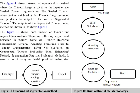
In this section a detailed architecture of the segmentation is presented. The proposed Tumour-Cut segmentation method consists of a few phases as shown in figure I. The figure I shows tumour cut segmentation method where the Tumour image is given as the input to the Seeded Tumour segmentation, The Seeded Tumour segmentation which takes the Tumour Image as input and produces the output in the form of Segmented Tumour7. The outputs of the Segmented Tumour under method are shown in the above figure I.
The figure II shows brief outline of tumour cut segmentation method. There are following steps: Seed Selection is marked based on Tumour Response Measurement Criteria, Adapting Transition Rule to Tumour Characteristics, Level Set Evolution on Constructed Tumour Probability Map, Enhancing/ Necrotic Segmentation Data and Evaluation Methods. It consists in choosing an initial pixel or region that
belongs to one object of interest, followed by an interactive process of neighbourhood analysis, deciding if whether each neighbouring pixel belongs or not to the same object. It consists in choosing an initial pixel or region that belongs to one object of interest, followed by an interactive process of neighbourhood analysis, deciding if whether each neighbouring pixel belongs or not to the same object3.
2.1 Cellular Automata In Image Segmentation
A cellular automaton is basically a computer algorithm that is discrete in space and time which operates on a lattice of cells. Since it was first proposed by Von Neumann and Ulam, Cellular Automata has attracted researchers from various fields in both physical and social sciences because of its simplicity, and potential in modelling complex systems. Each individual cell is in a specific state and changes synchronously depending on the states of some neighbours as determined by a local update rule8. They are parallel, local and homogeneous, since the state of any cell depends only on the states of the local neighbours at the previous time step and the update rules are same for every cell. Formally, a cellular automaton (CA) is a triple A=(S,N,£), Where S is a nonempty set, called the state set, N is the neighbourhood, and is the local transition function (rule) Sn , which is the argument of £: , indicates the states of the neighbourhood cells at a given time. While, which is its value, is the state of the central cell at the upcoming time step7. While this is usually defined as Cellular
User Input
Seeded Tumour–
cut Seg-mentation
Output
Automata is that in favour of a finite state set (discrete and bounded), continuous state sets in which the states are real numbers are also used in CA literature under the name “Coupled Map Lattices" or “Continuous CA” . The complete study in this and some of the issues that can arise while using a continuous state set on a finite machine8.The main advantage of using CA algorithm is its ability to obtain a multilabel solution in a simultaneous iteration. Another advantage is that the local transition rules are simple to explain, and it is possible to manipulate prior knowledge, specific to the problem, into the segmentation algorithm9.
The automata are initialized by assigning corresponding labels at seeds with a strength value between 0 and 1 where a higher value reflects a higher confidence in choosing the seed. 0’s are set to Strengths for unlabelled cells. where g is a pixel similarity function is bounded in the range [0, 1] depending on the image features i.e.
... (1) 2.2 Tumour-Cut Algorithm
Viewing an image as a weighted graph, this algorithm can be expressed by means of a common energy function on the differences between neighbouring nodes.
In the steps of the proposed cellular automata based tumour segmentation algorithm as follows. First, the user select a seed points over a MRI image tumour; one for foreground and one for background using this points, a VOI is selected; then tumour CA algorithm is run on the VOI for each two sets of seeds (for the foreground and background) to obtain strength maps for foreground and background at each voxel; two strength maps are combined together to obtain the tumour probability map i.e., PT and a level set surface is initialized at TT=0.5 and the map, TT is used to evolve the surface which converges to the final segmentation map. After the probability map is obtained and segmented the smoothing over a constructed tumour. Finally, the
necrotic regions of the tumour are segmented using a CA -based method with the chosen enhanced and necrotic seeds9. Steps which have been involved are as follows: Read the Tumour image. Determine the VOI, foreground seeds, background seeds. Determine the seed points of the Tumour drawn by the user. Run the CA algorithm twice, once for Tumour seeds and once for background seeds, to calculate the Tumour strengths (on the left) and the background strengths (on the right). Tumour probability map (PT) is obtained by combing strength maps are combined to obtain a PT=0.5 surface is the result of the CA algorithm without imposing smoothness (Figure 3a).A level set surface is initialized at PT=0.5 surface and evolved on the Tumour probability map to obtain the total tumour segmentation10.
2.3 Enhancing/Necrotic Segmentation
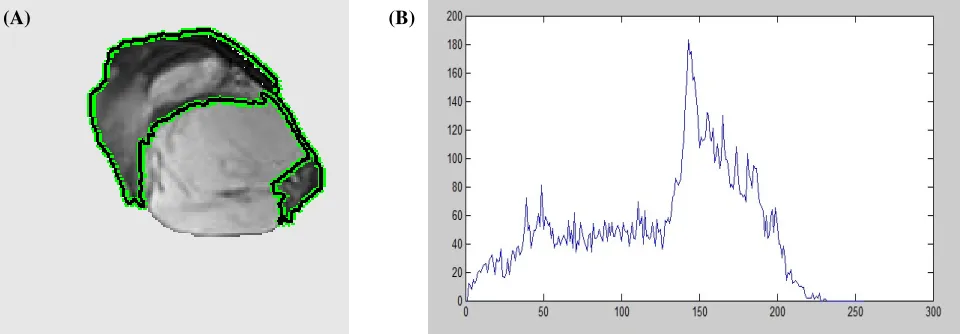
The necrotic region within the whole tumour is an important problem in assessment of the tumour progress. Delayed radiation necrosis, which typically occurs three months or more after treatment, is the primary risk associated with stereotactic radio surgery (Figure III).
equation (2)
Where H is the volume intensity histogram , and are rough volumes estimated with K-means clustering where K-means algorithm is one of the simplest non-supervised learning algorithms classes that solve the clustering segmentation problem. The method follows the usual steps to satisfy the primary objective that is clustering all the image objects into K distinct groups and and are the necrotic and enhanced thresholds respectively.
3. Results And Discussion
This chapter gives us detailed information of tumour images used in the experimental observations, number of
(A) (B)
the images used. The solid tumours with the minimal user interaction which is fast and robust practical tool for segmenting which will assist clinicians and researchers radiosurgery planning and assessment of the response to the therapy. Particularly, cellular automata (CA) based seeded tumour segmentation method on contrast enhanced T1 weighted magnetic resonance (MR) images, which standardizes the volume of interest (VOI) and seed selection, is suggested1. First, we establish the connection of the CA-based segmentation to the graph-theoretic methods to show that the iterative CA framework solves the shortest path problem (Table I) . In this consider we modify the state transition function of the CA to calculate the exact shortest path solution2. Furthermore, a sensitivity parameter is introduced to adapt to the heterogeneous tumour segmentation problem, and an indirect level set surface is evolved on a tumour probability map constructed from CA states to impose spatial smoothnes10. Sufficient knowledge to
initialize the algorithm is gathered from the user simply by a points drawn on the tumour, in points with the clinical practice. Moreover, an algorithm based on CA is presented to differentiate necrotic and enhancing tumour tissue content, gains importance for a detailed assessment of radiation therapy response. This demonstrate 90%– 99% Accuracy performance of the proposed algorithm with an emphasis on less sensitivity to seed initialization, healthiness with respect to different and heterogeneous tumour types, and its productivity in terms of computation time.
The active surface propagation over the constructed probability map aims at correction and improvement of the segmentation by smoothing out the tumour borders, and avoiding sharp protrusions, however, here due to the low intensity contrast between the tumour tissue and the gray matter, the Tumour-cut performed worse than expected. This low performance result obtained on the non-enhancing tumour reveals the limitation of the method by application to the tumours that are enhanced with the contrast agent (Table I, II). Tumour tissue depicts intensity levels close to those of the surroundings, and high, continuous gradients of the complex background near the right boundary leads to a spread-out probability map12,13.
We included all the results including low performance ones rather than reporting that the algorithm failed for some tumours and excluding them as outliers from the statistics. Without such “outlier” cases, the overall statistics would as expected indicate higher success rates.
Image Sl.
No. Recall Precision
F
Score Accuracy
IMAGE 1 1 0.95 0.9743 0.9948
IMAGE 2 1 0.9803 0.99 0.99
IMAGE 3 0.9898 1 0.9948 0.9949
IMAGE 4 0.99 1 0.995 0.995
IMAGE 5 1 0.99 0.995 0.995
Table I: Table of 100 images performance
Image
Count Recall Precision F Score Accuracy
100 0.9956 0.99385 0.99468 0.994868 Table II: Average data of 100 images performance
Tumour and necrotic seeds are chosen by applying a threshold to intensity histogram of the segmented region (Figure IV a & b).
4. Conclusion
In this work we present a segmentation algorithm for the problem of tumour representation which exhibit varying tissue characteristics. These can be widely used in clinical practices. However, the algorithm is dependent on the correctness of user marked labels.
We presented validation studies over 100 tumour images which are of highly heterogeneous collection. Precision and recall are the evaluation methods that’s has been used in evaluate our algorithm. Performance changes over varying initial seeds were reported as accuracy and F measure. This became important in calculating true robustness (strong and healthy) of the proposed algorithm in real application scenarios.
1In this work we were able to achieve accuracy of 99.53% average on 100 brain tumour images whereas previously it was reported that Menze et al.14 achieved 60% average on 25 glioma patients and Gooya et al.15 reported 74.5% average on 15 glioma patients with about 6–14 h of processing time is taken in grow cut segmentation, where as in tumour cut segmentation it is more applicable for medical image segmentation such as brain.
Conflict of interest
The author’s declares none.
References
1. Sinop A & Grady L. A seeded image segmentation framework unifying graph cuts and random walker which yields a new algorithm, ICCV, (2007) pp.1 - 8. 2. Popovici A. & Popovici D. Cellular automata in
image processing", Proceedings of 15th International Symposium on Mathematical Theory Networks and Systems (2002) 34 - 44.
3. Davatzikos C, Genc A, Xu D, & Resnick SM. Voxel-based morphometry using the RAVENS maps: methods and validation using simulated longitudinal atrophy. NeuroImage 14(2001) 1361–1369.
4. Liu J, Udupa JK, Odhner D, Hackney D, & Moonis G. A system for brain tumour volume estimation via
MR imaging and fuzzy connectedness. Computerized Medical Imaging and Graphics. 29 (2005) 21-34 5. Jolly MP & Boykov Y. Interactive graph cuts for
optimal boundary and region segmentation of objects in n-d images", Proc. ICCV, (2001)105 -112.
6. Vezhnevets V & Konouchine V. Growcut-interactive multi-label n-d image segmentation by cellular automata. In Proceedings of Graphicon (2005) 150-156.
7. Kari J. Theory of cellular automata: a survey, Theoretical Computer Science, 334 (2005) 3–33. 8. Armstrong CJ, Price BL, & Barrett WL. Interactive
segmentation of image volumes with live surface. Computers & Graphics. 31 (2007) 212–229.
9. Zou KH, Warfield SK, Bharatha A, Tempany CMC, Kaus MR, Haker SJ, Wells WM, Jolesz FA, & Kikinis R. Statistical validation of image segmentation quality based on a spatial overlap index. Academic Radiology. 11 (2004) 178–189.
10. Prastawa M, Bullitt E, Ho S & Gerig G. A brain tumour segmentation framework based on outlier detection. Medical Image Analysis, 8 (2004) 275-83. 11. Ho S, Bullitt E, & Gerig G. Level-set evolution with
region competition: Automatic 3-D segmentation of brain tumours. In Proceedings of International conference on Pattern Recognition, 1 (2002) 532-535.
12. Gonzalez RC, Woods RE & Eddins SL. Digital Image Processing Using MATLAB Published by Dorling Kindersley (India) Pvt. Ltd, (2009)
13. Bishop JM. The molecular biology of RNA tumour viruses: a physician's guide. The New England Journal of Medicine. 303 (1980) 675-682.
14. BH Menze, Leemput K Van , Lashkari D, Weber MA, Ayache N & Golland P. A generative model for brain tumor segmentation in multi-modal images. Medical Image Computing and Computer- Assisted Intervention. 13 (2010) 151-154.