Available online at www.JGTPS.com
Research Article
ISSN:2230-7346
Journal of Global Trends in Pharmaceutical Sciences
Vol.3, Issue 3, pp -763-771, July–September 2012
EXPRESSION AND PARTIAL PURIFICATION OF RECOMBINANT PDGF-BB EXPRESSED IN E.COLI
Polavarapu Srikanth1*, S.M.Jain1, R.C.Saxena1,Ramesh.B2
1P.G.Department of Zoology, S.S.L.Jain College, Barkatulla University, Bhopal. 2Department of Biotechnology, JNTUH, Hyderabad.
*Corresponding Author E-mail: [email protected]
ABSTRACT
Recombinant human PDGF is a 24.3 KDa disulfide linked homodimer of two B chains with 218 total amino acids is cloned into pET 23a vector and transformed into BL21 DE3 cells. The best expressing clone was selected; best suitable media was optimized for over expression of protein using different composition of media at 1 litre shake flask and fermentation process was optimized at 10 L scale. The culture was harvested and cells were lysed followed by centrifugation. The expressed protein was in the form of inclusion bodies which were solubilised and refolded. The purity of obtained refolded protein was about 80% and was identified by immunoblotting.
Key Words:Platelet Derived Growth Factor, expression, Fermentation, Inclusion bodies, wound healing.
INTRODUCTION:
Escherichia Coli is an important microorganism for the production of recombinant proteins used in many therapeutic applications like cancer, Diabetes, wound healing etc. E.coli has several advantages over other expression systems like yeast, mammalian cell culture like growth on inexpensive carbon source, rapid biomass accumulation and simple process for scale up for large scale production of high value recombinant therapeutic proteins. However, over expression of
conformation. The pET system is the most powerful for the cloning and expression of recombinant proteins in E.coli. The some of recombinant therapeutic proteins commercialized using E.coli are Insulin and its analogs, Granulocyte colony stimulating factor (GCSF), Interferon’s (IFNs), Streptokinase, Human Growth Hormone (HGH), Epidermal Growth Factor (EGF) and Platelet derived Growth Factor (PDGF) etc.
Platelet Derived Growth Factor (PDGF) is a 24.3 kDa disulfide linked homodimer of two B chains, 218 total amino acids (2) potent mitogen for a wide range of cell types including fibroblasts, smooth muscle and connective tissue. PDGF is an important mediator in wound healing. It plays a role in embryonic development, cell proliferation, cell migration and angiogenesis.
MATERIALS AND METHOD:
All the equipments and /instruments required for manufacturing or analysis are procured from reputed manufacturers. All the chemicals used were of analytical/reagent grade obtained from Merck or Qualigens. All the biochemicls, enzymes and other required were obtained from Sigma/Bio-Rad/Invitrogen/New England Biolabs / Novagen. Primers were procured from Bioserve Biotechnologies India Pvt Ltd.
Clone Construction and Expression of rhPDGF-BB:
Full length cDNA clone containing ORF of PDGF-BB was procured from RZPD, Germany. The PDGF-BB gene was amplified from cDNA by using specific primers: Forward primer: 5’-AGTTACCATATGAATCGCTGCTGG GC-3’; Reverse primer: 5’-GTATCACTCGAGTCACTAGGCTCC AAGGGTC-3’.
The amplified PDGF-BB gene and pET-23a vector (Novagen) were digested with NdeI and XhoI (New England Biolabs). The digested and gel purified PDGF-BB gene was cloned into digested pET-23a vector. PET23a-PDGF-BB clone was confirmed by colony PCR, restriction digestion and DNA sequencing. PET23a-PDGF-BB plasmid DNA was transformed into E.coli strain, BL-21(DE3)(3). For expression BL21 (DE3)/pET23a-PDGF-BB culture was grown in LB medium with ampicillin (100μg/ml) at 37°C, 180 rpm until OD reaches to 0.6 at 600 nm, the culture was induced with 1mM IPTG. Samples were collected before and after induction (~4 hours) for expression analysis, culture was centrifuged and the cell pellet was lysed using 1N sodium hydroxide. The sample were subjected to SDS-PAGE using 12% gel followed by coomassie staining and observe the bands and document using gel documentation system (Bio-Rad).
Shake Flask Studies:
Based on the SDS-PAGE results best expressing clone was selected and cell banks were prepared and stored at -80oC. One vial from cell bank was taken and grown in 10 mL of LB medium with100
μg/mL of ampicilin, different shake flask experiments were performed for the optimization of growth conditions different media compositions, different Inducers (Lactose/IPTG), different concentrations of IPTG (0.1, 0.5, 1, 1.5, 2mM).
Fermentation studies:
One vial of from cell bank was taken and
grown in LB medium with 100μg/ml of
ampicillin. Inoculum for fermenter was prepared in two stages. In stage one, 10 mL of grown culture was inoculated into 100 mL of the LB medium with
100μg/ml of ampicillin. The culture was
200 rpm for 12 hours at 37°C. In the second stage, the culture was transferred to 5 aliquots of 200 mL of TB medium (Difco) and grown at 37°C on orbital shaker at 200 rpm. This culture was used as the inoculums for 10L fermenter. The grown culture with OD600 of about
3.5-4.0 was inoculated into fermenter with 4.5 L of TB media.
Fermentation was carried out in fed-bath mode and was continued for 6 hours and feed was continued for another 10 hours, the culture was
induced with 1 mM isopropyl β-D-1 isopropyl thiogalactopyranoside (IPTG) and continued for 6 hours. Cell density was measured at 600nm with UV-visible spectrophotometer (Hitachi (U-2910). Glucose concentration (g/L) in the culture was determined using GOD-POD method. After completion of fermentation process culture was harvested.
Cell lysis & Recovery of Inclusion bodies:
The harvested culture was centrifuged by using high speed centrifuge (Kubota) at 4°C, 8000 rpm for 30 minutes. The supernatant was discarded and the cell pellet was resuspended with 10mM Tris, pH-8.0, 1mM EDTA and culture was lysed by passing through high pressure homogenizer (Niro Soavi) at 8000 bar by maintaining the culture temperature at 4oC. The resultant lysate was centrifuged at 8000 rpm, 2-8 degree centigrade for 30 minutes and the supernatant was discarded and the resultant pellet was collected and stored at -20oC until further process was carried out.
Separation of Soluble contaminants in IBs:
The inclusion bodies were isolated and washed with different wash buffers, different buffering conditions were optimized for separation of soluble contaminants by using detergents
(Triton-X100 (0.2,0.3,0.4 and 0.5%) and sodium deoxycholate (0.5,1.0.1.5 and 2%) at different incubation times
Solubilization and Refolding:
The inclusion bodies were solubilized with different concentrations of solubilzation buffer (6M guanidium hydrochloride or 8M urea, (10mM DTT and TE buffer) at 4°C for over night for refolding at different pH values, different protein concentrations (1 mg/mL,2 mg/mL and 3 mg/mL), different temperatures (4,15,20 and 25oC) and different concentrations of cysteine and cystine redox pairs (1 mM:2 mM, 1:5 mM) were used to optimized the refolding conditions.
ANALYTICAL METHODS: Electrophoresis:
PDGF gene amplification, cloning samples and restriction digestion, colony PCR samples were analyzed by horizontal electrophoresis and DNA samples were observed with ethidium bromide dye. Protein expression, purification and refolding process samples were analyzed by SDS-PAGE method with 12% gels, stained with coomassie stain.
Western Blotting:
The SDS-PAGE samples were electrophoretically transferred to 0.2µm PVDF membrane and western blotting was performed using primary and secondary antibodies developed with substrate against PDGF-BB.
RESULTS & DISCUSSION:
expressed was with molecular weight of about 24 kDa.
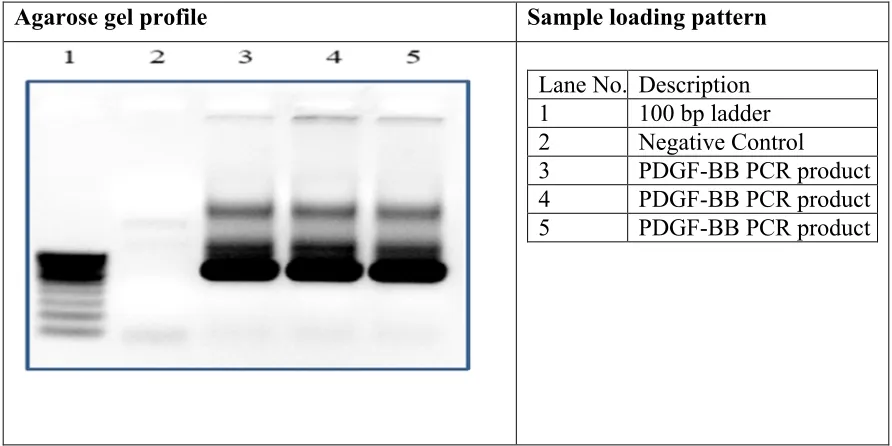
PCR reaction was performed and analyzed on 0.8% agarose gel. The gel image indicates that PDGF-B gene was
amplified and band was appeared between 600 & 700 bps which corresponds to the actual size of PDGF-BB gene. No amplification was seen in negative control.
Agarose gel profile Sample loading pattern
Lane No. Description 1 100 bp ladder 2 Negative Control
3 PDGF-BB PCR product
4 PDGF-BB PCR product
5 PDGF-BB PCR product
Figure 1:PDGF-B Gene Amplification. Colony PCR was performed for
transformants and analyzed for PCR products on 0.8% agarose gel. All the colonies showed PDGF-BB gene
amplification except negative control, which indicates that all colonies were positive clones.
Agarose gel profile Sample loading pattern
Lane No. Description 1 100 bp ladder 2 Negative Control
3 Colony-1
4 Colony-2
5 Colony-3
6 Colony-4
7 Colony-5
8 Colony-6
9 Colony-7
Restriction Digestion:
To conform the clone double digestion and single digestion was performed for Plasmid DNA using NdeI/XhoI and incubated at 37oC for 1 hour. After incubation DNA samples were loaded on 0.8% agarose gel and the
gel image indicated that in uncut sample open circular & super coiled DNA was observed. In NdeI/XhoI single digestion samples were linearized. The double digested DNA sample showed two bands, which indicates vector back bone and gene was released from the plasmid.
Agarose gel profile Sample loading pattern
Lane No. Description
1 1kb Ladder
2 Uncut
3 NdeI digestion
4 XhoI Digestion
5 NdeI+XhoI
Digestion
Figure 3:Analysis of Plasmid DNA Digestion samples by Agarose Gel Electrophoresis
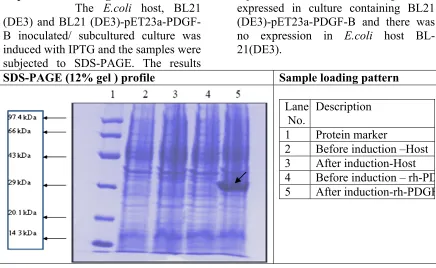
Expression Checking:
The E.coli host, BL21
(DE3) and BL21 (DE3)-pET23a-PDGF-B inoculated/ subcultured culture was induced with IPTG and the samples were subjected to SDS-PAGE. The results
represented that the PDGF-BB gene was expressed in culture containing BL21 (DE3)-pET23a-PDGF-B and there was no expression in E.coli host BL-21(DE3).
SDS-PAGE (12% gel ) profile Sample loading pattern
Lane No.
Description 1 Protein marker
2 Before induction –Host 3 After induction-Host
4 Before induction – rh-PDGF 5 After induction-rh-PDGF
Shake Flask Studies:
Shake flask studies were carried out using different media (LB and TB), , OD at 600 nm, growth curve was drawn pH and glucose concentrations were measured at different time intervals. From the results (Figure 5) it was concluded that, LB medium is not suitable for large scale fermentation due to inherent limitations in aiding growth of recombinant E.coli to high density to enable high level induction of a recombinant protein. Also, induction of recombinant protein in E.coli cultures grown in LB medium in shake-flask studies are best obtained when induced
at an OD600 value of 0.6-1.0 (Sambrook
and Russel, 2001), where as E.coli cultures grown in TB medium in shake-flask studies can be induced at much high cell densities to get good expression of recombinant protein.
It was observed that the pH in TB medium is maintained during the study (figure 5), where as pH in LB medium showed a drastic increase which indicates the depletion of energy source (glucose) which is not suitable for culture to be grown to high cell densities (Figure 5). Based on these results, TB medium is selected for further studies.
Growth pattern of PDGF-B OD and pH Values Time
Interval (h)
OD pH
LB TB LB TB
0 0.187 0.244 7.4 7.19
2 1.139 1.075 6.86 7.14
4 1.98 2.92 7.25 6.85
6 2.49 3.53 7.55 6.93
8 3.51 4.3 7.82 7.05
10 3.79 5.4 7.93 7.15
12 4.15 6.06 8.06 7.22
14 4.15 6.27 8.21 7.32
16 4.1 7.2 8.36 7.48
18 4.04 7.43 8.43 7.56
20 3.96 8.03 8.48 7.62
22 3.88 8.3 8.59 7.83
24 3.5 8.9 8.6 7.8
Fermentation studies:
Cell lysis:
The harvested culture was centrifuged at 8000 rpm, 2-8oC for 30 minutes, 1 mL of supernatant was collected and the remaining supernatant was discarded and the pellet was resuspended with lysis buffer and the culture was lysed using
high pressure homogenizer at 8000 bar and lysate is centrifuged. Soluble and insoluble fractions were analyzed by SDS-PAGE. The results revealed that the target protein was observed in insoluble fraction indicating that the protein will not be expressed in soluble form.
SDS-PAGE (12% gel ) profile Sample loading pattern
Lane No. Description
1 Protein Marker
2 Insluble fraction 3 Soluble fraction
Figure.6 -Probe Sonication sample analysis by SDS-PAGE
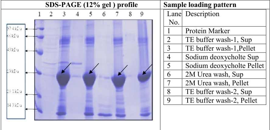
IBs washes:
The expressed PDGF-BB in the form of inclusion bodies were obtained from the harvested culture by lysing the cells using high pressure homogenizer.The collected inclusion bodies were washed with TE buffer, TE buffer with 1% triton, TE buffer with 0.02% sodium deoxycholate and 2M urea. The results indicated that the washing of inclusion bodies initially with TE Buffer, then TE Buffer with 0.02% sodium deoxycholate and then TE Buffer with 2M urea and consecutive washes is optimized for
washing IBs to facilitate the solubilization of IBs in the further steps.
Solubilisation and Refolding:
SDS-PAGE (12% gel ) profile Sample loading pattern
Lane
No. Description 1 Protein Marker
2 TE buffer wash-1, Sup 3 TE buffer wash-1,Pellet 4 Sodium deoxycholte Sup 5 Sodium deoxycholte Pellet 6 2M Urea wash, Sup
7 2M Urea wash, Pellet 8 TE buffer wash-2, Sup 9 TE buffer wash-2, Pellet
Figure.7 Inclusion bodies wash.
PROTEIN ANALYSIS AND CHARACTERIZATION:
Protein Estimation by Bradford’s method:
The solubilized protein concentration was estimated by Bradfords method (2). The results represented that the obtained concentration of solubilized PDGF-B protein is 8.52 mg/mL.
Western Blotting:
Refolded PDGF-B Protein samples were transferred from SDS-PAGE gel onto PVDF membrane and western blotting was proceeded as per the standard protocol using 10 antibodies raised against PGDF in rabbit and 20anti goat antibodies conjugated with HRP. The results after development highlighted the bands specific to PDGF-B and the protein is confirmed.
Figure 8: Immunoblot of PDGF
CONCLUSION:
Recombinant Platelet Derived Growth Factor producing clone was developed using established cloning and transformation techniques in E.coli cells. The shake flask, fermentation and partial purification process was optimized for
the production and purification of PDGF. The protein was characterized for its identification and protein concentration. The refolded protein was found to be greater than 85% pure and further purification process has to be optimized on order to get purity greater than 99% for clinical applications.
REFERENCES:
1. Ross R, Vogel A The platelet-derived growth factor. Cell (1978) 14:203–210
2. Rinas U, Bailey JE: Protein compositional analysis of inclusion bodies produced in E.coli. Appl Microbiol Biotechnol, (1992), 37:609-614. 3. Sambrook J., and Russell, D. W.
Molecular Cloning: A
Laboratory Manual. New York: Cold Spring Harbor Laboratory Press. (2001).
4. Carrio MM, Villaverde A: Protein aggregation as bacterial inclusion bodies is reversible. FEBS Lett (2001), 489:29-33. 5. Cejka J, Vodrazka Z, Salak J:
Carbamylation of globin in
electrophoresis and
chromatography in the presence of urea. Biochim Biophys Acra (1968), 154:589-591.
6. Raines EW, Ross R Purification of human platelet-derived growth factor. Methods Enzymol (1985) 109:749–773.
7. Rinas U, Bailey JE: Protein compositional analysis of inclusion bodies produced in E.coli. Appl Microbiol Biotechnol, (1992), 37:609-614. 8. Tsumoto K, Ejima D, Kumagai I,
Arakawa T: Practical considerations in refolding proteins from inclusion bodies. Protein Express purify (2003),28:1-8.