Dr. Bodhisattwa Chakraborty Department of Pharmacology,
Gupta College of Technological Sciences, Asansol, West Bengal, India.
E.mail: [email protected] Address for correspondence
Access this article online www.japer.in
Anti-inflammatory and Anti-asthmatic activities of
Alangium
lamarkii
(Alangiaceae)
INTRODUCTION
Inflammation in the body response to noxious or injurious stimuli, characterized by warmth, redness of the skin, pain, swelling and loss of function. Inflammation is a part of host defense mechanism. There are several tissue factors that are known to be involved in the inflammatoryreactions such as release of histamines, bradykinin and prostaglandins. [1, 2] Asthma is a chronic inflammatory disease of the respiratory tract that is characterized by increased airway hyper-responsiveness and mucus production that leads to episodes of wheezing, coughing and shortness of breath. Asthma is common among the individuals (up to 10% in adults and 35% in children), and as a result, large quantities of asthma medications are needed to stop the asthma prevalence increase [3]. Since generations, in India people are using the
extracts and leachates of different herbs in order to stimulate and promote the growth of specific herbs. The example of Alangium lamarkii is one of them. Traditionally, the leaves are useful in treatment of inflammations, blood disorders, burning sensation,spermatorrhoea, gleet, acute fever and lumbago [4, 5]. In case of intense pain due to gout, the patients are advised by the healers to apply the Ankol leaves in affected parts. The leaves are also used in treatment of asthma. The leaves are dried and put on fire. The patients are advised to inhale the fumes [6, 7] However this plant has not been studied for anti-inflammatory and anti-asthmatic activity. Based on this an attempt has been made to evaluate the inflammatory and anti-asthmatic potency of Alangium lamarkii with their phytoconstituents.
MATERIALS AND METHODS
Plant material
The leaves of Alangium lamarkii are collected form Asansol, West Bengal, India. A herbarium sheet was prepared and it was identified and authenticated (CNH/35/2011/TECH II/446) by the Botanical Survey To evaluate the anti-inflammatory and anti-asthmatic property with their phytoconstituents of the methanolic extracts of Alangium lamarkii leaves.
Alangium lamarkii (Family-Alangiaceae) commonly known as Ankora is found in western Africa, Madagascar, southern and eastern Asia. It is mainly used traditionally for inflammations, blood disorders, burning sensation, spermatorrhoea, gleet, acute fever and lumbago.
In the present study, methanolic extract of Alangium lamarkii was evaluated for anti-inflammatory and anti-asthmatic property with their phytoconstituents using carrageenan induced rat paw oedema and histamine induced bronchoconstriction respectively. Student’s t-test and Dennett’s test were used for statistical analysis.
The result showed the presence of alkaloid and steroids in the leaves and significant anti-inflammatory and anti-asthmatic property of methanolic extract of Alangium lamarkii. The methanolic extract of Alangium lamarkii leavesshowed potent anti-inflammatory and anti-asthmatic activity in comparison with the standard drugs diclofenac sodium and Pheniramine Maleate respectively, possibly due to may be presence of alkaloids and steroids in the leaves.
Keywords: Alangium lamarkii (Alangiaceae), Anti-inflammatory, Anti-asthmatic activity, Phytochemical study, Oedema, Histamine, Bronchoconstriction.
ABSTRACT ABSTRACT ABSTRACT ABSTRACT
Bodhisattwa Chakraborty*1, Uttam
Kumar Bhattacharyya2, Rana
Datta1, Manik Boral3, Bibek Laha4, ,
Subhangkar Nandy1
1Department of Pharmacology, Gupta
College of Technological Sciences, Asansol,West Bengal, India
2Department of Pharmaceutical
Chemistry, Gupta College of Technological Sciences, Asansol, West Bengal, India.
3Department of Pharmacognosy,
Gupta College of Technological Sciences, Asansol, West Bengal, India
4Department of Pharmaceutics, Gupta
College of Technological Sciences, Asansol,West Bengal, India
of India, Howrah, West Bengal, India. The leaves were dried in shade to avoid too many chemical changes occurring and made into a coarse powder. Methanol was used as solvent for extraction and extraction was performed in Soxhlet Apparatus.
Fig.1: Picture showing the Alangium lamarkii leaves
Preparation of extract
The air dried crushed leaves (1000g) were soaked for 12 hr in Methanol (3L) at room temperature. The residue was extracted with hot Methanol under reflux 3 times (each 1500 ml) after vacuum filtration. All solvent was evaporated under vacuum and extract was then lyophilized, to yield approximately 12% w/w/) of the residue, which was stored at 20ºC until use.
Experimental animals
Healthy male and female rats (Wistar albino) and Guinea pigs of 4-8 weeks old were selected after physical and behavioral veterinary examination from Institutional Animal House of Gupta College of Technological Sciences. The weight range was fall within ± 20% of the mean body for each sex at the time of initiation of treatment. All experiments involving animals complies with the ethical standards of animal handling and approved by Institutional Animal ethics committee (955/A/06/CPCSEA). Sixty young adult male Wistar rats (120–150 g) and Guinea pigs (350 to 400 g), were obtained from the Institutional Animal House of Gupta College of Technological Sciences. The rats were housed in polyethylene cages in the Animal House. The rats were housed in polyethylene cages, allowed one week of
acclimatization, and maintained on standard rat chow and standard laboratory conditions throughout the experiment.
Phytochemical Screening
The concentrated extracts were used for preliminary screening of various phytoconstituents viz. carbohydrate, amino acid, alkaloids, tannins and flavonoids were detected by usual methods prescribed in standard tests.The various physicochemical parameters such as total ash, acid insoluble ash, water soluble ash, water soluble extractive value and alcohol soluble extractive value were determined by the method reported by Sailor et al. 10 with slight modification. [8]
Acute toxicity test
Acute toxicity study was performed as per OECD guidelines 423. (Acute toxicity class method). [9]
Anti-inflammatory activity study
Carrageenan – induced rat paw edema
This is the simplest and most widely used model for studying the anti inflammatory activity of new compounds. The development of edema after a sub plantar injection of carrageenan in the animal is attributed to the release of histamine, serotonin, kinins and prostaglandins. [10]
The animals were weighed and numbered. The right hind paw just beyond tibio tarsal junction was marked, so that every time the paw was dipped into water column upto the fixed mark to ensure constant paw volume. The initial paw volume of each rat was measured by water displacement method. The animals were divided into five groups each comprising of six rats.
Group I: Control animals were received normal saline solution at the dose of 10ml/kg.
Group II: Animals received standard Diclofenac sodium at the dose of 10mg/kg body weight.
Group III: Animals received methanolic extract at the dose of 100mg/kg body weight.
Group V: Animals received methanolic extract at the dose of 400mg/kg body weight.
All groups received intra peritoneal injection. After 30 minutes 0.1 ml of 1% (w/v) caregeenan was injected in the plantar region of the right paw of each rat. The paw volume was measured after 30, 60, 120 and 180 minutes of administration of carrageenan. Compare the mean percentage change in paw volume in control, extract, and diclofenac treated animals and expressed as percent oedema inhibition.
Anti-asthmatic activity study
Histamine induced Bronchoconstriction
Twenty four guinea pigs of either sex (150-350) were divided into four groups containing six in each. To screen the sensitivity of guinea pigs were placed in a Plexiglas’s chamber (Histamine chamber) and sprayed with 0.1% histamine under the average pressure of 180 ± 30mmHg. The time to onset of respiratory distress (preconvulsive time) during the challenge with these agent was measured. The guinea pigs were randomly allotted to different groups with 6 per each group. The negative control received distilled water orally, and the positive control animals received Pheniramine Maleate by intra peritoneal, the other two groups were treated with methanol extract of
Alangium lamarkii (200, 400 mg/kg BW) doses respectively. All groups treated with a single dose daily for five days prior to the challenge, the last dose given 2 hrs before the challenge. The methods of challenge were the same as those of screening the sensitive guinea pigs. The delitescence of convulsion for each guinea pig and tumble numbers for each group during challenge within exposure period were recorded. Protection from anoxic convulsion was calculated as (1 – T1/T2) X 100 where T1 is the mean of pre-convulsion time before treatment and T2 is the mean of pre-convulsion time 5 days after treatment and % protection was expressed relative to control. [11]
Guinea pig Tracheal chain preparation
Guinea pigs of either sex (200-250g) were divided into 3groups. Each group contains 6 animals and is allowed to starve overnight and free access to water.
The animals were killed by blow on the head and exsanguinations. The isolated trachea was mounted in a 30ml organ bath containing Tyrode solution, maintained at 37± 1⁰C and gassed with air. The tissue was equilibrated for 45min during which the bath solution is replaced every 10min. At the end of the equilibrium period, histamine (0.5μg/ml) induced contraction as well as effect of extract (upto 800μg/ml) was recorded. A drug tissue contact time of 1min was maintained. The percent response of each group was calculated from the height of the peak obtained.
Statistical analysis
All the values ware statistically analyzed by one-way analysis of variance (ANOVA) followed by multiple comparison test. Comparison between control and drug treated groups were considered to be significant p<0.01, p<0.001. All values are expressed as mean ± SEM.
RESULTS AND DISCUSION
Phytochemical studies
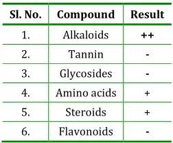
From the Phytochemical study, it has evaluated the presence of alkaloid, amino acid and steroid in leaves.
Table 1: Preliminary phytochemical screening of the various extracts of the leaves of Alangium lamarkii.
Sl. No. Compound Result
1. Alkaloids ++
2. Tannin -
3. Glycosides -
4. Amino acids +
5. Steroids +
6. Flavonoids -
++ means strongly present, + means present and – means absent
amounts of water soluble compounds than alcohol soluble compounds.
Table 2: Physico-chemical characters of the leaf powder of Alangium lamarkii.
S.
No. Parameter
Values (%)
1. Total Ash 20%
2. Acid insoluble ash 30%
3. Water Soluble Ash 10%
4. Determination of Water Soluble
Extractives 1.6%
5. Determination of Alcohol Soluble
Extractives 1.2%
Acute toxicity studies
The extracts of Alangium lamarkii did not show any sign of toxicity up to 2000 mg/kg body weight and hence it was considered to be safe.
Anti-inflammatory study
Methanolic Extract of Alangium lamarkii was evaluated for Anti-inflammatory activity. In Carrageenan induced paw oedema, the intraperitoneally administration of leaves extract produced a significant anti-inflammatory activity in a dose-dependent manner respectively (100, 200 and 400mg/kg body wt) in the rats. In 400mg/kg dose was showed highest anti-inflammatory potential comparing with the standard anti-inflammatory agent.
Table 3: Effect of Methanolic extract of Alangium lamarkii on carrageenan induced inflammation
Sl. No Dose Initial volume 30 min 60min 120min 180min
1. Control 0.6±0.02 0.8±0.01 0.94±0.02 1.2±0.05 1.32±0.07
2.
Standard Diclogenac Sodium
(10mg/kg b.w)
0.48±0.04 0.56±0.02a* (30%)
0.65±0.01a*
(30.85%)
0.58±0.02a*
(51.6%)
0.56±0.03a*
(57.5%)
3. Extract
(100mg/kg b.w) 0.50±0.03
0.58±0.02a*
(27.5%)
0.64±0.05a*
(31.9%)
0.83±0.03a*
(30.8%)
0.97±0.08a*
(26.51)
4. Extract
(200mg/kg b.w) 0.40±0.05
0.52±0.03a*
(35%)
0.60±0.02a*
(36%)
0.80±0.04a*
(33%)
0.67±0.03a*
(49%)
5. Extract
(400mg/kg b.w) 0.45±0.03
0.50±0.05a*
(37.5%)
0.63±0.03a*
(32.9%)
0.75±0.02a*
(37.5%)
0.60±0.03a*
(54%)
(The data are expressed as mean ± S.E.M. Significant differences in each group versus the control were as follows: * P < 0.05. ** P < 0.01).
Fig. 1: Graph showing anti-inflamatory activity at various doses with respect to time interval
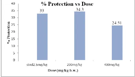
Anti-asthmatic study
The time of occurrence of PCD was significantly increased, suggestive of bronchodilating activity
significantly (P<0.001) after administration of the extract at doses of 200 and 400 mg/kg b.w. p.o.
Table 4: Effect of methanol extract of Alangium lamarkii on guinea pigs bronchoconstriction induced by Histamine
Group Dose (mg/kg)
Preconvulsive at 0 day (sec)
Preconvulsive time at 5th day
(secs)
% protection (1-T1/T2)X100
Control as
Distilled water 5ml/kg 93.0±0.5 90.0± 0.57 -
Standard (Pheniramine
Maleate ) 2.5 92.5±0.3 137.0± 0.61** 33
Extract 200 78.5±0.4* 104± 0.58** 24.51
400 90.5±0.5** 133.0± 0.44** 32.71
(Values are presented as means ± S.E.M, n = 6, ** P < 0.01 and * P < 0.05statistical significance against control. statistical test done by t-test)
The isolated guinea pig tracheal chain preparation showed dose dependent significant (P<0.001) inhibition of the contraction of the tracheal muscles induced by histamine as compared to control group.
Table 5: Percent inhibition of Alangium lamarkii of histamine induced contraction on isolated guinea pig
trachea
Treatment Conc.(μg/ml) Peak height
% inhibition
Histamine 0.5 3.21±0.11 -
Histamine+
MEAL 500 2.66±0.08** 17.134
Histamine+
MEAL 800 2.20±0.03** 31.4642
(MEAL=methanolic extract of Alangium lamarkii,
value are expressed as mean ±SEM, n=6, **P˂0.001 as compared to positive control (histamine induced
group).
Fig. 2: Graph showing anti asthmatic activity at different doses
DISCUSSION
Inflammation is a common phenomenon and it is a reaction of living tissues towards injury. An inflammatory response has been associated with various manifestations such as elevated body temperature and pain [12, 13, 14 and 15]. Preliminary pharmacological screening experiments were
conducted with crude Alangium lamarkii leaf extracts found to exhibit significant anti inflammatory activity whose effect is comparable to that of standard drug-Diclofenac reported in this study. Extract caused significant (P < 0.001) reduction in paw edema from the second hour at the 200mg/kg and 400mg/kg dose level, whereas significant (P< 0.001) reduction in paw edema was not observed from the second hour at the 100mg/kg dose level.
plays a major role in the development of airway inflammation and in the accompanying bronchial hyper reactivity [18, 19]. Neutrophils and monocytes play a pivotal role in the disease process as they are a source of variety of inflammatory mediators which are responsible for bronchial hyper responsiveness and airway inflammation [20, 21, 22 and 23].
The relaxant effects of MEAL on tracheal chains of guinea pigs might be produced by different chain mechanisms including stimulation of β-adrenergic receptors, inhibition of histamine H1receptors or an anticholinergic property of this plant. The relaxant effect of all concentrations of the extract of MEAL obtained where significantly lower than control group. These finding suggest probable β-adrenergic stimulatory, muscurinic or histaminic H1blocking properties of the plant extract.
Phytochemical screening showed presence of alkaloid, amino acid and steroid etc. Anti-inflammatory and anti-asthmatic activity may be due to presence of above constituents.
Our data suggest that the methanolic extract of the leaves of A. lamarkii possesses significant anti-inflammatory and anti-asthmatic activity. Further studies are needed to establish molecular mechanism and to isolate and characterise the active principles which are responsible for inflammatory and anti-asthmatic property.
Conflict of interest:
There is no conflict of interest associated with the authors of this paper.
ACKNOWLEDGEMENTS
The authors are thankful to Prof. Debesh Chandra Mazumder (Chairman, Trinity Trust, Asansol, W.B., India), Dr. Kalyan Kumar Sen (Principal, Gupta College of Technological Sciences, Asansol, W.B., India) and Dr. Somashree Roy (Professor in Pharmaceutics, Gupta College of Technological Sciences, Asansol, W.B., India).
REFERENCES
1. Taranalli AD, Thimmaiah NV, Srinivas S, Saravanan
E., Anti inflammatory, analgesic and anti ulcer
activity of certain thiazolidinones. Asian Journal of
Pharmaceutical and Clinical Research, 2009; 2: 79-83.
2. Georgewill OA, Georgewill UO, Nwankwoala RNP.,
Antiinflammatory effects of Morninga oleifera lam
extract in rats. Asian Pac J Trop Med, 2010; 3: 133-35.
3. Annesi-Maesano, Epidemiologie de l’asthme. Revue
du Praticien, 2005; 55: 1295–98.
4. Ghosh MN., Fundamentals of Experimental
Pharmacology. Hilton & Company, Kolkata, 2005.
5. Mukherjee P., Quality Control of Herbal Drugs.
Bussiness Horizons Pharmaceutical Publishers, New
Delhi, 2007
6. Pankaj Oudhia. Botanical.com Research Note-2001,
2002, 2003.
7. Itoh A, Ikuta Y, Tanahashi T and Nagakura N., Title. J
Nat Prod 2000; 63: 723-5.
8. Sailor GU, Ghanshyam P, Ashvin VD, Seth NR, Seth
AK., Pharmacognostical and Preliminary
Phytochemical Investigation of Leucas cephalotes
(Roth) Spreng. International Journal of
Pharmaceutical Research, 2010; 2: 14-21.
9. Mohamed Saleem TK, Azeem AK, Dilip C, Sankar C,
Prasanth NV, Duraisami R., Anti-inflammatory
activity of the leaf extacts of Gendarussa vulgaris
Nees. Asian Pacific Journal of Tropical Biomedicine,
2011; 1: 147-49.
10. Kannan K, Ortmann RA, Kimpel D., Animal models of
rheumatoid arthritis and their relevance to human
disease. Pathophysiology, 2005; 12: 167-81.
11. Hazekamp A, Verpoorte R, Panthong A., Isolation of a
bronchodilator flavonoid from the Thai medicinal
plant. Clerodendrum petasites. Journal of
ethnopharmacology,2001; 78: 45-49.
12. Sheetal SC, Sanjay RC, Machindra JC., Analgesic,
anti-inflammatory and anti-arthritic activity of Cassia
uniflora Mill. Asian Pacific Journal of Tropical
Biomedicine, 2012; 2: 181-86.
13. Stanley PL, Steiner S, Havens M., Tramposch KM.
Mouse skin inflammation induced by multiple topical
applications of 12-O
-tetradecanoylphorbol-13-acetate. Skin Pharmacol, 1991; 4: 262–71.
14. David Osvaldo SS, Maribel HR, Salud P, Enrique JF,
Hautriwaic Acid Isolated from Dodonaea viscosa
Leaves. Molecules 2012; 17: 4292-4299..
15. Jude EO, Anwanga EU, Samuel GF, Louis UA.,
Anti-inflammatory and analgesic activities of Melanthera
scandens. Asian Pacific Journal of Tropical
Biomedicine, 2012; 1: 144-148.
16. Masaru N, Masaki F, Yoshitaka O, Shinji N., Effect of
montelukast in a guinea pig model of cough variant
asthma. Pulmonary Pharmacology & Therapeutics,
2007; 1: 101-6.
17. Church MK, Bradding P, Walls AF. Allergy and
allergic diseases-Human mast cells and basophils.
Oxford, 1997.
18. Azzwi M, Bradley B, Jeffery PK, Frew AJ, Wardlaw AJ,
Knowles G., Identification of activated T lymphocytes
and eosinophils in bronchial biopsies in stable atopic
asthmatics. Am Rev Respir Dis, 1990; 142:
1407-1413.
19. Barnes PJ, Chung KF, Page CP., Inflammatory
mediators of asthma: An update. Pharmacol Rev,
1998; 50: 515-596.
20. Corriagan CJ, Kay AB., T cells and eosinophils in
pathogenesis of asthma. Immunol Today, 1992; 13:
501-507.
21. Dnyaneshwar JT, RavindraYP., Antiasthmatic activity
of Ricinus communis L. roots. Asian Pacific Journal of
Tropical Biomedicine 2011; 1: 13-16.
22. Chhaya R, Dutt KR, Shobharam S, Lokesh D.,
Antiasthmatic activity of the methanolic extract of
Physalis angulata Linn. Journal of Medicinal Plants
Research, 2011; 5: 5351-55.
23. Raju.D, Chitra.V, Hari DK, Silambu JP, Shankari M.,
Materials and methods Plant material. Evaluation of
Anti-asthmatic Activity of Aqueous Extract of
Achillea mellifolium Linn Flowers. Archives of
Applied Science Research, 2009; 1: 287-93.
How to cite this article: Bodhisattwa Chakraborty*1, Uttam
Kumar Bhattacharyya2, Rana Datta1 , Manik Boral3, Bibek
Laha4, Subhangkar Nandy1; inflammatory and
Anti-asthmatic activities of Alangium lamarkii (Alangiaceae); J. Adv. Pharm. Edu. & Res. 2013: 3(4): 524-530.