A
LICJAK
ĘDZIA1, M
ARCINZ
IAJKIEWICZ1, A
NNAS
EREDYN1, K
RZYSZTOFD
UDEK2Computer Morphometric Analysis of the Palmaris
Longus Muscle in the Fetal Period
Komputerowa analiza mięśnia dłoniowego długiego
w okresie prenatalnym w aspekcie klinicznym
1Normal Anatomy Department, Wroclaw Medical University, Poland
2Institute of Machines Design and Operation, Wroclaw University of Technology, Poland Adv Clin Exp Med 2009, 18, 5, 437–447
ISSN 1230−025X
ORIGINAL PAPERS
© Copyright by Wroclaw Medical University
Abstract
Objectives. The goal of this study was to assess the palmaris longus muscle (musculus palmaris longus) in the fetal period.
Material and Methods.The material consisted of 170 upper extremities originating from 85, 4− to 7−month−old fetuses (26 females) with crown−rump lengths of 95–220 mm. The methods applied were anthropological, prepa− rational, computer measurements with a Scion for Windows system, IrfanView, and statistical methods. Image acquisition was performed with a high−resolution camera.
Results.Symmetry, sexual dimorphism, and growth in particular weeks were examined. The fetal somatic para− meters v−pl, v−tub, body mass, forearm length, length and width of the palmaris longus belly and tendon, and the height of palmar aponeurosis divergence in relation to the flexor retinaculum of the hand were measured (850 mea− surements in all). Statistically significant asymmetry of the palmaris longus muscle was observed. The palmaris longus muscle of the right hand was 1 mm longer although both extremities had the same length.
Conclusions.However, no significant difference was found in palmaris longus muscle length between male and female fetuses (Adv Clin Exp Med 2009, 18, 5, 437–447).
Key words: palmaris longus muscle, growth, fetal period.
Streszczenie
Cel pracy.Ocena morfologii mięśnia dłoniowego długiego w okresie płodowym.
Materiał i metody.Materiał stanowiło 170 kończyn górnych 85 płodów (w tym 26 żeńskich) o długości ciała
v−tub95–220 mm, tj. w wieku 4–7 miesiąca życia płodowego. Zastosowane metody badań to: antropologiczna, preparacyjna, komputerowe pomiary w systemie Scion for Windows, Irfan−View oraz metody statystyczne z wy− korzystaniem pakietu Statistica (testy normalności Shapiro−Wilka i Kołmogorowa−Smirnowa, test U Manna− −Whitneya, test Wilcoxona). Akwizycja obrazu odbywała się za pośrednictwem aparatu fotograficznego o wyso− kiej rozdzielczości.
Wyniki.Analizowano symetrię, dymorfizm płciowy i przyrosty w poszczególnych tygodniach. Wykonano pomia− ry: somatyczne płodów: v−pl, v−tub, masy ciała, długości przedramienia, długości i szerokości brzuśca i ścięgna mu− sculus palmaris longus, wysokości odejścia aponeurosis palmarisw odniesieniu do troczka zginaczy (w sumie 850 pomiarów). Zaobserwowano statystycznie istotną asymetrię długości mięśnia dłoniowego długiego. Musculus palmaris longusręki prawej jest dłuższy o ok. 1 mm, mimo jednakowej długości obu kończyn.
Wnioski.Nie stwierdzono natomiast istotnej różnicy między długością mięśnia dłoniowego długiego płodów żeń− skich i męskich (Adv Clin Exp Med 2009, 18, 5, 437–447).
Słowa kluczowe: mięsień dłoniowy długi, wzrastanie, okres prenatalny.
The palmaris longus muscle (musculus pal−
maris longus) is a superficial forearm muscle char− acteristic for its morphological variability. The available literature provides reports concerning
[9–11]. Abnormal morphology of the palmaris longus muscle plays an important role in damage to the median nerve [12, 13], which ensures hand prehensility. Only recently is the palmaris longus used in reparatory procedures as a donor for flex− or long muscles of the thumb and fingers, in facial nerve surgery after paralysis and in ble− pharospasm, eyelid ptosis, lip and cheek recon− struction [14], ophthalmology [15], and otolaryn− gology [3, 16, 17]. Hand surgery should be pro− ceeded by MRI [18] and USG [8] examinations. Available literature does not provide any reports on analysis of this muscle in the fetal period. Observed lesions are of congenital character. According to Stecco [19] and Caughall [20], the palmar aponeurosis is an individual anatomical unit. Moss [1] states that palmaris longus absence is not connected with a lack of the plantaris mus− cle, which is also a donor in surgery. Available lit− erature contains only one paper with results based on histological examinations carried out on 33 fe− tuses [20] aged 5–40 weeks. Although some of the fetuses lacked the palmaris longus tendon, the pal− mar aponeurosis was always present.
Material and Methods
The material consisted of 170 upper extremities originating from 85 4− to 7−month−old fetuses (26 fe− males) with crown−rump lengths of 95–220 mm. The methods applied in the study were anthropological, preparational, computer measurements with a Scion for Windows system, IrfanView, as well as statistical methods using the Statistica package (Shapiro− −Wilk’s test, Kołmogorov−Smirnov’s test, Mann− −Whitney’sUtest, Wilcoxon’s test). Image acquisi− tion was performed with a high−resolution camera.
Results
The basic statistics of the somatic features are presented in Table 1. The following measurements were performed: forearm length, belly length and width, palmaris longus tendon length and width, as well as palmar aponeurosis divergence height in relation to the flexor retinaculum (Fig. 1). The results are collected in Tables 2–5. Symmetry and sexual dimorphism as well as their weekly increases were analyzed. The left and right sides were compared and the palmaris longus muscle length was measured.
Statistically significant asymmetry of the pal− maris longus muscle was observed. The right pal−
Table 1.Descriptive statistics (mean ± standard devia−
tion) of fetal somatic features
Tabela 1.Statystyki opisowe (średnia ± odchylenie stan−
dardowe) cech somatycznych płodów
Parameter Month Sex (N) x±SD (Parametr) (Miesiąc) (Płeć)
Body length IV F (2) 98.0 ±9.9 v−tub – mm M (4) 104.5 ±3.3
(Długość Σ(6) 102.3 ±±6.1
ciała v−tub V F (12) 146.8 ±16.0
– mm M (19) 139.1 ±13.7
Σ(31) 142.1 ±±14.9
VI F (15) 185.3 ±9.5 M (13) 176.6 ±14.2
Σ(28) 181.3 ±±12.5
VII F (3) 220.0 ±10.0 M (19) 205.3 ±18.9
Σ(22) 207.3 ±±18.5
Body length IV F (2) 118.0 ±42.4 v−tub– mm M (4) 145.0 ±10.0 (Długość ciała Σ(6) 136.0 ±±24.8
v−pl– mm) V F (12) 210.0 ±43.7 M (19) 202.9 ±21.3
Σ(31) 205.8 ±±31.4
VI F (15) 266.7 ±15.0 M (13) 252.5 ±21.9
Σ(28) 260.1 ±±19.6
VII F (3) 320.0 ±13.2 M (19) 300.1 ±29.8
Σ(22) 302.8 ±±28.8
Body mass – g IV F (2) 51.5 ±16.3 (Masa ciała – g) M (4) 58.2 ±12.6
Σ(6) 56.0 ±±12.7
V F (12) 202.3 ±85.1 M (19) 177.1 ±58.5
Σ(31) 186.8 ±±69.8
VI F (15) 426.8 ±76.6 M (13) 341.8 ±143.6
Σ(28) 387.3 ±±118.6
VII F (3) 658.7 ±86.6 M (19) 569.8 ±144.5
Σ(22) 581.9 ±±139.9
maris longus muscle was 1 mm longer in spite of both extremities having the same length (Fig. 2). However, no significant difference was found between male and female fetuses in palmaris longus muscle length (Fig. 4).
belly lengths and widths (Table 4) in all the age groups were similar on the left and right side and in male and female fetuses (Figs. 7, 8).
All analyzed sizes of the tendon, belly, and aponeurosis correlated significantly with fetal age (p < 0.05) and their values can be calculated on the basis of the linear regression models presented in the diagrams (Fig. 11). Tendon length (1.34 mm/ /week) and belly length (1.06 mm/week) revealed the most intensive growth and tendon width increased most slowly (0.04 mm/week). The ten− don and aponeurosis length/width indexes in the analyzed period remained stable and amounted to
3.0 and 55.5%, respectively. However, poor posi− tive correlation was observed between age and the belly length/width index. This index’s weekly growth amounted to 0.3%.
On the basis of the test, linear growth of the palmaris longus muscle was observed as well as its asymmetry and absence of sexual dimorphism. The length of the muscle with aponeurosis seems to increase most intensively (3.05 mm/week). Anomalies in the palmaris longus muscle were observed and their variety can be both quantitative and qualitative. Quantitative variation included muscle duplication in 2 cases and muscle lack in
Table 2.Descriptive statistics (mean ± standard deviation) of palmar muscle length in the periods of fetal life
Tabela 2.Statystyki opisowe (średnia ± odchylenie standardowe) długości mięśnia dłoniowego długiego w analizowanych
okresach życia płodowego
Parameter Month Sex (N) Left side Right side Left vs. right side
(Parametr) (Miesiąc) (Płeć) (Strona lewa) (Strona prawa) (Strona lewa vsprawa)
Forearm length – mm IV F (2) 22.1 ±2.8 22.2 ±2.5 p= 0.676
(Długość przedramienia M (4) 20.2 ±2.4 20.2 ±2.4
– mm) Σ(6) 20.8 ±±2.4 20.8 ±±2.4
V F (12) 31.7 ±4.8 32.5 ±4.7 p= 0.047
M (19) 32.3 ±4.5 32.8 ±4.9
Σ(31) 32.1 ±±4.6 32.7 ±±4.7
VI F (15) 41.7 ±6.0 40.7 ±5.8 p= 0.119
M (13) 38.8 ±6.0 38.9 ±6.0
Σ(28) 40.4 ±±6.1 39.9 ±±5.9
VII F (3) 45.1 ±1.0 48.7 ±5.7 p= 0.891
M (19) 46.7 ±6.2 46.3 ±6.3
Σ(22) 46.5 ±±5.8 46.6 ±±6.2
Muscle length without IV F (2) 20.1 ±3.1 20.6 ±4.0 p= 0.922
aponeurosis – mm) M (4) 20.3 ±3.6 20.0 ±3.5
(Długość mięśnia bez Σ(6) 20.2 ±±3.1 20.2 ±±3.2
rozcięgna – mm) V F (12) 30.3 ±4.6 29.6 ±4.6 p= 0.401
M (19) 29.0 ±4.3 28.7 ±4.8
Σ(31) 29.5 ±±4.4 29.1 ±±4.7
VI F (15) 39.9 ±6.3 39.5 ±6.5 p= 0.452
M (13) 37.4 ±7.1 37.2 ±7.3
Σ(28) 38.8 ±±6.7 38.5 ±±6.9
VII F (3) 46.4 ±3.2 42.9 ±2.8 p= 0.077
M (19) 45.3 ±6.6 44.0 ±6.6
Σ(22) 45.5 ±±6.2 43.9 ±±6.2
Muscle length with IV F (2) 26.3 ±3.1 26.9 ±3.9 p= 0.567
aponeurosis – mm M (4) 26.1 ±3.7 25.9 ±3.5
(Długość mięśnia Σ(6) 26.2 ±±3.2 26.3 ±±3.2
z rozcięgnem – mm) V F (12) 39.3 ±4.9 38.8 ±4.7 p= 0.476
M (19) 39.4 ±5.5 38.4 ±5.6
Σ(31) 39.3 ±±5.2 38.5 ±±5.2
VI F (15) 50.3 ±6.6 49.7 ±6.9 p= 0.447
M (13) 48.0 ±8.9 48.0 ±9.0
Σ(28) 49.2 ±±7.7 48.9 ±±7.9
VII F (3) 63.0 ±6.4 58.8 ±1.6 p= 0.365
M (19) 57.7 ±8.2 56.4 ±8.2
Fig. 2. Left and right forearm length comparison and Wilcoxon’s test results
Ryc. 2.Porównanie długości przedramienia ręki lewej i prawej oraz wynik testu Wilcoxona
Wilcoxon's test: Z= 0.747; p= 0.456
right/prawa left/lewa
side/strona
35.5 36.0 36.5 37.0 37.5 38.0 38.5 39.0 39.5 40.0
mean mean ±SE mean ±1.96*SEmean ±1.96*SE
forearm
length
d³ugoœæ
przedramienia
mm
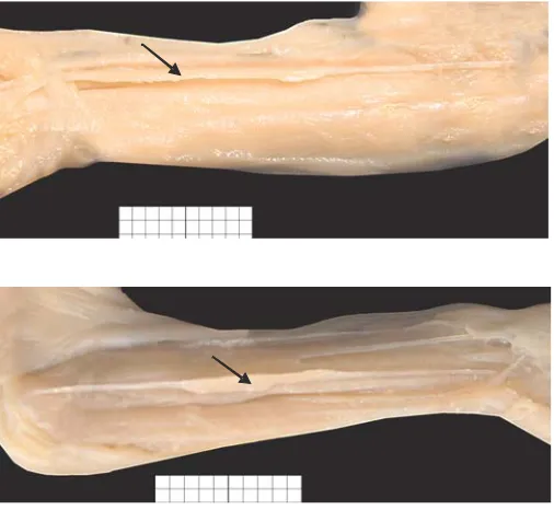
12: in 5 cases bilaterally and in 7 cases unilateral− ly. Palmaris longus muscle inversion was present in 1 case, belly presence in the mid forearm in 1 case, and tendon ramification in the distal seg− ment in 2 cases.
Fig. 4. Male and female palmaris longus muscle length comparison and Mann−Whitney’s Utest results
Ryc. 4. Porównanie długości mięśnia dłoniowego długiego płodów żeńskich i męskich oraz wynik testu
U Manna−Whitneya
Mann-Whitney's test: Z= 0.322; p= 0.616
F M
sex/p³eæ 10
20 30 40 50 60 70 80
median 25–75% min–max
muscle
length
with
aponeurosis
d³ugoœæ
miêœnia
z
rozciêgnem
mm
Wilcoxon's test: Z= 2.882; p= 0.004
right/prawa left/lewa
33 34 35 36 37 38 39
mean mean ±SE mean ±1.96*SE
side/strona
muscle
length
without
aponeurosis
d³ugoœæ
miêœnia
bez
rozciêgna
mm
Fig. 3. Left and right palmaris longus muscle length comparison and Wilcoxon’s test results
Ryc. 3. Porównanie długości mięśnia dłoniowego długiego ręki lewej i prawej oraz wynik testu Wilcoxona
Wilcoxon's test: Z= 2,452; p= 0,014
right/prawa left/lewa
43 44 45 46 47 48 49 50
mean mean ±SE mean ±1,96*SE
side/strona
muscle
length
with
aponeurosis
d³ugoœæ
miêœnia
z
rozciêgnem
mm
Fig. 1.Palmar long muscle. Arrows denote the length measurements of: a) the whole muscle, b) belly, c) ten− don, d) aponeurosis palmaris, e) forearm
Discussion
An image transformation computer system, when used to measure linear sizes (length and width) of forearm structures on digital pictures, allows reproducing the results elicited by various research workers and does not damage necroscop− ic material. The present authors’ earlier observa− tions showed the applicability of the Scion for
Windows program [21]. Available literature dis− cussing the palmaris longus muscle is very broad, but it concerns mainly its variability in maturity [1–16] as well as its practical use in hand surgery, plastic surgery, neurosurgery, and neurology [4, 14–16]. A thorough knowledge of its morphology and topography is of great importance in patho− logical studies because in special cases, compres− sion and median nerve damage may lead to pre−
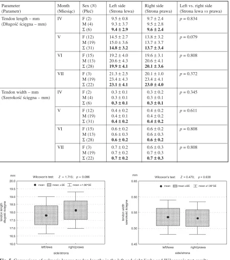
Table 3.Descriptive statistics (mean ± standard deviation) of the length and width of the palmaris longus tendon
Tabela 3.Statystyki opisowe (średnia ± odchylenie standardowe) długości i szerokości ścięgien musculus palmaris longus
Parameter Month Sex (N) Left side Right side Left vs. right side
(Parametr) (Miesiąc) (Płeć) (Strona lewa) (Strona prawa) (Strona lewa vsprawa)
Tendon length – mm IV F (2) 9.5 ±0.8 9.7 ±2.4 p= 0.834
(Długość ścięgna – mm) M (4) 9.3 ±3.7 9.5 ±2.8
Σ(6) 9.4 ±±2.9 9.6 ±±2.4
V F (12) 14.5 ±2.7 13.8 ±3.2 p= 0.079
M (19) 15.0 ±3.6 13.7 ±3.7
Σ(31) 14.8 ±±3.2 13.7 ±±3.4
VI F (15) 19.2 ±4.0 19.6 ±3.1 p= 0.808
M (13) 20.6 ±4.3 20.6 ±4.1
Σ(28) 19.9 ±±4.1 20.1 ±±3.6
VII F (3) 21.3 ±2.5 20.1 ±1.0 p= 0.372
M (19) 23.4 ±4.3 23.4 ±4.1
Σ(22) 23.1 ±±4.1 23.0 ±±4.0
Tendon width – mm IV F (2) 0.3 ±0.1 0.3 ±0.2 p= 0.345
(Szerokość ścięgna – mm) M (4) 0.3 ±0.1 0.3 ±0.1
Σ(6) 0.3 ±±0.1 0.3 ±±0.1
V F (12) 0.4 ±0.2 0.4 ±0.2 p= 0.611
M (19) 0.4 ±0.1 0.4 ±0.2
Σ(31) 0.4 ±±0.2 0.4 ±±0.2
VI F (15) 0.6 ±0.2 0.6 ±0.2 p= 0.808
M (13) 0.6 ±0.3 0.6 ±0.3
Σ(28) 0.6 ±±0.2 0.6 ±±0.2
VII F (3) 0.7 ±0.2 0.6 ±0.3 p= 0.808
M (19) 0.7 ±0.2 0.7 ±0.3
Σ(22) 0.7 ±±0.2 0.7 ±±0.3
Wilcoxon's test: Z= 1.715; p= 0.086
right/prawa left/lewa
side/strona 16.0
16.5 17.0 17.5 18.0 18.5 19.0 19.5 20.0
mean mean ±SE mean ±1.96*SE
tendon
length
d³ugoœæ
œciêgna
mm
Wilcoxon's test: Z= 0.470; p= 0.638
0.45 0.50 0.55 0.60 0.65
mean mean ±SE mean ±1.96*SE
right/prawa left/lewa
side/strona
tendon
width
szerokoϾ
œciêgna
mm
Fig. 5. Comparison of palmaris longus tendon lengths in the left and right limbs and Wilcoxon’s test results
hensile ability impairment of the hand. The avail− able literature does not provide any material dis− cussing the palmaris longus muscle based on fetal material with the use of image digital inscription and computerized quantitative analysis. Caughall [20], in his paper describing dissimilar structures
in the palmaris longus muscle and palm aponeuro− sis based on 33 fetuses aged 5 weeks to 9 months, denied the presence of any correlation between the anatomical structures of these two entities. In his histological surveys he observed that the aponeu− rosis becomes visible at the earliest in the 5th week Mann-Whitney's test: Z= –1.232; p= 0.218
F M
sex/p³eæ 0
5 10 15 20 25 30 35
median 25–75% min–max
tendon
length
d³ugoœæ
œciêgna
mm
Mann-Whitney's test: Z= -0.709; p= 0.479
F M
0.0 0.2 0.4 0.6 0.8 1.0 1.2 1.4 1.6
median 25–75% min–max
sex/p³eæ
tendon
width
szerokoϾ
œciêgna
mm
Fig. 6.Comparison of tendon palmaris longus muscle length and width in male and female fetuses as well as Mann−Whitney’s Utest results
Ryc. 6. Porównanie długości i szerokości ścięgna mięśnia dłoniowego długiego płodów żeńskich i męskich oraz wynik testu U Manna−Whitneya
Table 4.Descriptive statistics (mean ± standard deviation) of belly sizes
Tabela 4.Statystyki opisowe (średnia ± odchylenie standardowe) wymiarów brzuśca
Parameter Month Sex (N) Left side Right side Left vs. right side
(Parametr) (Miesiąc) (Płeć) (Strona lewa) (Strona prawa) (Strona lewa vsprawa)
Belly length – mm IV F (2) 12.7 ±0.7 10.9 ±1.6 p= 0.345
(Długość brzuśca – mm) M (4) 11.0 ±0.4 10.5 ±1.5
Σ(6) 11.6 ±±1.0 10.6 ±±1.4
V F (12) 16.5 ±4.0 15.8 ±2.9 p= 0.959
M (19) 14.7 ±3.7 15.1 ±3.0
Σ(31) 15.4 ±±3.9 15.4 ±±2.9
VI F (15) 21.0 ±5.6 20.9 ±5.2 p= 0.737
M (13) 17.1 ±5.1 16.6 ±5.2
Σ(28) 19.2 ±±5.6 18.9 ±±5.6
VII F (3) 23.1 ±1.5 22.8 ±3.4 p= 0.069
M (19) 22.4 ±4.5 20.4 ±4.7
Σ(22) 22.7 ±±4.3 20.7 ±±4.6
Belly width – mm IV F (2) 1.0 ±0.1 1.0 ±0.1 p= 0.590
(Szerokość brzuśca – mm) M (4) 1.0 ±0.3 1.1 ±0.6
Σ(6) 1.0 ±±0.3 1.0 ±±0.4
V F (12) 1.7 ±0.5 1.7 ±0.3 p= 0.456
M (19) 1.8 ±0.6 1.8 ±0.5
Σ(31) 1.8 ±±0.5 1.7 ±±0.4
VI F (15) 2.5 ±0.5 2.6 ±0.5 p= 0.267
M (13) 2.2 ±0.5 2.3 ±0.5
Σ(28) 2.4 ±±0.5 2.5 ±±0.5
VII F (3) 2.6 ±0.3 2.5 ±0.4 p= 0.948
M (19) 3.0 ±1.1 2.9 ±0.7
of pregnancy. In his material, the palmaris longus tendon was invisible in 5 fetuses (15%). By light microscopy he showed that the palmar aponeuro− sis consists of longitudinal and transverse fibers. In the 5thweek, these two layers are very thin and usually cellular. The aponeurosis is situated super− ficially and runs from the flexor retinaculum to the metacarpophalangeal joints. In the 12th week, the longitudinal and transverse fibers start to develop. The palmaris longus tendon becomes visible only from the 6thweek of pregnancy, which points to its different developmental flow. The tendon is con− nected with the longitudinal fibers of the palmar aponeurosis and is situated superficially between the medial nerve and the forearm fascia. At the wrist level and at the site of transition into the palmar aponeurosis, it is situated over the flexor retinaculum and is flattened. The literature does not include any paper dealing with these tissues’
biomorphology, so the results of the present authors’ surveys cannot be compared with other reports.
There are, however, papers discussing mor− phological variety, such as a lack of the palmaris longus muscle, its morphological anomalies, and its location instability. There are surveys of mater− ial consisting of living adults and sectional mater− ial [1–4, 6, 7, 9–11]. The problem of absence of the muscle has been discussed by many authors [2, 9–11]. The studies were carried out using clinical methods on living individuals randomly selected from the population. Their results confirm the pre− sent authors’ observations of frequent variability in the palmaris longus muscle; however, they do not contribute much to understanding its morphol− ogy, but only demonstrate its prevalent instability. The results of the present study concerning the morphological instability of the palmaris longus
Wilcoxon's test: Z= 1.761; p= 0.078
right/prawa left/lewa
16.0 16.5 17.0 17.5 18.0 18.5 19.0 19.5 20.0
mean mean ±SE mean ±1.96*SE
side/strona
belly
length
d³ugoœæ
brzuœca
mm
Wilcoxon's test: Z= 0.322; p= 0.747
right/prawa left/lewa
side/strona 2.0
2.1 2.2 2.3 2.4 2.5
mean mean ±SE mean ±1.96*SE
belly width
szerokoœæ brzuœca
mm
Fig. 7.Comparison of palmaris longus muscle belly length and width in the left and right limbs and Wilcoxon’s test results
Ryc. 7.Porównanie długości i szerokości brzuśca m. palmaris longusręki lewej i prawej oraz wynik testu Wilcoxona
Mann-Whitney's test: Z= 1.232; p= 0.218
F M
8 12 16 20 24 28
32 median 25–75% min–max
belly
length
d³ugoœæ
brzuœca
sex/p³eæ
mm Mann-Whitney's test: Z= 0.040; p= 0.968
F M
0 1 2 3 4 5 6 7
median 25–75% min–max
sex/p³eæ
belly
width
szerokoϾ
brzuœca
mm
Fig. 8.Comparison of palmaris longus muscle belly length and width in male and female fetuses with Mann− −Whitney’s Utest results
Ryc. 8.Porównanie długości i szerokości brzuśca m. palmaris longuspłodów żeńskich i męskich oraz wynik testu
muscle (inversion, variable location, uni− or bilat− eral absence) are consistent with other authors’ results from autopsic examinations [6, 9, 20]. In turn, anomalies such as a tendon−belly−tendon structure or threadlike tendon have not been described in any paper. Such a muscular structure is very rare, and only due to very numerous mate− rial has it been detected and described.
Symmetry is another element of the present study and it is also discussed in Derkowski’s paper [22]. It requires individual discussion because of the very interesting observations. The present sur− vey incorporating computer analysis revealed asymmetry in the structure of the palmaris longus muscle. The asymmetry was very clearly in favor of the right side. Derkowski [22] also discusses the
Table 5.Descriptive statistics of the palmaris longus aponeuroses length and width
Tabela 5.Statystyki opisowe długości i szerokości rozcięgien musculus palmaris longus
Parameter Month Sex (N) Left side Right side Left vs. right side
(Parametr) (Miesiąc) (Płeć) (Strona lewa) (Strona prawa) (Strona lewa vsprawa) Aponeurosis length – mm IV F (2) 6.7 ±0.6 6.4 ±0.2 p= 0.893
(Długość rozcięgna – mm) M (4) 5.9 ±0.9 5.9 ±1.0
Σ(6) 6.1 ±±0.9 6.0 ±±0.8
V F (12) 9.9 ±1.8 9.8 ±1.9 p= 0.066
M (19) 10.5 ±2.4 9.7 ±1.6
Σ(31) 10.3 ±±2.2 9.7 ±±1.7
VI F (15) 10.4 ±2.2 10.2 ±2.2 p= 0.515
M (13) 10.7 ±2.9 10.8 ±3.0
Σ(28) 10.5 ±±2.5 10.5 ±±2.5
VII F (3) 16.3 ±3.7 16.1 ±3.4 p= 0.778
M (19) 12.5 ±2.6 12.5 ±2.6
Σ(22) 13.0 ±±3.0 13.0 ±±2.9
Aponeurosis width – mm IV F (2) 4.1 ±0.2 4.1 ±0.3 p= 0.068 (Szerokość rozcięgna – mm) M (4) 4.0 ±0.2 4.2 ±0.4
Σ(6) 4.0 ±±0.2 4.2 ±±0.3
V F (12) 5.4 ±0.9 5.5 ±1.1 p= 0.677
M (19) 5.4 ±1.0 5.4 ±0.8
Σ(31) 5.4 ±±0.9 5.4 ±±0.9
VI F (15) 5.4 ±0.9 5.6 ±0.8 p= 0.213
M (13) 5.3 ±0.8 5.3 ±0.9
Σ(28) 5.3 ±±0.8 5.5 ±±0.8
VII F (3) 7.3 ±0.7 7.0 ±0.3 p= 0.733
M (19) 6.8 ±1.3 6.9 ±1.4
Σ(22) 6.8 ±±1.2 6.9 ±±1.3
Wilcoxon's test: Z= 1.520; p= 0.129
right/prawa left/lewa
9.5 10.0 10.5 11.0 11.5 12.0
mean mean ±SE mean ±1.96*SE
side/strona aponeurosis length d³ugoœæ rozciêgna
mm
Mann-Whitney test's: Z= –0.850; p= 0.395
F M
0 5 10 15 20 25
median 25–75% min–max
sex/p³eæ aponeurosis length d³ugoœæ rozciêgna
mm
Fig. 9.Comparison of palmaris longus muscle aponeurosis length in the left and right limbs with Wilcoxon’s test result and in male and female fetuses with Mann−Whitney’s Utest results
Fig. 10.Comparison of the palmaris longus muscle aponeu− rosis width in male and female fetuses and Mann−Whitney’s
Utest result
Ryc. 10. Porównanie szerokości rozcięgna m. palmaris longuspłodów żeńskich i męskich oraz wynik testu
U Manna−Whitneya Mann-Whitney's test: Z= –0.666;p= 0.505
F M
2 3 4 5 6 7 8 9 10
median 25–75% min–max
sex/p³eæ aponeurosis width szerokoœæ rozciêgna
mm
Fig. 11.Diagrams and models of size increases in the palmaris longus muscle
Ryc. 11.Diagramy i modele wzrastania wymiarów mięśnia dłoniowego długiego tendon length = 11.3 + 1.34 * week–
d³ugoœæ œciêgna = 11.3 + 1.34 * tydzieñ–
15 16 17 18 19 20 21 22 23 24 25 26 27 0
5 10 15 20 25 30 35
median 25 75%– non-outlier range outliers
tendon width = 0.4 + 0.04 * week– szerokoœæ œciêgna = 0.4 + 0.04 * tydzieñ–
15 16 17 18 19 20 21 22 23 24 25 26 27 0.0
0.3 0.6 0.9 1.2 1.5
median 25–75% non-outlier range outliers
belly length = 5.5 + 1.06 * week– d³ugoœæ brzuœca = 5.5 + 1.06 * tydzieñ–
15 16 17 18 19 20 21 22 23 24 25 26 27 5
10 15 20 25 30 35
median 25 75%– non-outlier range outliers
belly width = 1.8 + 0.18 * week– szerokoœæ brzuœca = 1.8 + 0.18 * tydzieñ–
15 16 17 18 19 20 21 22 23 24 25 26 27 0
1 2 3 4 5 6 7
median 25 75%– non-outlier range outliers extremes
aponeurosis length = –3.8 + 0.67 * week d³ugoœæ rozciêgna = 3.8 + 0.67 * tydzieñ–
15 16 17 18 19 20 21 22 23 24 25 26 27 3
6 9 12 15 18 21
24 median 25 75%– non-outlier range outliers
aponeurosis width = –0.7 + 0.29 * week szerokoœæ rozciêgna = 0.7 + 0.29 * tydzieñ–
15 16 17 18 19 20 21 22 23 24 25 26 27 2
3 4 5 6 7 8 9 10
median 25 75%– non-outlier range outliers extremes
GA – weeks/wiek – tydzieñ GA – weeks/wiek – tydzieñ
GA – weeks/wiek – tydzieñ GA – weeks/wiek – tydzieñ
GA – weeks/wiek – tydzieñ GA – weeks/wiek – tydzieñ
aponeurosis length d³ugoœæ rozciêgna
belly length
d³ugoœæ brzuœca
tendon length
d³ugoœæ œciêgna
aponeurosis szerokoœæ rozciêgna
width
belly
brzuœca
width
szerokoϾ
tendon width
œciêgna
szerokoϾ
mm
mm
mm
mm
mm
problem of asymmetry in his paper. In his exami− nations he found a high symmetry level of the cra− nium base in relation to the body’s median plane. Asymmetry between the hemispheres is a rather rare case which somehow supports the theory that asymmetry is a subsequent phenomenon in human life and is very rarely detected in the fetal period. In the present material, asymmetry was found both
in normally built palmaris longus muscle and in anomalies. However, no paper could be found which considered the problem of the palmaris longus muscle. The present examinations show the presence of considerable asymmetry in upper limb structure even in the fetal period. However, no sexual dimorphism was observed, although it has been discussed in other papers [22].
Fig. 12.Diagrams and models of forearm and palmaris longus muscle size increases
Ryc. 12.Diagramy i modele wzrastania wymiarów przedramienia i mięśnia dłoniowego długiego forearm length = –15.5 + 2.44 * week
d³ugoœæ przedramienia = 15.5 + 2.44 * tydzieñ–
15 16 17 18 19 20 21 22 23 24 25 26 27 10
15 20 25 30 35 40 45 50 55 60 65
median 25–75% non-outlier range outliers extremes
muscle length = 16.5 + 2.39 * week– d³ugoœæ miêœnia = 16.5 + 2.39 * tydzieñ–
15 16 17 18 19 20 21 22 23 24 25 26 27 10
20 30 40 50 60 70 80
median 25–75% non-outlier range outliers extremes
muscle length = –20.1 + 3.05 * week d³ugoœæ miêœnia = 20.1 + 3.05 * tydzieñ–
15 16 17 18 19 20 21 22 23 24 25 26 27 10
20 30 40 50 60 70 80
median 25–75% non-outlier range outliers GA – weeks/wiek – tydzieñ
GA – weeks/wiek – tydzieñ GA – weeks/wiek – tydzieñ
muscle length without d³ugoœæ miêœnia bez rozciêgna
aponeurosis
muscle length with d³ugoœæ miêœnia z rozciêgnem
aponeurosis
forearm length
d³ugoœæ przedramienia
mm
mm mm
Fig. 13.Anomalies in photograph: a) palmaris longus muscle inversion, the belly is situated in the forearm’s distal segment; b) in the mid forearm
References
[1] Moss ALH:Is There an Association Between an Absence of Palmaris Longus Tendon and an Absence of Plantaris Tendon? Eur J Plast Surg 1988 11, 32–34.
[2] Thompson NW, Mockford BF, Cran GW:Absence of the palmaris longus muscle: a population study. Ulster Med J 2001, 70, 22–4.
[3] Roohi SA, Choon−Sian L, Shalimar A, Tan GH, Naicker AS: Study on the Absence of Palmaris Longus in a Multiracial Population.Malaysian Orthop J 2007, 1, 1, 26–28.
[4] Sebastin SJ, Lim AYT, Wong HB:Clinical Assessment of Absence of the Palmaris Longus and its Association With Other Anatomical Anomalies – A Chinese Population Study. Ann Acad Med Singapore 2006, 35, 249–245. [5] Sassoli Fazan VP: Reversed Palmaris Longus Muscle median nerve relationship. Case report and literature
revive. Braz J Morphol Sci 2007, 24, 2, 88–91.
[6] Ninkovic M, Hefel L, Ohler K:Acute median nerve compression produced by reversed palmaris longus muscle. Eur J Plast Surg 1995, 18, 129–130.
[7] Paraskevas G, Tzaveas A, Natsis K, Kitsoulis P, Spyridakis I:Failure of palmaris longus muscle duplication and its clinical application. Folia Morphol (Warsz) 2008, 67, 2, 50–153.
[8] Kącik W, Wienzek K:Carpal tunnel isthmus syndrom caused by palmaris longus muscle anatomical anomaly. Locomotor Syst Surg Orthopaedics Pol, 1991, LVI. Chir Narz Ruchu Ortop Pol 1991, LVI.
[9] Koo CC and Roberts AHN:The palmaris longus tendon. Another variation in its anatomy. J Hand Surg [Br] 1997, 22 (1), 138–139.
[10] Machado AB, DiDio LJ:Frequency of the musculus palmaris longus studied in vivoin some Amazon Indians. Am J Phys Anthrop 1967, 27, 11–20.
[11] Żebrowski P:Badania mięśnia dłoniowego długiego na ludziach żywych. Folia Morphol 1934, 5, 80–91. [12] Lorenzo JS, Canada M, Diaz L, Sarasua G: Compression of the median nerve by an anomalous palmaris longus
tendon: A case report. J Hand Surg [Am] 1996, 21 (5), 858–860.
[13] Sterry Ashby B:Hypertrophy of the palmaris longus muscle. Report of a case. J Bone Hand Surg 46, 21964, 230–232.
[14] Sudhir K Kapoor, Akshay Tiwari:Clinical relevance of palmaris longus agenesis; Common anatomical aberra− tion. Anat Sci Int 2008, 83, 45–48.
[15] Bachelor EP, Jobe RP: The absent lateral canthal tendon: Reconstruction using a Y graft of Palmaris longus ten− don. Ann Plast Surg 1980, 5, 362–368.
[16] Song IC, Bromberg BE:Pharyngo−palatoplasty with free transplantation of the Palmaris longus. Br J Plast Surg 1974, 27, 337–43.
[17] Kayode AO, Olamide AA, Blessing IO, Victor OU:Incidence of Palmaris Longus Muscle Absence in Nigerian Population.Int J Morphol 2008, 26 (2), 305–308.
[18] Zeiss J, Guilliam−Haidet L:MR demonstration of anomalous muscle about the volar aspect of the wrist and fore− arm. Clin Imaging 1996, 20(3), 219–221.
[19] Stecco C, Lancretto L, Porzionato A, Macchi V, Tiengo C, Parenti A:The Palmaris Longus and its Relations with the Antebrachial Fascia and the Palmar Aponeurosis. Clin Anat 2009, 22, 221–229.
[20] Caughall KA:Developmental anatomy of the palmar aponeurosis and its relationship to the Palmaris longus ten− don. J Hand Surg 1988, 13 (4), 485–93.
[21] Kędzia A, Woźniak J, Dudek K, Ziajkiewicz M:Model matematyczny wzrostu kości długich kończyny górnej w okresie prenatalnym [Mathematical model of upper−limb long bone growth in fetal period]. XV Kowban 2008, 209–216.
[22] Derkowski W, Kędzia A, Glonek M:. Cranium symmetry and asymmetry in foetal period on the basis of image computer analysis. Kowban XV, 197–201.
Address for correspondence:
Alicja KędziaDepartment of Normal Anatomy Wroclaw Medical University Chałubińskiego 6a
50−368 Wrocław Poland
Tel.: +48 71 784 00 80
E−mail: [email protected]
The authors concluded that considerable indi− vidual variety in palmaris longus muscle morphol− ogy was detected which is of great clinical impor− tance. In the fetal period, the determined asymme−
try was in favor of the right side and no sexual dimorphism was observed. The Scion for Windows system proved very useful in morpho− logical surveys.
Conflict of interest: None declared