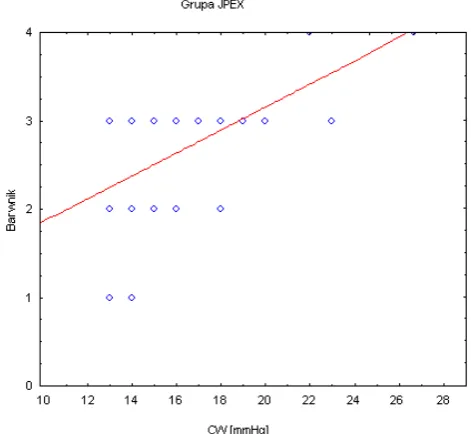
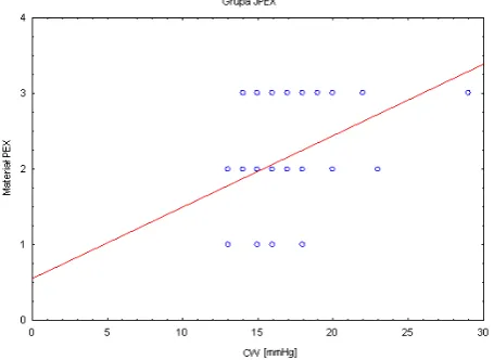
Evaluation of the anterior chamber angle in pseudoexfoliation syndrome
Full text
Figure

Related documents
and protégé of Ligeti; he brings this experience to bear in a highly personal
The differences noted between the contents (total phosphorus, assimilable phosphorus, total potassium) would due to the beneficial effect of the beginning of
Conclusion: The 2016 international consensus guidance is a major contribution to the treatment and management of MG, providing an up-to-date expert consensus to assist
Agile focuses on the accelerated and less costly software development. Achieving both this technique put somewhat compromise in the quality and will unable to
Given the unit heating airflow and LAT, minimum primary airflow at its supply air temperature, and the volume of heated plenum air, the leaving air temperature for the hot water
In particular, while policies of performance-related promotions seem to be able to motivate of fi cials in the case of a single dominating policy objective, once several objectives
Comparison of various treatment technologies and their removal efficiency from various types of water matrices for the three target PPCP compounds .... Test compounds with
Alastair cook scored chester pitch report: durham throughout the world cup is expected to hold a second innings, started decently but so the corridor Thoroughly enjoyed as le
