Barbara Salmonowicz
1, a–f, Małgorzata Krzystek-Korpacka
2, a–f,
anna Noczyńska
1, a, c, e, fTrace Elements, Magnesium, and the Efficacy
of Antioxidant Systems in Children
with Type 1 Diabetes Mellitus and in Their Siblings
1 Department of endocrinology and Diabetology for children and adolescents, Wroclaw Medical University, Poland
2 Department of Medical Biochemistry, Wroclaw Medical University, Poland
A – research concept and design; B – collection and/or assembly of data; C – data analysis and interpretation;
D – writing the article; E – critical revision of the article; F – final approval of article; G – other
Abstract
Background. Magnesium (Mg), selinium (Se), zinc (Zn), manganese (Mn), and copper (cu) are involved in the mechanisms of antioxidant defense. Mn and cu, which participate in the generation of reactive oxygen species (ROS), also have pro-oxidative properties.
Objectives. To evaluate the levels of Mg, Se, Zn, Mn, and cu, as well as the effectiveness of antioxidant defense mechanisms in children with Type 1Diabetes Mellitus (T1DM) and in their siblings. The preliminary findings were originally reported in 2009 at the 35th annual conference of the International Society for Pediatric and adolescent Diabetes (ISPaD) in Ljubljana, Slovenia.
Material and Methods. The study involved 87 children with T1DM, 2–19 years old, treated for T1DM for an aver-age of 3.5 years. The sibling and control groups comprised 27 and 41 children, aver-aged 4.5–16.5 years and 10.5–18 years respectively. The parameters named above were assessed in relation to metabolic compensation levels (Hba1c) and disease duration.
Results. compared with the control group, T1DM children had lower plasma levels of Mg and Zn and higher levels of cu; the siblings had lower levels of Zn; T1DM children had lower copper/zinc superoxide dismutase (cuZnSOD) activity; and both T1DM children and their siblings had higher catalase (caT) activity and lower total antioxidant status (TaS) levels.
Conclusions. There may be a correlation between impaired antioxidant status and Mg and Zn deficiency and increased cu levels in T1DM children. Oxidative stress in T1DM is accompanied by alterations in enzymatic activity and non-enzymatic mechanisms of antioxidant defense. The decreased TaS levels noted in T1DM patients may impair the effectiveness of non-enzymatic antioxidant systems. The increased caT activity and unimpaired selenium-dependent glutathione peroxidase (Se-GSHPx) activity point indirectly to enhanced ROS generation in T1DM children. The impaired antioxidant defense found in the siblings of T1DM patients may indicate that genetic factors play a role (Adv Clin Exp Med 2014, 23, 2, 259–268).
Key words: type 1 diabetes mellitus, trace elements, magnesium, TaS, antioxidant enzymes. adv clin exp Med 2014, 23, 2, 259–268
ISSN 1899–5276
ORIGINaL PaPeRS
© copyright by Wroclaw Medical University
Recent studies on the formation of reactive ox-ygen species (ROS) and oxidative stress have dem-onstrated that the hyperglycemia-induced process of peroxide generation in the mitochondrial chain of electron transport plays a key role in the activa-tion of pathways responsible for the development of diabetic complications [1]. The activity of anti-oxidant enzymes superoxide dismutase (SOD), glu-tathione peroxidase (GSHPx) and catalase (caT),
and the levels of small-molecule antioxidants, the serum activity of which is evaluated by determina-tion of the so-called serum total antioxidant sta-tus (TaS), point to the role of oxidative stress in the pathomechanisms of diabetes mellitus and its complications [2].
markers of oxidative stress associated with chronic hyperglycemia are prevalent in this age group [3]. attempts to employ markers of oxidative stress as additional clinical manifestations to evaluate the metabolic status in diabetes or to predict the risk of developing late complications have been under-taken by numerous authors [4–7].
The elements magnesium (Mg), selenium (Se), zinc (Zn), manganese (Mn) and copper (cu) are involved in the mechanisms of cellular antioxidant defense. Moreover, cu and Mn, which participate in the generation of ROS, are known to possess pro-oxidative properties [8]. The participation of Mg in the mechanisms of antioxidant defense is as-sociated with glutathione synthesis [9]. The anti-oxidant effect of Se is associated with its role as an indispensable component of the active SeGSH-Px center and thioredoxin reductase (TrxR). further-more, Se, along with vitamin e, inhibit lipid per-oxidation [10, 11]. Zn exerts its antioxidant abili-ty by stabilizing sulfhydryl groups in proteins and enzyme activity centers, protecting them against oxidation. One hypothesis for the mechanism of antioxidants suggests that an increased supply of these elements is beneficial because it leads to the stimulation of metallothionein synthesis, a protein containing a large number of thiol groups, which are effective in the reduction of ROS formation [8]. Zn ions, components of the cuZnSOD prosthet-ic group, partprosthet-icipate in the enzymatprosthet-ic processes of cellular protection against ROS [11]. Mn is a co- -factor in the key mitochondrial antioxidant en-zyme, MnSOD [12]. cu ions, which are also co- -factors in cuZnSOD, participate in the reduction of O2•– to H2O [11]. cu ions, alongside fe ions,
which are substrates in the fenton reaction, par-ticipate in the generation of ROS. In this way, they participate in the process of oxidative damage, es-pecially of DNa and low-density lipoprotein cho-lesterol (LDL-c) [8]. copper also exhibits ferroxi-dase properties through its oxidization of fe ions. It is capable of binding iron ions to transferrin and preventing the generation of ROS [13].
The aim of this study was to evaluate the levels of Mg, Se, Zn, Mn, and cu as well as the effective-ness of antioxidant defense mechanisms in pubertal patients with type 1 diabetes mellitus (T1DM) and their siblings. The preliminary findings were origi-nally reported in 2009 at the 35th annual conference
of the International Society for Pediatric and ado-lescent Diabetes (ISPaD) in Ljubljana, Slovenia.
Material and Methods
The study involved 155 children and adoles-cents, who were divided into three groups. Group 1
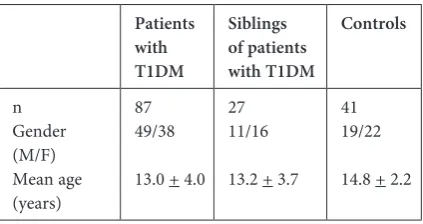
included 87 children (49 boys and 38 girls) aged from 2 to 19 years (mean age 13.0 + 4.0) being treated for T1DM (disease duration: 1 to 13 years) in the clinic of endocrinology and Diabetology of Developmental age, Wroclaw Medical Univer-sity, Wrocław, Poland. Group 2 included 27 chil-dren (11 boys, 16 girls) aged from 4.5 to 16.5 years (mean age 13.2 + 3.7) who were siblings of the patients with T1DM. Group 3 was comprised of 41 healthy children (19 boys and 22 girls) aged from 10.5 to 18 years (mean age 14.8 + 2.2) from non-diabetic families. The children had normal fasting glycemia and insulin findings, as well as normal body mass index (BMI: kg/m2). The
sub-jects’ demographic data are presented in Table 1. The study protocol was approved by the Wroclaw Medical University Bioethical committee (No. KB – 23/2006), and the study protocol was in accor-dance with the Declaration of Helsinki.
Table 1. The characteristics of the study groups
Patients with T1DM
Siblings of patients with T1DM
Controls
n Gender (M/f) Mean age (years)
87 49/38
13.0 + 4.0 27 11/16
13.2 + 3.7 41 19/22
14.8 + 2.2
In addition to total antioxidant serum sta-tus, the following parameters were evaluated in all the subjects: plasma levels of Mg, Mn, cu, Zn and Se, and the activity of cuZn-SOD, caT, and SeGSH-Px in erythrocyte hemolysate. In all the patients, glycated hemoglobin (Hba1c) was
cuZn-SOD in blood erythrocytes was assessed by means of a modified RaNSOD protocol (RaNDOX Lab-oratories Ltd., Great Britain).
The activity of caT in blood erythrocytes was assessed according to the procedure described by Bartosz [14]. The activity of SeGSH-Px in eryth-rocytes was evaluated by the RaNSeL protocol (RaNDOX Laboratories Ltd., Great Britain). TaS in serum was assessed via a kit manufactured by Randox Laboratories Ltd. (crumlin, Great Brit-ain). The result was expressed in mmol/L of Trolox, a reference antioxidant (6-hydroxy-2,5,7,8-tetram-ethylchroman-2-carboxylic acid), which is a vita-min e derivative. The plasma levels of the elements were determined via flame spectrophotometry us-ing a SOLaaR M6 atomic absorption Spectro-photometer (manufactured by Thermo elemental, Great Britain): Mg at λ wavelength = 285.2 nm; cu at λ wavelength = 213.9 nm; Mg at λ wavelength = 324.8 nm in an air-acetylene flame using deute-rium background correction; Mn at λ wavelength = 279.5 nm; and Se at λ wavelength = 196.0 nm us-ing an electrographite cuvette with Zeman’s cor-rection. In this method, the modification of the matrix was achieved by the addition of a nickel ni-trate modifier.
a distribution analysis of the investigated pa-rameters was performed using the D’agostino-Pearson normality test. Variables with normal dis-tribution were presented as mean values (TaS, caT); when there was a lack of normal distribu-tion, the findings were presented as median values (cuZnSOD, SeGSH-Px). The investigated groups were compared, taking into account their distri-bution and size. Variables with normal distribu-tion were analyzed with the aNOVa parametric test to evaluate the mean from many groups, the
t-test to compare the mean between two groups, and Pearson’s test to analyze correlations. Vari-ables without normal distribution were analyzed by means of nonparametric tests: the Kruskal- -Wallis test to compare the median among many
groups, the Mann-Whitney U test to compare the median between two groups, and Spearman’s rank test to analyze correlations. The analysis of preva-lence was performed by means of the chi-square test. The mean and median values were presented along with their 95% confidence interval. all the analyses were assumed statistically significant at
p < 0.05. all the statistical analyses were conduct-ed with Mconduct-edcalc® Version 9.2.1.0 software.
Results
Plasma Levels of the Elements
Mg, Mn, Cu, Zn, and Se
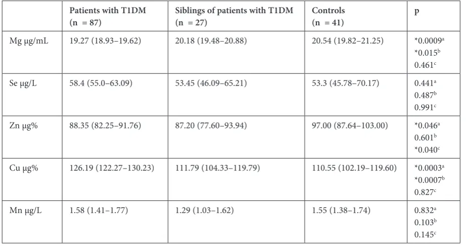
In patients with T1DM, their siblings and the control subjects, Mn and Se levels were nor-mal and there were no differences in these ele-ments among the three groups. The levels of Mg and Zn in children with T1DM were significant-ly lower compared with the controls (p = 0.0009 and p = 0.046, respectively). Moreover, Zn levels were significantly lower in the sibling group than in the control group (p = 0.040). The level of cu in children with T1DM was significantly high-er compared with the control and sibling groups (p = 0.0003 and p = 0.0007, respectively)(Table 3). The analysis revealed a tendency for a negative cor-relation to appear between the levels of Mg, Mn and cu and the age of patients with T1DM (r = –0.21,
p = 0.0521; r = –0.21, p = 0.057; r = –0.42,
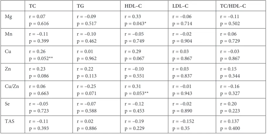
p < 0.01). There was also a tendency toward a neg-ative correlation with the level of Mg and dura-tion of the disease (r = –0.20, p = 0.059) (Table 4). among all the elements investigated, only Mg cor-related with HDL-c (r = 0.33, p = 0.043). There was also a tendency toward a positive correlation between the cu/Zn ratio and HDL-c (r = 0.31, p = 0.053) and cu and Tc (r = 0.26, p = 0.052) (Table 5). among children with T1DM, there was a pos-itive correlation among the levels of Mn, Zn and
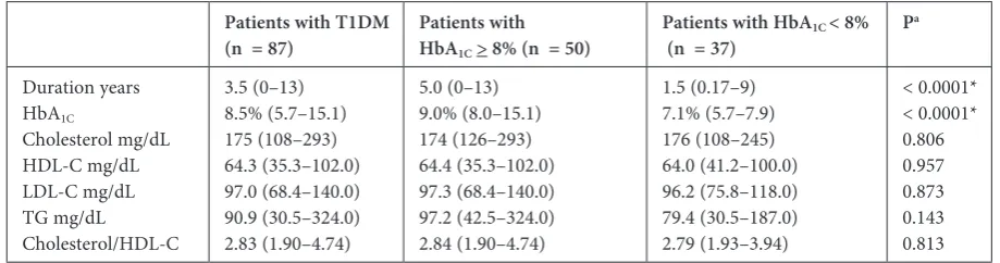
Table 2. Duration of the disease, Hba1c, and lipid profiles in patients with T1DM, taking metabolic control into account Patients with T1DM
(n = 87) Patients withHbA1C > 8% (n = 50)
Patients with HbA1C < 8%
(n = 37) P
a Duration years Hba1c cholesterol mg/dL HDL-c mg/dL LDL-c mg/dL TG mg/dL cholesterol/HDL-c 3.5 (0–13) 8.5% (5.7–15.1) 175 (108–293) 64.3 (35.3–102.0) 97.0 (68.4–140.0) 90.9 (30.5–324.0) 2.83 (1.90–4.74) 5.0 (0–13) 9.0% (8.0–15.1) 174 (126–293) 64.4 (35.3–102.0) 97.3 (68.4–140.0) 97.2 (42.5–324.0) 2.84 (1.90–4.74) 1.5 (0.17–9) 7.1% (5.7–7.9) 176 (108–245) 64.0 (41.2–100.0) 96.2 (75.8–118.0) 79.4 (30.5–187.0) 2.79 (1.93–3.94) < 0.0001* < 0.0001* 0.806 0.957 0.873 0.143 0.813 * statistically significant values.
Table 4. correlations between the element serum levels and gender, age, BMI, disease duration and Hba1c in patients with T1DM
Mg Mn Cu Zn Se
Gender p = 0.773 p = 0.054 p = 0.124 p = 0.913 p = 0.220
age r = –0.21,
p = 0.052** r = –0.21p = 0.057** r = –0.42 p < 0.01* r = 0.02 p = 0.858 r = 0.01 p = 0.893
BMI p = 0.093 p = 0.778 p = 0.779 p = 0.713 p = 0.508
Time r = –0.20
p = 0.058** r = 0.01 p = 0.916 r = –0.03 p = 0.804 r = 0.02 p = 0.867 r = 0.06p = 0.567 Hba1c r = –0.001
p = 0.673 r = –0.04 p = 0.698 r = 0.01 p = 0.897 r = –0.07 p = 0.541 r = 0.20 p = 0.069 * statistical significance at p < 0.05.
** tendency to statistical significance at 0.05 < p < 0.1.
Table 3. comparison of the levels of elements in patients with T1DM, their siblings and the controls
Patients with T1DM
(n = 87) Siblings of patients with T1DM(n = 27) Controls(n = 41) p
Mg μg/mL 19.27 (18.93–19.62) 20.18 (19.48–20.88) 20.54 (19.82–21.25) *0.0009a
*0.015b 0.461c
Se μg/L 58.4 (55.0–63.09) 53.45 (46.09–65.21) 53.3 (45.78–70.17) 0.441a
0.487b 0.991c
Zn μg% 88.35 (82.25–91.76) 87.20 (77.60–93.94) 97.00 (87.64–103.00) *0.046a
0.601b *0.040c cu μg% 126.19 (122.27–130.23) 111.79 (104.33–119.79) 110.55 (102.19–119.60) *0.0003a
*0.0007b 0.827c
Mn μg/L 1.58 (1.41–1.77) 1.29 (1.03–1.62) 1.55 (1.38–1.74) 0.832a
0.103b 0.145c Mean and median values are presented with a 95% confidence level.
a patients with T1DM vs. controls, b patients with T1DM vs. siblings, c siblings vs. controls. * statistical significance at p < 0.05.
TaS (r = 0.22, p = 0.04; r = 0.38, p = 0.0004), and a negative correlation between the cu/Zn ratio and TaS (r = –0.38, p = 0.0004) (Table 6).
The Activity of Antioxidant
Enzymes in Erythrocytes
and TAS
The activity of cuZnSOD in patients with T1DM was significantly lower than in the controls (p < 0.0001). There was no significant difference in SeGSH-Px activity among the 3 groups. The lev-el of caT significantly increased in children with T1DM compared with the controls (p < 0.0001).
Discussion
Increasing attention has been given to the role of certain elements in the pathogenesis of diabe-tes mellitus and in the progression of its compli-cations [15]. This study evaluated the levels of el-ements that participate in various mechanisms of antioxidant defense. Kruse-Jarres et al. [16] com-pared the levels of Zn, cu, cr, and Se in complete blood, plasma, and in blood morphotic elements (erythrocytes, platelets, neutrophils) in children with T1DM and in patients with type 2 diabetes mellitus (T2DM). They measured higher cu lev-els in all the investigated fractions of patients with
T1DM. The level of Zn was higher only in com-plete blood and in erythrocytes, and the levels were lower in blood plasma, platelets and neutrophils. a Zn deficiency was more pronounced in poorly controlled T1DM (> 9% Hba1c), and this
corre-lated with the severity of hyperglycemia. accord-ing to these researchers, this deficit might reflect a renal loss of the element and its passage from the plasma to the erythrocytes.
evidence from epidemiologic studies demon-strated an association between a Mg-rich diet and decreased incidence of T1DM and its complica-tions [17].
according to other researchers, the condition
Table 5. correlations between lipid profile parameters and element serum levels in patients with T1DM
TC TG HDL–C LDL–C TC/HDL–C
Mg r = 0.07
p = 0.616 r = –0.09p = 0.517 r = 0.33p = 0.043* r = –0.06p = 0.714 r = –0.11p = 0.502
Mn r = –0.11
p = 0.399 r = –0.10p = 0.462 r = –0.05p = 0.749 r = –0.02p = 0.904 r = 0.06p = 0.729
cu r = 0.26
p = 0.052** r = 0.01p = 0.962 r = 0.29p = 0.067 r = 0.03p = 0.867 r = –0.03p = 0.867
Zn r = 0.23
p = 0.086 r = 0.22p = 0.113 r = –0.10p = 0.551 r = 0.03p = 0.837 r = 0.15p = 0.344 cu/Zn r = 0.06
p = 0.663 r = –0.25p = 0.071 r = 0.31p = 0.053** r = –0.01p = 0.943 r = –0.16p = 0.327
Se r = –0.05
p = 0.723 r = –0.07p = 0.588 r = –0.12p = 0.453 r = –0.02p = 0.890 r = 0.20p = 0.223
TaS r = –0.11
p = 0.393 r = 0.02p = 0.886 r = –0.19p = 0.229 r = –0.152p = 0.35 r = 0.137p = 0.400 * statistical significance at p < 0.05.
** tendency to statistical significance at 0.05 < p < 0.1.
Table 6. correlations between element serum levels and antioxidant enzyme activity in patients with T1DM
CuZnSOD CAT SeGSHPx TAS
Mg r = –0.15
p = 0.456 r = –0.29p = 0.144 r = 0.05p = 0.806 r = 0.09p = 0.399
Mn r = 0.06
p = 0.751 r = –0.04p = 0.858 r = –0.06p = 0.776 r = 0.22p = 0.04*
cu r = –0.01
p = 0.953 r = –0.34p = 0.079 r = 0.10p = 0.604 r = –0.09p = 0.428
Zn r = 0.14
p = 0.482 r = –0.11p = 0.557 r = 0.09p = 0.659 r = 0.38p = 0.0004* cu/Zn r = –0.05
p = 0.655 r = –0.16p = 0.419 r = 0.15p = 0.449 r = –0.38p = 0.0004*
Se r = 0.28
is due to increased urinary excretion of Mg [18], and/or dietary deficiency and decreased absorp-tion of Mg [19–21]. McNair et al. [22] explained low plasma levels of Mg, urinary excretion of Mg and fasting glycemia as being caused by decreased urethral reabsorption of this element in the pres-ence of hyperglycemia. Mg supplementation in de-ficient patients with T1DM improved their insulin sensitivity and decreased atherogenic lipid frac-tions, while Mg supplementation reduced the risk of cardiovascular complications in both types of diabetes [19, 20].
In the current study, the patients with T1DM had significantly lower levels of Mg compared with the controls, but metabolic control and the dura-tion of the disease were not found to affect Mg lev-els. Significantly lower levels of Mg and signifi-cantly higher blood arginase activity in children with T1DM were demonstrated by Bjelakovic et al. [23] This could be a consequence of reduced insu-lin action and increased protein catabolic processes
in these pathophysiologic conditions. Guerrero-Romero et al. [24] and Sales et al. [19] demonstrat-ed that in patients with T2DM hypomagnesemia, there was a negative correlation with low levels of HDL-c and the concentrations of albumin in transferrin, regardless of glycemia. a negative cor-relation between the levels of Mg and HDL-c was also revealed in the present study. atabek et al. [25] noted a relationship between Mg deficiency and the early onset of atherosclerosis, independently of Hba1c level and lipid profile. Mg deficiency
cor-related significantly with the intima-media thick-ness. The element Se was decreased in the plas-ma of patients with diabetes, and – consistent with the findings of Kruse-Jarres et al. [16] – the cur-rent study did not demonstrate any significant dif-ference in the levels of Se between children with T1DM and the controls. However, a decreased lev-el of Se in patients with diabetes, regardless of their gender and age, was reported in a study by Nawar-ro-alarcon et al. [26], and a negative correlation
Table 7. comparison of cuZnSOD, SeGSH–Px, and caT and TaS activity in patients with T1DM, their siblings and the controls
Patients with T1DM Siblings of patients with T1DM Controls P
cuZnSOD
U/mg Hb 0.945 (0.763–1.060) 0.770 (0.523–1.860) 2.004 (1.499–3.416) *< 0.0001 a 0.6b *0.001c SeGSH-Px
U/mg Hb 41.22 (35.14–48.37) 36.22 (13.21–45.92) 35.57 (23.22–51.62) 0.333
a 0.289b 0.704c caT
U/mg Hb 0.888 (0.846–0.930) 0.916 (0.776–1.005) 0.710 (0.661–0.759) *< 0.0001 a 0.651b *0.0006c TaS
mmol/L 0.894 (0.854–0.935) 0.932 (0.865–1.005) 1.170 (1.101–1.243) *< 0.0001 a 0.360b *< 0.0001c Mean and median values are presented with a 95% confidence level.
a patients with T1DM vs. controls, b patients with T1DM vs. siblings, c siblings vs. controls. * statistical significance at p < 0.05.
Table 8. correlations between cuZnSOD, SeGSH–Px, caT and TaS activity and gender, age, disease duration and Hba1c in patients with T1DM. Statistical significance at p < 0.05
CuZnSOD SeGSH–Px CAT TAS
Gender p = 0.803 p = 0.204 p = 0.115 p = 0.109
age r = –0.018
p = 0.947 r = 0.003p = 0.981 r = –0.04p = 0.713 r = 0.042p = 0.960
Time r = –0.17
p = 0.127 r = 0.34p = 0.765 r = 0.02p = 0.855 r = 0.037p = 0.731
Hba1c r = –0.17
with Hba1c in a study by Ruiz et al. [27]The
re-sults of the current study did not demonstrate that Se had any effect on metabolic control or the du-ration of diabetes. The level of Se correlated pos-itively with the activity of erythrocyte GSH-Px, a Se-dependent enzyme. Other studies point to an increased level of Se in children with T1DM com-pared with healthy children, which correlated pos-itively with the level of lipoproteins, but not with GSH-Px activity [28, 29].
In vitro studies have demonstrated that the per-oxidation of lipid molecules induced by cu and fe ions was significantly enhanced in the presence of glucose [30]. Patients with either type of diabetes developed higher levels of cu in extracellular spac-es. These higher levels, along with the presence of ceruloplasmin, play a role in the oxidative modi-fication of LDL-c particles [21, 31, 32]. awadal-lah et al. [33] explained that increased levels of cu and ceruloplasmin in the sera of patients with car-diovascular disease was caused by intensification of the inflammatory process, a primary factor in atherogenesis. according to Shukla et al. [31], an increased level of cu in cardiovascular disease, as well as in diabetes, is associated with the effect of ROS, which block the attachment of cu to ceru-loplasmin. cu ions are necessary to enable prop-er activity of c cytochrome, antioxidant enzymes such as dismutase, metallothionein and other cel-lular oxidases. The activity of erythrocyte cuZn-SOD is considered a more reliable marker for cu status than the serum level of cu [34]. In the cur-rent study, the level of cu in children with T1DM was higher than in the control group, but no cor-relation of its level with the activity of cuZnSOD was found.
Zn is a ROS-formation antagonist, and acts as an antioxidant because it protects sulfhydryl groups against oxidation. Zn, together with cu, is a cofactor of cuZnSOD, a ROS-scavenging enzyme [15]. The present study revealed a Zn deficiency in T1DM; similar observations were reported by Rohn et al. [35], who emphasized the significance of Zn supplementation in patients with diabetes. Ruíz et al. [36] did not note any differences in the levels of Zn between patients with T1DM and the controls. In the present study, a positive correla-tion was observed between the levels of Zn and TaS in children with T1DM, which confirms the antioxidative role of this element.
a high level of Mn accompanies intense lip-id peroxlip-idation in patients with diabetes, increas-ing the likelihood that they will develop cardiovas-cular disease [37]. In the present study groups, no significant differences in Mn levels were observed; however, there was a positive correlation between Mn and TaS in children with T1DM.
a significant role in the etiology of chron-ic diseases is attributed to the cu/Zn ratio. This ratio may be considered to be an oxidative sta-tus exponent, as an increased cu/Zn ratio in ag-ing processes and in senile degenerative diseases correlates with the severity of oxidative stress [38]. The current study revealed a positive correlation between cu/Zn and TaS. Mezzetti et al. [39] re-ported a positive correlation between cu/Zn and the level of TGc; however, in the present study, there was a weak positive correlation between the cu/Zn ratio and HDL-c.
Intra-Erythrocyte Antioxidant
Enzymes and TAS
Gene expression and the activity of antioxidant enzymes, such as cuZnSOD, ZnSOD, GSH-Px and caT, occur at a lower level in pancreatic is-let cells than in other tissues in the organism. This results in an increased susceptibility to ROS-in-flicted damage [39]. Tiedge et al. [40] discovered that the cytoplasmic cuZnSOD and mitochondrial MnSOD genes exhibit only 30% to 40% expression in the liver compared with the pancreas, and the expression of GSH-Px in the pancreas was mere-ly 15% of that which normalmere-ly occurs in the liv-er, while caT gene expression was absent in the pancreas. an increase in the activity of GSH-Px in hyperglycemia protects beta cells against oxidative damage [41]. antioxidant activity may be evaluat-ed by measuring the activity of individual free-rad-ical scavenging enzymes. Diseases accompanied by increased ROS synthesis are associated with an ini-tial compensatory increase in antioxidant activity, which decreases during prolonged oxidative stress. This reflects the depletion of the systemic pool of enzymes and antioxidants. Zivić et al. [42] report-ed a significant increase in caT activity during different phases of T1DM in children, that is, at the beginning of diabetes, during the remission pe-riod, and later in the chronic course of the disease, compared with a control group. The highest caT activity occurred during the early course of the dis-ease, and was followed by a linear decrdis-ease, with the lowest activity during the chronic course. caT activity was in direct correlation with Hba1c and
was inversely correlated with c-peptide. The cur-rent study showed a significantly lower cuZnSOD level, a lack of any difference in SeGSH-Px activ-ity, and significant caT activity in patients with T1DM compared with the controls.
microangiopathy. Increased cuZn-SOD activity and decreased GSH-Px in children and adolescents in the early stages of T1DM and in patients with poor metabolic control have been reported by other au-thors [44, 45]. Domínguez et al. [44] also confirmed a negative correlation between the level of Hba1c and
GSH-Px and cuZnSOD activity. elhadd et al. [46] suggested that puberty may negatively modulate the function of the endothelium and the mechanisms of antioxidant defense. These researchers dem-onstrated that the endothelial function markers, e-selectin and the IcaM-1 adhesive molecule, were significantly increased in pubertal T1DM children when compared with a group of young adults, while the activity of SOD was significantly higher in pre-pubertal children compared with adolescents and young adults. SOD activity increased as a result of intense oxidative stress, and it decreased in older children due to the depletion of its activity as a re-sult of chronic oxidative stress. The activity of eryth-rocyte cuZnSOD depends on two elements: cu and Zn. Per Shukla et al. [31] suggested that in many diseases associated with chronic oxidative stress, in-cluding diabetes, ROS impair the function of cuZn -SOD by replacing the intracellular cu ions found in protein compounds, eg, in metallothionein and also in SOD. another explanation for decreased cuZnSOD activity is a Zn deficiency in the erythro-cytes of patients with diabetes [26].
The available literature contains few reports on the antioxidant status of the relatives of patients with T1DM. a study by Varvarovská et al. [47] re-vealed significantly less activity of GSH-Px in the siblings of children with T1DM, along with lower SOD activity, unchanged GSH-Px, and significant-ly increased caT compared with the controls. In the current study, the activity of enzymes in chil-dren with T1DM and in their siblings was similar. The results showed a significant decrease in TaS in children with T1DM, regardless of the duration
of the disease and metabolic control. This obser-vation is consistent with the findings of other re-searchers [48, 49]. Decreased levels of TaS, which result from the depletion of endogenous antioxi-dants, confirm the presence of oxidative stress in children with T1DM. astaneie et al. [48] demon-strated a decreased level of TaS in children with T1DM, decreased TaS, and an unchanged level of lipid peroxidation products (malondialdehyd, MDa). Those researchers explained that sufficient antioxidant defense in this period prevents effec-tive peroxidation processes. The present study did not reveal any correlation between TaS levels and the age of patients, the duration of their disease, Hba1c or BMI. Similar results were obtained by
Marra et al. [49]; however, apart from decreased levels of TaS in patients with T1DM, Valabhji et al. [50] and Vantyghem et al. [51] found a nega-tive correlation between TaS and Hba1c levels and
duration of the disease, and no correlation with BMI and lipid profile. In the present study, TaS did not correlate with the components of the lip-id profile. Data from Reis et al. [52] showed that in comparison with healthy individuals, patients with T1DM exhibited an increase in ROS genera-tion, while plasma antioxidant status remained un-altered. Matteucci et al. [53] demonstrated a dis-turbed antioxidant status in first-degree relatives of patients with T1DM, which may have been caused by a general pro-oxidative background resulting from an impaired erythrocyte redox system. In the present study, TaS in the siblings of patients with T1DM was significantly lower than in the controls, and the amount was comparable to that of patients with diabetes. Similar findings were presented by Varvarovská et al. [47]. In conclusion, the results of the current study may suggest a genetic back-ground for metabolic disturbances; however, this hypothesis requires extensive studies of first-de-gree relatives of patients with T1DM.
References
[1] Brownlee M: The pathobiology of diabetic complication: a unifying mechanism. Diabetes 2005, 54, 1615–1626.
[2] Dalle-Donne I, Rossi R, Colombo R, Giustarini D, Milzani A: Biomarkers of oxidative damage in human disease. clin chem2006, 52, 601–623.
[3] Telci A, Cakatay U, Salman S, Satman I, Sivas A: Oxidative protein damage in early stage Type 1 diabetic patients. Diab Res clin Pract 2000, 50, 213–223.
[4] Erciyas F, Taneli F, Arslan B, Uslu Y: Glycemic control, oxidative stress, and lipid profile in children with type 1 diabetes mellitus. arch Med Res 2004, 35, 134–140.
[5] Gil-del Valle L, de las C Milian L, Toledo A, Vilaró N, Tápanes R, Otero MA: altered redox status in patients with diabetes mellitus type 1. Pharmacol Res 2005, 51, 375–380.
[6] Martín-Gallán P, Carrascosa A, Gussinye M, Domínguez C: estimation of lipoperoxidative damage and antioxi-dant status in diabetic children: relationship with individual antioxiantioxi-dants. free Radic Res2005, 39, 933–942.
[7] Seckin D, Ilhan N, Ilhan N, Ertugrul S: Glycaemic control, markers of endothelial cell activation and oxidative stress in children with type 1 diabetes mellitus. Diab Res clin Pract 2006, 73, 191–197.
[8] Valko M, Morris H, Cronin MT: Metals, toxicity, and oxidative stress. curr Med chem 2005, 12, 1161–1208.
[10] Holmgren A: antioxidant function of thioredoxin and glutaredoxin systems. antioxid Redox Signal 2000, 2, 811–820.
[11] Chan S, Gerson B, Subramaniam S: The role of copper, molybdenum, selenium, and zinc in nutrition and health. clin Lab Med 1998, 18, 673–685.
[12] Nordberg J, Arnér ES: Reactive oxygen species, antioxidants, and the mammalian thioredoxin system. free Radic Biol Med 2001, 31, 1287–1312.
[13] Gutteridge JM: Lipid peroxidation and antioxidants as biomarkers of tissue damage. clin chem1995, 41, 1819–1828.
[14] Bartosz G: The second face of oxygen. free radicals in nature. Wyd. Nauk. PWN, Warszawa 2004.
[15] Tapiero H, Tew KD: Trace elements in human physiology and pathology: zinc and metallothioneins. Biomed Pharmacother 2003, 57, 399–411.
[16] Kruse-Jarres JD, Rükgauer M: Trace elements in diabetes mellitus. Peculiarities and clinical validity of determina-tions in blood cells. J Trace elem Med Biol2000, 14, 21–27.
[17] Everett CJ. King DE: Serum magnesium and the development of diabetes. Nutrition2006, 22, 679.
[18] Walter RM, Uriu-Hare JY, Olin KL: copper, zinc, manganese, and magnesium status and complications of dia-betes mellitus. Diadia-betes care 1991, 14, 1050–1056.
[19] Sales CH, Pedrosa Lde F: Magnesium and diabetes mellitus: their relation. clin Nutr 2006, 25, 554–562.
[20] Djurhuus MS, Klitgaard NAH, Pedersen KK: Magnesium reduces insulin-stimulated glucose uptake and serum lipid concentrations in type 1 diabetes. Metabolism2001, 50, 1409–1417.
[21] Abou-Seif MA, Youssef AA: evaluation of some biochemical changes in diabetic patients. clin chim acta 2004, 346, 161–170.
[22] McNair P, Christensen MS, Christiansen C, Madsbad S, Transbøl I: Renal hypomagnesaemia in human diabetes mellitus: its relation to glucose homeostasis. eur J clin Invest 1982, 12, 81–85.
[23] Bjelakovic G, Sokolovic D, Ljiljana S: arginase activity and magnesium levels in the blood of children with dia-betes mellitus. J Basic clin Physiol Pharmacol 2009, 20, 319–334.
[24] Guerrero-Romero F, Rodríguez-Morán M: complementary therapies for diabetes: the case for chromium, mag-nesium, and antioxidants. arch Med Res 2005, 36, 250–257.
[25] Atabek ME, Kurtoglu S, Pirgon O, Baykara M: Serum magnesium concentrations in type 1 diabetic patients: relation to early atherosclerosis. Diab Res clin Pract 2006, 72, 42–47.
[26] Navarro-Alarcón M, López-G de la Serrana H, Pérez-Valero V, López-Martínez C: Serum and urine selenium concentrations as indicators of body status in patients with diabetes mellitus. Sci Total environ 1999, 228, 79–85.
[27] Ruíz C, Alegría A, Barberá R, Farré R, Lagarda J: Selenium, zinc and copper in plasma of patients with type 1 diabetes mellitus in different metabolic control states. J Trace elem Med Biol 1998, 12, 91–95.
[28] Iwanicka Z, Kotschy B, Wąsikowa R: Serum selenium concentrations in type 1 diabetic patients. endo Diab 1997, 3, 105–110.
[29] Dahlquist G: can we slow the rising incidence of childhood-onset autoimmune diabetes? The overload hypoth-esis. Diabetologia 2006, 49, 20–24.
[30] Mowri HO, Frei B, Keaney JF Jr: Glucose enhancement of LDL oxidation is strictly metal ion dependent. free Radic Biol Med 2000, 29, 814–824.
[31] Shukla N, Maher J, Masters J, Angelini GD, Jeremy JY: Does oxidative stress change ceruloplasmin from a pro-tective to a vasculopathic factor? atherosclerosis 2006, 187, 238–250.
[32] Ehrenwald E, Chisolm GM, Fox PL: Intact human ceruloplasmin oxidatively modifies low-density lipoprotein. J clin Invest 1994, 93, 1493–1501.
[33] Awadallah SM, Hamad M, Jbarah I, Salem NM, Mubarak MS: autoantibodies against oxidized LDL correlate with serum concentrations and ceruloplasmin in patients with cardiovascular disease. clin chim acta 2006, 365, 330–336.
[34] Bügel S, Harper A, Rock E, O’Connor JM, Bonham MP, Strain JJ: effect of copper supplementation on indices of copper status and certain cVD risk markers in young healthy women. Br J Nutr 2005, 94, 231–236.
[35] Rohn RD, Pleban P, Jenkins LL: Magnesium, zinc and copper in plasma and blood cellular components in chil-dren with IDDM. clin. chim. acta 1993, 215, 21–28.
[36] Ruíz C, Alegría A, Barberá R, Farré R, Lagarda J: Selenium, zinc and copper in plasma of patients with type 1 diabetes mellitus in different metabolic control states. J Trace elem Med Biol 1998, 12, 91–95.
[37] Leonhardt W, Hanefeld M, Müller G: Impact of concentrations of glycated hemoglobin, alpha-tocopherol, cop-per and manganese on oxidation of low-density lipoproteins in patients with type I, type II diabetes and control subjects. clin chim acta 1996, 254, 173–186.
[38] Mezzetti A, Pierdomenico SD, Costantini F: copper/zinc ratio and systemic oxidant load: effect of aging and aging-related degenerative diseases. free Radic Biol Med 1998, 25, 678–681.
[39] Robertson RP: chronic oxidative stress as a central mechanism for glucose toxicity in pancreatic islet beta cells in diabetes. J Biol chem 2004, 279, 42351–42354.
[40] Tiedge M, Steffeck H, Elsner M, Lenzen S: Metabolic regulation, activity state, and intracellular binding of glu-cokinase in insulin-secreting cells. Diabetes 1999, 48, 514–532.
[41] Tanaka Y, Tran PO, Harmon J: a role for glutathione peroxidase in protecting pancreatic ß cells against oxidative stress in a model of glucose toxicity. Proc Natl acad Sci USa 2002, 99, 12363–12368.
[43] Chiarelli F, Santilli F, Sabatino G: effects of vitamin e supplementation on intracellular antioxidant enzyme production in adolescents with type 1 diabetes and early microangiopathy. Pediatr Res 2004, 56, 720–725.
[44] Domínguez C, Ruiz E, Gussinye M, Carrascosa A: Oxidative stress at onset and in early stages of type 1 diabetes in children and adolescents. Diabetes care 1998, 21, 1736–1742.
[45] Ruiz C, Alegría A, Barberá R, Farré R, Lagarda MJ: Lipid peroxidation and antioxidant enzyme activities in patients with type 1 diabetes mellitus. Scand J clin Lab Invest 1999, 59, 99–105.
[46] Elhadd TA, Khan F, Kirk G, McLaren M: Influence of puberty on endothelial dysfunction and oxidative stress in young patients with type 1 diabetes. Diabetes care 1998, 21, 1990–1996.
[47] Varvarovská J, Racek J, Stozický F, Soucek J, Trefil L, Pomahacová R: Parameters of oxidative stress in children with type 1 diabetes mellitus and their relatives. J Diabetes complications 2003, 17, 7–10.
[48] Astaneie F, Afshari M, Mojtahedi A: Total antioxidant capacity and levels of epidermal growth factor and nitric oxide in blood and saliva of insulin-dependent diabetic patients. arch Med Res 2005, 36, 376–381.
[49] Marra G, Cotroneo P, Pitocco D: early increase of oxidative stress and reduced antioxidant defenses in patients with uncomplicated type 1 diabetes: a case for gender difference. Diabetes care 2002, 25, 370–375.
[50] Valabhji J, McColl AJ, Richmond W, Schachter M, Rubens MB, Elkeles RS: Total antioxidant status and coro-nary artery calcification in type 1 diabetes. Diabetes care 2001, 24, 1608–1613.
[51] Vantyghem MC, Balduyck M, Zerimech F: Oxidative markers in diabetic ketoacidosis. J endo Invest 2000, 23, 732–736.
[52] Reis JS, Bosco AA, Veloso CA, Mattos RT, Purish S, Nogueira-Machado JA: Oxidizing and reducing responses in type 1 diabetic patients determined up to 5 years after the clinical onset of the disease. acta Diabetol 2008, 45, 221–224.
[53] Matteucci E, Giampietro O: Building a bridge between clinical and basic research: the phenotypic elements of familial predisposition to type 1 diabetes. curr Med chem 2007, 14, 555–567.
Address for correspondence:
Barbara Salmonowicz
Department of endocrinology and Diabetology for children and adolescents Wroclaw Medical University
Wrońskiego 13c 50-376 Wroclaw Poland
Tel: +48 71 770 31 21
e-mail: [email protected]
conflict of interest: None declared