Marta Wasilewska
A–D, F, Rajmund Adamiec
A–C, E, FCerebral Regulation of Insulin Secretion
and the Development of Insulin Resistance
in Type 2 Diabetes
Mózgowa regulacja wydzielania insuliny
a rozwój insulinooporności w cukrzycy typu 2
Department of Angiology, Hypertension and Diabetology, Wroclaw Medical University, Poland
A – research concept and design; B – collection and/or assembly of data; C – data analysis and interpretation;
D – writing the article; E – critical revision of the article; F – final approval of article; G – other
Abstract
Type 2 diabetes (T2DM) is a metabolic disorder that still constitutes a significant clinical problem due to its nume-rous micro- and macroangiopathic complications. Although many pathogenic factors for T2DM have been estab-lished to date, along with many methods of control and compensation, diabetes is still a subject of intense research aimed at finding new therapeutic regimens. Established protocols of management are based on the modification of risk factors and the administration of hypoglycemic agents, which act at the level of the pancreas or in target tissues for insulin. However, in recent years research has more and more frequently been centered upon the superior role of the central nervous system in the maintenance of the widely understood energy balance of the body, including carbohydrate metabolism. In this review the authors present current evidence confirming an association between the central nervous system (CNS) and insulin release, and discuss the potential risk of developing insulin resis-tance, obesity and diabetes in states of impaired CNS function. The key point of this review is to provide an analysis of a system of selected neuropeptides of central origin that act both at the level of the brain and in the periphery, playing an important role in the control of energy balance. The conclusions derived from the experimental studies and clinical trials discussed in this review undoubtedly suggest that impairment of the hereby presented system of central regulators can result in metabolic disorders (Adv Clin Exp Med 2012, 21, 6, 695–703).
Key words: diabetes, insulin resistance, obesity, neuropeptides, brain.
Streszczenie
Cukrzyca typu 2 jest schorzeniem metabolicznym będącym nadal istotnym problemem klinicznym z uwagi na licz-ne powikłania mikro- oraz makroangiopatyczlicz-ne. Mimo że dotychczas poznano wiele czynników patogelicz-netycznych tej choroby oraz metod ją kontrolujących lub wyrównujących, cukrzyca niezmiennie pozostaje przedmiotem inten-sywnych badań naukowców poszukujących nowych schematów terapii. Znane programy leczenia opierają się na modyfikacji czynników ryzyka oraz stosowaniu leków hipoglikemizujących o mechanizmach działania na poziomie trzustki lub tkanek docelowych dla insuliny. W ostatnich latach coraz częściej podkreśla się nadrzędną rolę ośrod-kowego układu nerwowego w utrzymaniu szeroko pojętej homeostazy energetycznej organizmu, w tym gospodarki węglowodanowej. W pracy przedstawiono dane naukowe potwierdzające związek ośrodkowego układu nerwo-wego z uwalnianiem insuliny, a także zagrożenie rozwojem insulinooporności, otyłości i cukrzycy w stanach jego zaburzonego funkcjonowania. Podstawowym punktem są rozważania nad systemem wybranych neuropeptydów pochodzenia centralnego, które działając zarówno na poziomie mózgu, jak i w obszarze obwodowym, odgrywają istotną rolę w kontroli równowagi energetycznej. Wnioski z przedstawionych badań eksperymentalnych, a także klinicznych jednoznacznie wskazują na możliwość powstawania schorzeń metabolicznych w przypadku wystąpie-nia uszkodzeń w układzie opisywanych ośrodkowych regulatorów (Adv Clin Exp Med 2012, 21, 6, 695–703).
Słowa kluczowe: cukrzyca, insulinooporność, otyłość, neuropeptyd, mózg.
Adv Clin Exp Med 2012, 21, 6, 695–703 ISSN 1899–5276
EDIToRIAl
Insulin exerts its systemic action through spe-cific receptors present on the cellular membranes in classical target tissues, such as muscle, adipose and liver. The results of studies performed over the last 40 years have confirmed the presence of insulin and its receptors in the brain, document-ing the significant role of cerebral regulation in the maintenance of the body’s energy balance.
The presence of insulin in the brain suggests the possible transfer of this peripherally synthesized hormone across the blood-brain barrier, as well as the de novo synthesis of small amounts of insulin in nervous tissue [1, 4]. Acting through specific re-ceptors, insulin participates in the brain signaling involved in the regulation of various vital processes and is responsible for the proper function of the cen-tral nervous system [1–4]. Therefore, insulin exerts its effects by several mechanisms, enabling the con-trol of such processes as: cognitive function, repro-ductive function, energy homeostasis of the body (maintaining a balance between energy require-ments and energy consumption), appetite, body mass, metabolism of “cerebral” glucose, release of neurotransmitters, synaptic plasticity, growth, dif-ferentiation and function of neurons [1–2].
The availability and activity of insulin, and the mechanism of its action at the level of the CNS are still not fully understood. Undoubtedly, these pro-cesses occur at various levels, whether peripheral (where the amounts of insulin reaching the brain are determined) or central. These processes in-volve many factors, such as hormones, peptides, neurotransmitters, etc. [1, 4]. Understanding these mechanisms can stimulate research to develop novel therapeutic protocols, based on the associ-ation between insulin resistance and disorders in regulation occurring at the level of the brain.
Insulin Receptors
in the Central
Nervous System
Cerebral insulin receptors have been detected both in animals and in humans, mostly in neurons [1–3]. Additionally, to a markedly lesser extent, they have been observed in glial cells. It has been revealed that both the structure of these central receptors and their mechanism of action closely resemble those characteristics of peripheral in-sulin receptors. They have a tetrameric structure comprised of two alpha subunits and two beta sub-units. After the formation of the insulin-receptor complex, tyrosine kinase is activated, which initia-tes a cascade of intracellular events similar to those observed at the periphery [1, 3]. There are cerebral
regions with a particularly high concentration of insulin receptors, enabling the study of the poten-tial function of these receptors. Such regions were precisely localized in rats, and include the fol-lowing (starting from the highest insulin receptor concentration): olfactory bulb, vascular plexus, ce-rebral cortex, hypothalamus (particularly the arcu-ate nucleus, paraventricular nucleus, and supraop-tic nucleus), limbic system, cerebellum, brainstem, mesencephalic structures, thalamus [3].
The results of experimental studies have elu-cidated most of the functional aspects of insulin receptors located in the CNS and the systemic consequences of their blockage. In the case of NIRKo mice with neuron-specific insulin recep-tor “knockout”, the blockage was reflected by an increased appetite, and an increased adipose tis-sue content and body mass with resultant obesity. Additionally, they were characterized by increased serum levels of insulin and leptin and an altered lipid metabolism with elevated triglycerides and normal cholesterol levels [1–2]. In another study including rats [1, 4], central insulin receptors were blocked with antisense oligodeoxynucleotides directed against the insulin receptor precursor protein. The blockage in this study also resulted in increased food consumption and the develo-pment of insulin resistance and obesity. In both rats and primates, chronic intracerebral infusion of insulin exerted anorectogenic and leptogenic effects, manifested by decreased appetite and re-duced body mass [1].The presence of insulin and a high concentration of insulin receptors were also observed in the hippocampus, a cerebral structure responsible for cognitive function, memory and the ability to learn (including the memorization of signals associated with eating) [1, 3]. The involve-ment of insulin in these processes was confirmed in rats. They memorized various experiences re-lated to eating, and this process was associated with a relative increase in insulin concentrations and increased expression of insulin receptors in the hippocampus and hypothalamus [1, 3].
in-jury. Insulin present in the CNS can be considered as a kind of mediator, participating in the control of a number of vital functions. The impairment of insulin signaling can have similar consequen-ces to impaired pancreatic secretion of insulin or the defective action of this hormone at the level of peripheral receptors [4]. This was confirmed dur-ing postmortem studies of humans with diabetes or obesity. A decreased expression of neuronal in-sulin receptors was observed in the brains of such individuals, along with lower levels of insulin.
Neurotransmitters,
Neuropeptides and Their
Effects on Insulin Secretion
and the Development of
Insulin Resistance
A variety of neurotransmitters have been iden-tified in the central nervous system, and postulated to be involved in control of the hypothalamus – the cerebral structure containing the hunger and satiety centers. By influencing these centers, neurotrans-mitters can stimulate or inhibit appetite, and there-fore control body mass [5]. Simultaneously, many
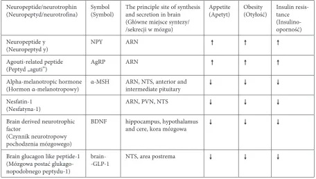
of these neurotransmitters can be involved in the maintenance of peripheral glucose homeostasis, the regulation of peripheral tissue sensitivity to insulin, and even the modulation of insulin secretion. Cen-tral transmitters involved in the maintenance of the body‘s energy balance have been classified into two basic groups [5]: orexigenic transmitters (increasing appetite) and anorexigenic transmitters (decreasing appetite). This first group includes: neuropeptide Y (NPY), agouti-related peptide (AgRP), orexin-A and -B, and endocannabinoids. The group of anorexigen-ic transmitters, in turn, includes: alpha-melanotropanorexigen-ic hormone (α-MSH), and cocaine and amphetamine regulated transcript (CART). Recently, newly iden-tified compounds regulating the energy state of the body have been increasingly researched and include: brain glucagon-like peptide 1 (GlP-1), nesfatin-1, and brain-derived neurotrophic factor (BDNF). In an era of dynamic research on the role of the incretin axis in the development of T2DM, these factors seem to play an important role in the central regulation of energy and carbohydrate homeostasis (Table 1).
Neuropeptide Y
Neuropeptide Y is a compound that strongly stimulates appetite. Chronic, intraventricular
in-Table 1. Anorexigenic and orexigenic factors
Tabela 1. Czynniki anoreksygeniczne i oreksygeniczne
Neuropeptide/neurotrophin
(Neuropeptyd/neurotrofina) Symbol(Symbol) The principle site of synthesis and secretion in brain (Główne miejsce syntezy/ /sekrecji w mózgu)
Appetite
(Apetyt) obesity(otyłość) Insulin resis-tance (Insulino- oporność) Neuropeptide y
(Neuropeptyd y) NPY ARN
Agouti-related peptide
(Peptyd „aguti”) AgRP ARN
Alpha-melanotropic hormone
(Hormon α-melanotropowy) α-MSH ARN, NTS, anterior and intermediate pituitary Nesfatin-1
(Nesfatyna-1) ARN, PVN, NTS
Brain derived neurotrophic factor
(Czynnik neurotropowy pochodzenia mózgowego)
BDNF hippocampus, hypothalamus
and cere, kora mózgowa
Brain glucagon like peptide-1 (Mózgowa postać glukago-nopodobnego peptydu-1)
brain-
-GlP-1 NTS, area postrema
ARN – arcuate nucleus of the hypothalamus; NTS – nucleus of the solitary tract; PVN – paraventricular nuclei of the hypo-thalamus; – reduces appetite/obesity/insulin resistance; – stimulates appetite/obesity/insulin resistance.
fusion of NPY in rats has resulted in an increased appetite and the development of obesity [5, 6]. The hypothalamus, and particularly its arcuate nucleus, constitutes the principle site of synthesis and secretion of NPY. The activity of neurons se-creting neuropeptide Y can be modulated by vari-ous factors. Decreased release of this transmitter is observed in the presence of increased levels of insulin, leptin and glucose, i.e. during the so-called “satiety state” of body. By contrast, NPY neurons are stimulated whenever the body is “fasting”, i.e. when the levels of these aforementioned substances are low [6]. Also, obesity is associated with higher levels of neuropeptide Y. Although the levels of leptin are high in obese individuals, its signaling is impaired due to leptin-resistance [6]. Finally, glu-cocorticoids have also been revealed to stimulate NPY neurons.
In animals that were administered neuro-peptide Y intraventricularly, a gradual decrease in liver sensitivity to insulin was observed, and consequently, a limited ability to inhibit glucose synthesis [6]. The same study revealed that neuro-peptide Y exerts these effects via the efferent sym-pathetic nerves supplying the liver. The authors of this study concluded that therapy including NPY receptor blockers could be a potential treatment method in diabetes and insulin resistance [6]. Another potential therapeutic modality could be targeted on the selective blocking of the respective sympathetic fibers innervating the liver [6].
AgRP
Agouti-related peptide is another orexigenic factor, i.e. a compound that stimulates appetite [5]. Similar to neuropeptide Y, the main source of AgRP includes neurons located in the arcuate nucleus of the hypothalamus. The release of AgRP is inhibited by leptin and stimulated by ghrelin [5]. AgRP influences energy homeostasis of the body mostly by acting through the melanocortin system, and particularly by way of type 3 and 4 melanotro-pic receptors (MC3 and MC4 receptors) [8]. Act-ing antagonistically on this system, AgRP can lead to increased appetite and a resultant development of obesity. Central administration of AgRP results in an inhibited metabolism, increased appetite and gain in body mass [9]. Increased plasma concen-trations of agouti-related peptide were observed in a study of obese males [7]. This parameter was positively correlated with body mass index (BMI), fat tissue mass, plasma levels of α-MSH and leptin (both being increased in obese individuals), and with fasting concentrations of insulin [7].
POMC
Proopiomelanocortin (PoMC) is a precursor polypeptide for such active peptides as adreno-corticotropic hormone (ACTH), β-lipoproteins and melanotropic hormones (α-MSH, β-MSH, γ-MSH). The derivatives of PoMC play various roles in the body: they modulate secretion by the adrenal cortex, regulate sexual behaviors and lac-tation, affect skin pigmenlac-tation, carry out immune functions, and are involved in the maintenance of energy balance [12]. In the central nervous system, PoMC neurons are localized predominantly in the hypothalamus (particularly in the arcuate nucleus) and in the nucleus of the solitary tract; addition-ally, they are found in the anterior and interme-diate pituitary [9, 12]. Therefore, PoMC neurons are found in regions that are particularly involved in the energy control of the body. PoMC-null mice are characterized by minimal adrenal gland size, undetectable serum concentrations of gluco-corticoids, and obesity, with these features being more pronounced in homozygotic individuals. Melanocortin peptides, especially alpha-MSH, ap-pear to have a critical role in the regulation of body weight, despite the absence of corticosteroids [9, 13]. Although the prevalence of diabetes was not increased significantly among these animals, they were characterized by some disorders of carbohy-drate metabolism regulation [9, 13]. Most of these disorders pertained to the impaired secretion of glucagon under hypoglycemic conditions [9, 13]. The lack of glucocorticoids could explain normal levels of insulin and glucose as well as defect of the counterregulatory response in insulin-induced hy-poglycemia. However, long-term supplementation with those hormones didn‘t lead to hyperglycemia, although it did prevent hypoglycemia. [9, 13] The relative effects of lack of melanocortins versus lack of glucocorticoids on appetite, body weight ho-meostasis and glucose hoho-meostasis is still unclear. It is known that the regulation of that homeostasis requires the integration of both central and pe-ripheral melanocortin signaling systems [9, 13].
receptor and obesity was observed in humans [9]. Recently, autoantibodies against the MC4 receptor were identified in obese individuals [9]. Knowing the function of these receptors, researchers have tried to understand their mechanism of action. Thus far, the activation of MC4R has been proven to be associated with the release of other factors involved in the control of energy balance, such as: BDNF, oxytocin, and corticotropin-releasing hormone (CRH) [9]. Additionally, an increased expression of nesfatin-1 was observed during the stimulation of MC4 receptors [9].
Nesfatin-1 is one of the more recently analy-zed elements of melanocortin pathway activation [9]. As mentioned above, this pathway is inhibited by agouti-related peptide, one of the orexigenic factors which acts antagonistically on type 3 and 4 melanotropic receptors [8–9].
Recent research has been centered upon the possible association of the melanotropic system (particularly α-MSH) with insulin resistance and diabetes. Impaired signaling of alpha-melano-tropic hormone at the level of the central ner-vous system (resulting from a defect of the MC4 receptor and to a lesser extent the MC3 receptor) is reflected by an increase in insulin resistance, while intracerebral administration of this peptide is associated with reduced insulin resistance [10]. Additionally, improved insulin sensitivity was observed in MC4 receptor-deficient mice during the peripheral administration of melanocortin system agonists [10]. Moreover, previous studies confirmed the protective influence of α-MSH on pancreatic islets. Alpha-MSH protected pancreatic islets from pro-inflammatory cytokines, enabling their proper endocrine function in terms of insulin secretion [9, 11].
Nesfatin-1
In 2006, Japanese researchers identified anoth-er intanoth-eresting compound in the central nanoth-ervous sys-tem, designated nesfatin-1. Numerous recent stud-ies have cleared up most of the questions dealing with the physiological role of this factor. Nesfatin-1 was revealed to be another regulator of the energy homeostasis of the body [14–16]. It is comprised of 82 amino acids, and originates from nucleobindin 2 protein (NUCB2) [15]. Expression of nesfatin-1 is observed in the brain, and particularly in regions that are significantly involved in the control of appetite and body mass. These regions include the hypotha-lamus (particularly the arcuate and paraventricular nuclei) and the brain stem (especially the nucleus of the solitary tract) [14–16]. Nesfatin-1 can cross the blood-brain barrier bidirectionally [14–15].
Authors of these studies assumed that besides the central form, there also probably exists a periphe-ral form of this polypeptide (as in the case of GlP-1), and searched for this form with a focus on the alimentary tract. Finally, they found nesfatin-1 in the endocrine cells of the stomach and duodenum (gastric form) [14–15]. Additionally, a pancreatic form of this peptide was identified, with expres-sion in the pancreatic islets of langerhans (both in murine and rat β cells, and in human β cells) [14]. Recently, both the expression of mRNA for NUCB2 precursor protein and the expression of nesfatin-1 itself were found in adipose tissue of animals and humans. In the case of clinical studies, experimental material was obtained by biopsy performed during scheduled surgeries. In the same studies, plasma lev-els of nesfatin-1 were determined in humans with no significant gender-related differences observed. A significant association was observed, however, between nesfatin-1 levels and higher values of BMI in humans. Additionally, an increased expression of this compound was observed in the adipose tissue of animals that were switched onto a fat-rich diet. In these animals, the levels of nesfatin-1 in fat tissue were positively correlated with an increase in body mass resulting from the excessive supply of energy [20]. Therefore, authors of this study suggested that nesfat1 might act as some kind of adipokine in-volved in the development of obesity. The activity of nesfatin-1 was compared to leptin as well, an-other factor released by fat tissue cells, an increased expression of which is also detected in obese indi-viduals [20].
not require the presence of leptin [16]. This finding raises a possibility for the future treatment of obese individuals with leptin resistance and impairment of some signaling pathways based on anorexigenic neurotransmitters [16].
Recently, interesting findings were published by Chinese researchers who analyzed the plasma levels of nesfatin-1 in patients with type 1 and type 2 diabetes. Similar to previous research, this study did not reveal any gender-related differences in nesfa-tin-1 concentration. However, a slight increase in nesfatin-1 levels was observed in type 1 diabetic patients when compared to healthy controls, while type 2 diabetic individuals were characterized by a significant decrease in this parameter [21]. This study provided further evidence that levels of nesfa-tin-1 can change with various pathological stages. Although the mechanisms behind the action of this factor and its role in the regulation of various vital functions are still not fully understood, the results of previous studies suggest that it is significantly associated with metabolic disorders. Therefore, be-sides understanding the role of nesfatin-1 in reduc-ing obesity, studies on its influence on carbohydrate metabolism seem another important direction of further research [21–22].
GLP-1
Glucagon like peptide-1 is a hormone released by the l cells of the alimentary mucosa, and partic-ularly by the cells of the small intestine. Addition-ally, a cerebral form of GlP-1 (brain glucagon like peptide-1) has been identified, which is released in the central nervous system and plays the role of a neuropeptide [5, 18, 19]. In the brain, GlP-1 was found predominantly in cells of the nucleus of the solitary tract and in the area postrema [18–19]. It was revealed that not only the peripheral form of GlP-1 is involved in the regulation of the pancre-atic secretion of insulin [19]. During experimental intraventricular infusion of a central GlP-1 re-ceptor agonist (exendin Ex4), a four-fold increase in insulin secretion was observed, along with en-hanced hepatic glycogenesis and reduced muscular glycogenesis [18]. These processes were observed under hyperglycemic conditions obtained by means of intravenous infusion of glucose. By con-trast, intraventricular infusion of a GlP-1 receptor antagonist (exendin Ex9), resulted in the decreased pancreatic secretion of insulin, along with a lower deposition of glycogen in the liver [18].
Therefore, brain glucagon like peptide-1 modulates the release of insulin from pancreatic β cells [18-19]. Additionally, this neuropeptide is involved in the utilization of glucose, stimulating
liver deposition of glycogen [18]. GlP-1 exerts its effects under hyperglycemic conditions, in order to prevent postprandial hypoglycemia [18].
The presence of glucose in the alimentary tract is a kind of stimulus inducing the pancrea-tic release of insulin. This process involves GlP-1 and is known as the incretin effect. Stomach and intestinal “detectors” of glucose are connected to the brain via the autonomic nervous system [18]. Central impairment of the action of cerebral GlP-1 may lead to the blockage of the peripheral incretin effect. This relationship was confirmed in mice during the intraventricular administration of a central GlP-1 receptor antagonist (exendin Ex9) under hyperglycemic conditions stimulated by the intravenous or intragastric administration of glu-cose. A similar degree of reduction in plasma in-sulin levels was observed irrespective of the route of glucose administration [18].
BDNF
metabo-lism, decreasing the blood concentration of gluco-se, improving its tolerance and insulin resistance [23–28]. Decreased plasma levels of BDNF have been observed in obesity and type 2 diabetes (also in cases of diabetes with no concomitant over-weight or obesity). This aforementioned relation-ship was confirmed both in animal studies and in humans [23–25, 27]. However, contrary find-ings were published by the Japanese authors; they found markedly elevated serum levels of BDNF in women with newly diagnosed type 2 diabetes, who did not previously receive any anti-diabetic treat-ment (either non-pharmacological managetreat-ment or hypoglycemic agents) [26, 27]. Additionally, the same clinical trial revealed a positive correla-tion between serum BDNF levels and body mass index [26]. Authors of this study concluded that the increased serum level of BDNF might result from an enhanced release of BDNF from blood platelets during the early stages of the disease. This could be some kind of protective or compensatory mechanism (particularly in light of the fact that blood platelets constitute the main depository of circulating BDNF). These mechanisms are most likely impaired due to the metabolic disorders tak-ing place in the later stages of diabetes and obesity, which are reflected by decreased serum levels of this neutrophin [26].
BDNF can cross the blood-brain barrier; its transfer from the central level to the periphery can be inhibited under hyperglycemic conditions [23]. It is probable that the concentration of circulating BDNF depends on the serum glucose concentra-tion, but is not related to the blood level of insulin. This relationship was also confirmed in humans in a study by Krabbe et al. [25]. They observed that peripheral secretion of BDNF was decreased by elevated blood levels of glucose. No significant changes in the levels of BDNF that crossed the blood-brain barrier were observed in the case of hyperinsulinemia with coexisting normoglycemia [25]. In the same study, the authors assumed that inflammation is involved in the etiopathogenesis of type 2 diabetes, and analyzed the association be-tween BDNF levels and CRP concentration. They found no significant relationship between these two parameters and therefore confirmed that the level of BDNF depends on disorders of carbohy-drate metabolism irrespective of concomitant in-flammation [25].
In view of the established function of BDNF, including its neuroprotective properties, involve-ment in body metabolism, and influence on car-bohydrate metabolism, it is currently considered a common pathogenic factor for such diseases as type 2 diabetes and neuropsychiatric and neurode-generative disorders [23, 25].
It is a well-known fact that diabetes is an inde-pendent risk factor for stroke. This may be caused by the inhibition of neurotrophin secretion and a reduced neuroprotection. Decreased expression of BDNF in microvascular brain endothelial cells was observed in a study of rats with streptozoto-cin-induced diabetes. This is probably associated with an increased accumulation of advanced gly-cation end products in microvessels as the result of prolonged hyperglycemia [32].
Recently, participation of BDNF in neuronal damage in diabetic retinopathy has also been em-phasized. Knowing its important role in the survival of neurons, researchers are considering its potential role in the rescue of photoreceptors via treatment in combination with ciliary neurotrophic factor (CNTF). In this case, the mechanism of action of BDNF may depend on its regulation of glutamine synthetase and increased glutamate uptake [31].
References
[1] Gerozissis K: Brain Insulin: Regulation, Mechanisms of Action and Functions. Cell Mol Neurobioly 2003, 23, 1–25.
[2] Brüning JC, Gautam D, Burks DJ, Gillette J, Schubert M,Orban PC, Klein R, Krone W, Müller-Wieland D, Kahn CR: Role of Brain Insulin Receptor in Control of Body Weight and Reproduction. Science 2000, 22, 2122– –2125.
[3] Schulingkamp RJ, Pagano TC, Hung D, Raffa RB: Insulin receptors and insulin action in the brain: review and clinical implications. Neurosci Biobehav Rev 2000, 24, 855–872.
[4] Pagotto U: Where Does Insulin Resistance Start? Diabetes Care 2009, 32, 174–177.
[5] Kocełak P, Zahorska-Markiewicz B, Olszanecka-Glinianowicz M: Hormonalna regulacja przyjmowania pokar-mu. Endokrynol Pol 2009, 60, 296–301.
[6] Hoek AM, Heijningen C, Schro¨der-van der Elst JP, Ouwens DM, Havekes LM, Romijn JA, Kalsbeek A, Pijl H: Intracerebroventricular Administration of Neuropeptide Y Induces Hepatic Insulin Resistance via Sympathetic Innervation. Diabetes 2008, 57, 2304–2310.
[7] Katsuki A, Sumida Y, Gabazza EC, Murashima S, Tanaka T, Furuta M, Araki-Sasaki R, Hori Y, Nakatani K, Yano Y, Adachi Y: Plasma levels of Agouti-Related Protein Are Increased in obese Men. J Clin Endocrinol Metab 2001, 86, 1921–1924.
[8] Li-Ying Fu, Pol AN: Agouti-Related Peptide and MC3/4 Receptor Agonists Both Inhibit Excitatory Hypothalamic Ventromedial Nucleus Neurons. J Neurosci 2008, 28, 5433–5449.
[9] Mountjoy KG: Functions for pro-opiomelanocortin-derived peptides in obesity and diabetes. Biochem J 2010, 428, 305–324.
[10] Costa JL, Hochgeschwender U, Brennan M: The role of melanocyte-stimulating hormone in insulin resistance and type 2 diabetes mellitus. Treat Endocrinol. 2006, 5, 7–13.
[11] Jung EJ, Han DJ, Chang SH, Lim DG, Wee YM, Kim JH, Kim YH, Koo SK, Choi M, Kim SC: Protective effect of alpha-melanocyte-stimulating hormone on pancreas islet cell against peripheral blood mononuclear cell-mediated cytotoxicity in vitro. Transplant Proc 2007, 39, 1604–1606.
[12] Millington G WM: The role of proopiomelanocortin (PoMC) neurones in feeding behaviour. Nutr Metab 2007, 4, 18.
[13] Hochgeschwender U, Costa JL, Reed P, Bui S, Brennan MB: Altered Glucose Homeostasis in Proopiomelanocortin-Null Mouse Mutants lacking Central and Peripheral Melanocortin. Endocrinology 2003, 144(12), 5194–5202.
[14] Zhang AQ, Li XL, Jiang CY, Lin L, Shi RH, Chen JD, Oomura Y: Expression of nesfatin-1/NUCB2 in rodent digestive system. World J Gastroenterol 2010, 16, 1735–1741.
[15] Stengel A, Goebel M, Yakubov I, Wang L, Witcher D, Coskun T, Taché Y, Sachs G, Nils SG, Lambrecht WG:
Identification and Characterization of Nesfatin-1 Immunoreactivity in Endocrine Cell Types of the Rat Gastric oxyntic Mucosa. Endocrinology 2009, 150, 232–238.
[16] Maejima Y, Sedbazar U, Suyama S, Kohno D, Onaka T, Takano E, Yoshida N, Koike M, Uchiyama Y, Fujiwara K,
Yashiro T, Horvath TL, Dietrich MO, Tanaka S, Dezaki K, Oh-I S, Hashimoto K, Shimizu H, Nakata M, MoriM,
Yada T: Nesfatin-1-Regulated oxytocinergic Signaling in the Paraventricular Nucleus Causes Anorexia through a leptin-Independent Melanocortin Pathway. Cell Metab 2009, 10, 355–365.
[17] Voss U, Riva M, Dekker Nitert M, Ling C, Lindqvist A, Wierup N: Nesfatin-1 stimulates insulin secretion, inhibits glucagon secretion and is expressed in human and rodent beta cells. Diabetologia 2010, 53, 548.
[18] Knauf C, Cani PD, Perrin C, Iglesias MA, Maury JF, Bernard E, Benhamed F, Grémeaux T, Drucker DJ, Kahn CR, Girard J, Tanti JF, Delzenne NM, Postic C, Burcelin R: Brain glucagon-like peptide-1 increases insulin secretion and muscle insulin resistance to favor hepatic glycogen storage. J Clin Invest 2005, 115, 3554–3563.
[19] Sandoval D: CNS GlP-1 Regulation of Peripheral Glucose Homeostasis. Physiol Behav. 2008, 94, 670–674.
[20] Ramanjaneya M, Chen J, Brown JE, Tripathi G, Hallschmid M, Patel S, Kern W, Hillhouse EW, Lehnert H, Tan BK, Randeva HS: Identification of Nesfatin-1 in Human and Murine Adipose Tissue: A Novel Depot-Specific Adipokine with Increased levels in obesity. Endocrinology 2010, 150, 3169–3180.
[21] Li QC, Wang HY, Chen X, Guan HZ, Jiang ZY: Fasting plasma levels of nesfatin-1 in patients with type 1 and type 2 diabetes mellitus and the nutrient-related fluctuation of nesfatin-1 level in normal humans. Regul Peptides 2010, 159, 72–77.
[22] García-Galiano D, Navarro VM, Gaytan F, Tena-Sempere M: Expanding roles of NUCB2/nesfatin-1 in neu-roendocrine regulation. J Mol Endocrinol 2010, 45, 281–290.
[23] Żełobowska K, Gumprecht J, Grzeszczak W: Neurotropowy czynnik pochodzenia mózgowego (BDNF) – udział w patogenezie insulinooporności i cukrzycy typu 2. Przew lek 2007, 4, 79–83.
[24] Pedersen BK, Pedersen M, Krabbe KS, Bruunsgaard H, Matthews VB, Febbraio MA: Role of exercise-induced brain-derived neurotrophic factor production in the regulation of energy homeostasis in mammals. Exp Physiol 2009, 94, 1153–1160.
[25] Krabbe KS, Nielsen AR, Krogh-Madsen R, Plomgaard P, Rasmussen R, Erikstrup C, Fischer CP, Lindegaard B, Petersen AMW, Taudorf S, Secher NH, Pilegaard H, Bruunsgaard H, Pedersen BK: Brain-derived neurotrophic factor (BDNF) and type 2 diabetes. Diabetologia 2007, 50, 431–438.
[27] Żołądź JA, Pilc A: The effect of physical activity on the brain derived neurotrophic factor: from animal to human studies. J Physiol Pharmacol 2010, 61, 533–541.
[28] Noble EE, Billington CJ, Kotz CM, Wang C: The lighter side of BDNF. Am J Physiol Regul Integr Comp Physiol 2011, 300, 1053–1069.
[29] Wang M, Chan Y, Lee H, Hong L: Regulation of the Intracerebroventricular Administration of Brain-Derived Neurotrophic Factor on Baroreflex Function and Insulin Sensitivity in Rats. Chin J Physiol 2012, 55, 184–191.
[30] Pedersen B: Muscles and their myokines. J Exp Biol 2011, 214, 337–346.
[31] Ola MS, Nawaz MI: Cellular and Molecular Mechanism of Diabetic Retinopathy. In: Diabetic Retinopathy. Eds.: ola MS, InTech 2012, 1–30.
[32] Navaratna D, Guo S, Hayakawa K, Wang X, Gerhardinger C, Lo EH: Decreased Cerebrovascular Brain-Derived Neurotrophic Factor-Mediated Neuroprotection in the Diabetic Brain. Diabetes 2011, 60, 1789–1796.
Address for correspondence:
Marta Wasilewska
Department of Angiology, Hypertension and Diabetology Wroclaw Medical University
Borowska 213 50-556 Wrocław Poland
Tel. +48 71 733 22 00 E-mail: [email protected]
Conflict of interest: None declared