R E S E A R C H
Open Access
Validation of a wireless dry electrode
system for electroencephalography
Sarah N Wyckoff
1*, Leslie H Sherlin
1,2,3, Noel Larson Ford
1,3and Dale Dalke
1Abstract
Background:Electroencephalography (EEG) is a widely used neuroimaging technique with applications in healthcare, research, assessment, treatment, and neurorehabilitation. Conventional EEG systems require extensive setup time, expensive equipment, and expertise to utilize and therefore are often limited to clinical or laboratory settings. Technological advancements have made it possible to develop wireless EEG systems with dry electrodes to reduce many of these barriers. However, due to the lack of homogeneity in hardware, electrode evaluation, and methodological procedures the clinical acceptance of these systems has been limited.
Methods:In this investigation the validity of a wireless dry electrode system compared to a conventional wet electrode system was assessed, while addressing methodological limitations. In Experiment 1, the signal output of both EEG systems was examined at Fz, C3, Cz, C4, and Pz using a conductive head model and generated test signals at 2.5 Hz, 10 Hz, and 39 Hz. In Experiment 2, two-minutes of eyes-closed and eyes-open EEG data was recorded simultaneously with both devices from the adjacent electrode sites in a sample of healthy adults. Results:Between group effects and frequency*device and electrode*device interactions were assessed using a mixed ANOVA for the simulated and in vivo signal output, producing no significant effects . Bivariate correlation coefficients were calculated to assess the relationship between electrode pairs during the simultaneous in vivo recordings, indicating a significant positive relationship (allp's < .05) and larger correlation coefficients (r>±0.5) between the dry and wet electrode signal amplitude were observed for theta, alpha, beta 1, beta 2, beta 3, and gamma in both the eyes-closed and eyes-open conditions.
Conclusions:This report demonstrates preliminary but compelling evidence that EEG data recorded from the wireless dry electrode system is comparable to data recorded from a conventional system. Small correlation values in delta activity were discussed in relation to minor differences in hardware filter settings, variation in electrode placement, and participant artifacts observer during the simultaneous EEG recordings. Study limitations and impact of this research on neurorehabilitation were discussed.
Keywords:Dry electrode, Electroencephalography, Signal generator, Signal quality, Validity, Wireless EEG
Background
Non-invasive electroencephalography (EEG) is a neuro-imaging technique that measures cortical electrical activ-ity of the brain with applications in healthcare, research, assessment, treatment, and neurorehabilitation. Digital (conventional) EEG systems are considered the established guideline for clinical EEG acquisition; recording voltage fluctuations using wired electrodes, digital amplifiers, and a direct connection to a laptop or desktop computer for
data storage and analysis [1]. In clinical settings, a reg-istered electroneurodiagnostic technologist and clinical electroencephalographer facilitate the acquisition and interpretation of clinical EEG recordings, while trained EEG technicians working under the supervision of a qualified electroencephalographer may facilitate data col-lection and analysis of EEG recordings from research and non-clinical populations in a laboratory setting [2]. The standard procedure for data collection requires accurate identification of recording sites (International 10–20 sys-tem), electrode site preparation with abrasive cleaners, electrode application/fixation (single lead with conductive * Correspondence:[email protected]
1SenseLabs, Mesa, Arizona, Atascadero, CA, USA
Full list of author information is available at the end of the article
paste or electrode cap system with injected conductive gel), and proper ground and reference electrode placement [3]. The cumbersome nature of conventional EEG systems and the need for assistive application make it difficult to conduct research outside of controlled clinical and labora-tory settings, limiting in vivo and ambulalabora-tory research opportunities. Additionally, these limitations, as well as the high cost of conventional systems, create barriers for providers and individuals interested in utilizing EEG-based applications such as neuropsychological assessment, neurofeedback, or brain-computer interface for restorative or assistive neurorehabilitation or treatment monitoring.
In recent years, wireless technology and advancements in conductive materials have led to the development of several wireless EEG dry electrode systems for research and commercial use. Several validation studies have directly compared the signal output of dry and wet (pasted/gelled) electrode systems (review, see [4–6]). However this body of research has been criticized due to the lack of homogeneity in hardware and electrode evaluation procedures and statistical methodology. In a recent review, Gargiulo and colleagues [5] highlight several problems associated with current validation procedures; recommending researchers provide a comparative assessment of the proposed device with a reference device, thorough quantitative measure-ment and characterization of the electrical circuit of study devices, qualitative evaluations of physiological signals, re-port of compliance with technical standards, and long-term monitoring and multicenter studies to facilitate clin-ical acceptance. In their review of dry electrode validation research, Lopez-Gordo and colleagues [6] emphasize that heterogeneity in evaluation procedures limit the com-parison of results between investigations and suggest mandatory reporting of the following study related charac-teristics: mechanical, electrical, evaluation, and usability.
The current investigation evaluates the validity of a wireless dry electrode system compared to a conventional wet electrode system, while addressing methodological limitations and complying with recommended reporting practices to standardize EEG system validation research. In a series of experiments, quantitative and qualitative aspects of the study related hardware and electrode per-formance were evaluated using simulated and in vivo re-cording techniques.
Methods
Ethics
All participants were provided written informed consent in accordance with the ethical conditions set forth as part of a larger data collection study overseen by the Western IRB (#20141246). Participants provided written consent to allow their data to be stored in a large database, de-identified, and published.
Study devices
For this investigation, study devices included the Versus wireless dry electrode system (SenseLabs, Mesa, AZ & Atascadero, CA, USA) and the Mitsar-201 conventional wet electrode system (Mitsar Ltd, St. Petersburg, Russia). Figure 1 provides a visual representation of the study de-vices. Table 1 provides the system specifications of the study devices. The Versus wireless headset with EEG Stream software features five embedded 15-prong carbon-silicon dry electrodes at the following International 10–20 locations (Fz, C3, Cz, C4, Pz) with an integrated double-sided reference-ground earclip. Figure 2 provides a dia-gram of the Versus headset dry electrode and electrical circuit. The Mitsar-201 was selected as the reference de-vice in the investigation, as it is a widely used, laboratory-based, EEG amplifier with 510 K (K143233) approval from the FDA. The Mitsar-201 amplifier with WinEEG software allows for the input of 19 EEG, 2 reference (A1 and A2), and 1 ground electrode using individual DIN style elec-trodes or an electrode cap with a serial port connector. For this investigation, single lead 9 mm flat DIN style gold electrodes were used for comparative testing with the con-ventional wet electrode system. Both study devices were in compliance with the guidelines set forth by the Ameri-can Clinical Neurophysiology Society for the technical re-quirements for recording of EEG [7, 8] and other published technical standards [9]. The wireless technology utilized by the Versus headset is equivalent to that of a cell phone or wireless cellular headset and compliant with and eligible for the "low power exclusion" under the COMAR [10] standards for exposure to radio frequency devices.
Description of experiments
In experiment 1, study devices were compared using a signal generator to apply multiple test signals to a conduct-ive head model with wet and dry electrodes attached at Fz, C3, Cz, C4, and Pz, with the Versus reference/ground elec-trode attached to the left ear (A1), and the Mitsar refer-ences and ground electrodes connected to the left and right ear and forehead, A1, A2, and FPz - respectively. In a series of 5-min serial EEG recordings, a 2.5 Hz, 10 Hz, and 39 Hz test signal was applied under a low and high resist-ance condition. The low resistresist-ance condition simulated proper electrode connection and the high resistance condi-tion simulated poor electrode conneccondi-tion.
and a five-minute eyes-open resting-state condition. Due to the fixed electrode positioning of the Versus headset, dry electrodes were located at Fz, C3, Cz, C4, and Pz with the reference and ground electrodes fixed to the left ear-lobe (A1). Under the supervision of the EEG technician, participants were instructed to place the headset on their
head and gently rock the device back and forth to allow the flexible carbon-silicon electrode protuberances to make contact with their scalp. Participants then clipped the reference/ground electrode to their left ear. Application of the Versus headset took approximately 2-min follow-ing the demonstration and required minimal assistance
Fig. 1Study devices . Graphic representation ofaVersus wireless electrode system with carbon-silicon dry electrodes andbMitsar 201 amplifier with DIN style gold wet electrodes
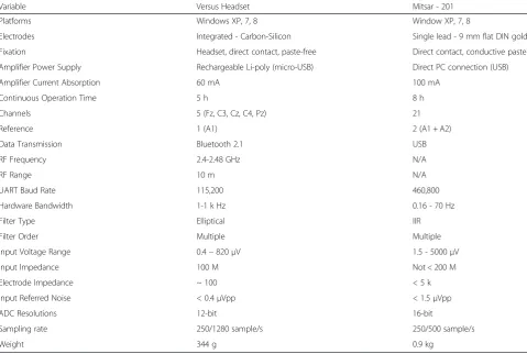
Table 1System specifications for study devices
Variable Versus Headset Mitsar - 201
Platforms Windows XP, 7, 8 Window XP, 7, 8
Electrodes Integrated - Carbon-Silicon Single lead - 9 mm flat DIN gold
Fixation Headset, direct contact, paste-free Direct contact, conductive paste
Amplifier Power Supply Rechargeable Li-poly (micro-USB) Direct PC connection (USB)
Amplifier Current Absorption 60 mA 100 mA
Continuous Operation Time 5 h 8 h
Channels 5 (Fz, C3, Cz, C4, Pz) 21
Reference 1 (A1) 2 (A1 + A2)
Data Transmission Bluetooth 2.1 USB
RF Frequency 2.4-2.48 GHz N/A
RF Range 10 m N/A
UART Baud Rate 115,200 460,800
Hardware Bandwidth 1-1 k Hz 0.16 - 70 Hz
Filter Type Elliptical IIR
Filter Order Multiple Multiple
Input Voltage Range 0.4–820μV 1.5 - 5000μV
Input Impedance 100 M Not < 200 M
Electrode Impedance ~ 100 < 5 k
Input Referred Noise < 0.4μVpp < 1.5μVpp
ADC Resolutions 12-bit 16-bit
Sampling rate 250/1280 sample/s 250/500 sample/s
from the EEG technician. Single lead wet electrodes were fixed to the scalp using conductive paste by the EEG technician. The wet electrodes were placed poster-ior to the C3, Pz, and C4 dry electrodes, and to slightly to the left posterior of the Fz and Cz dry electrode with the references and ground electrodes connected to the left and right ear and forehead, A1, A2, and FPz - re-spectively. The site preparation and application of the 8 single lead electrodes took approximately 10-min. The placements of dry and wet electrodes for the simultan-eous recordings are depicted in Fig. 3.
Signal processing
For both experiment 1 and 2, data collected using the Versus headset was exported in ASCII (.txt) format and data collected using the wet electrode system was re-referenced to A1 and exported in EDF format. All data files were imported and processed using the Brain Vision Analyzer software (version 1.05, Brain Product GmbH, Germany). Each record was down-sampled to 128sps, bandpass filtered from 1.5-45 Hz, and synchronization markers were manually applied based on participant arti-facts prompted at the onset of each task condition. Two minutes of continuous non-artifacted data from each con-dition (signal generator test, eyes-closed, and eyes-open) were segmented into 1 s epochs and subjected to Fast Fourier Transform (FFT) analysis (full spectrum, power, 0.5 Hz resolution, no windowing). Mean amplitude values (uV) for delta (1.5-3.5 Hz), theta (4–7.5 Hz), alpha (8–12 Hz), beta 1 (13–16 Hz), beta 2 (13–21 Hz), beta 3 (21–32 Hz), and gamma (35–45 Hz) frequency bands at each electrode site (Fz, C3, Cz, C4, and Pz) was exported for statistical analysis using IBM SPSS Statistics (Version 20).
Statistical analysis
The following hypothesis was tested in experiment 1. Hypothesis 1: mean amplitude values for delta, alpha, and gamma frequency bands at Fz, C3, Cz, C4, and Pzwill not be significantly different between the Versus dry electrode system and the Mitsar wet electrode system during the signal generator testing protocol. The following hypotheses
Fig. 2Diagram of the Versus headset dry electrode system
were tested in experiment 2. Hypothesis 2: mean amplitude values for delta, theta, alpha, beta 1, beta 2, beta3, and gamma frequency bands at Fz, C3, Cz, C4, and Pzwill not be significantly between the Versus dry electrode system and the Mitsar wet electrode system during the eyes-closed and eyes-open in vivo participant testing protocols. Hypothesis 3: mean amplitude values for delta, theta, alpha, beta 1, beta 2, beta3, and gamma frequency bands willbe significantly correlated between the Versus dry elec-trode system and the Mitsar wet elecelec-trode system during the eyes-closed and eyes-open in vivo participant testing protocols.
Hypotheses 1 and 2 were assessed using a mixed ANOVA, targeting the between-subjects effect of device and within-subjects interactions and pairwise comparisons of frequency*device and electrode*device. As multivariate analysis of variance (MANOVA) is not dependent upon the assumptions of sphericity, the Wilks’ Lambda multi-variate test statistics are reported when applicable. Hypoth-esis 3 was assessed by calculating the bivariate correlation coefficient between the dry and wet electrode system for each frequency band during the eyes-closed and eyes-open in vivo recording protocol. As the directional nature of the correlations was hypothesized, one-tailed probabilities were reported for all correlations.
Results
Participants
A convenience sample of nine right-handed healthy adults (3 female, 6 male), ages 18–64 years (M =47.11, SD = 15.19), volunteered to participate in the in vivo protocol of the current investigation.
Experiment 1
In the analysis of the signal generator protocol, the between-subjects effect of device produced a non-significant main
effect,F(1, 2) = 4.155,p= .178,ηρ2= .675. Due to insufficient residual degrees of freedom, only the multivariate test statistic for the frequency*device interaction could be produced, indicating a non-significant interaction effect, Wilks' λ= .004, F(1, 2) - 114.366, p= .066, η ρ2 = .996. Figure 4 displays the grand-average power spectral density plot of signal generator test protocol.
Experiment 2
In the analysis of the in vivo participant protocol, the be-tween-subjects effect of device produced a non-significant main effect for the eyes-closed recordings, F(1, 16) = .338, p= .569,ηρ2 = .021, and the eyes-open recordings,F(1, 16) = .061, p= .808, η ρ2 = .004. The multivariate test statistic for the frequency*device, Wilks' λ= .682, F(6, 11) = .856, p= .554, η ρ2 = .318, and electrode*device, Wilks' λ= .701, F(4, 13) = 1.388, p= .292, η ρ2 = .299, re-vealed non-significant interaction effects in the eyes-closed data. Similarly, the multivariate test statistic for the frequency*device, Wilks'λ= .835,F(6, 11) = .362, p= .888,
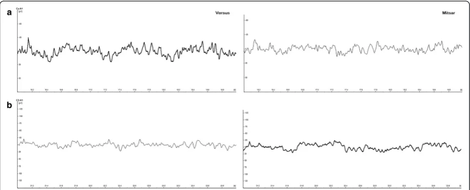
ηρ2= .165, and electrode*device, Wilks'λ= .629,F(4, 13) = 1.918,p= .167,ηρ2= .371, for the eyes-open data revealed non-significant interaction effects. Figure 5 provides a five-second sample of eyes-open simultaneous EEG traces taken from both dry/wet electrode systems for a single participant. Figure 6 provides the grand-average power spectral density plots for the participant sample, showing alpha wave attenuation at Pz during the eyes-open and eyes-closed recording conditions.
In the correlation analysis, a significant positive relation-ship (allp's < .05) between the dry and wet electrode signal amplitude was observed for theta, alpha, beta 1, beta 2, beta 3, and gamma in both the closed and eyes-open conditions. Additionally, large correlation coeffi-cients (r>± 0.5) were observed for these measures. A large correlation coefficient was observed for eyes-closed
delta activity,r= .580,p= .051, and a small correlation coefficient was observed for eyes-open delta activity, r= .278, p= .236, however, these positive relationships did not reach the level of significance. Table 2 provides a numeric summary of the Pearson's r and significance values. Correlations of dry and wet electrode frequency activity averaged across electrode sites for each record-ing condition are displayed in Fig. 8.
Discussion
In this study, serial simulated and simultaneous in vivo evaluation methods were employed to compare the signal output of EEG data recorded from a wireless dry electrode
system (Versus) and a conventional wet electrode sys-tem (Mitsar-201). electrodes. Hypothesis 1 was con-firmed, as no between-group effects for device and no frequency*device or electrode*device interaction effects were observed in delta, alpha, or gamma amplitudes for the comparison of signal generated activity at 2.5 Hz, 10 Hz, and 39 Hz. Hypothesis 2 was also confirmed, as no between-group effects for device and no frequency*device or electrode*device interaction effects were observed in delta, theta, alpha, beta 1, beta 2, beta 3, or gamma ampli-tudes for the comparison of simultaneous eyes-closed and eyes-open recordings in a sample of healthy adults. Hy-pothesis 3 was partially confirmed, as 85 % of the signal
Fig. 5Representative five-second sample of simultaneous EEG traces taken from pairs of dry/wet electrode combinations for a single participant. aUpper panel indicates wet electrode system signal [Mitsar] output.bLower panel indicates dry electrode system signal [Versus] output.Note. Sampling rate: 128 Hz, Filters: bandpass [1.5 Hz - 45 Hz] Gain: 50 uV
output correlations between devices produced a significant positive relationship (p> .05) under the eyes-closed and eyes-open conditions (Fig. 7), with 92 % demonstrating a large effect size (r> .5). Non-significant correlations and reduced effect sizes where observed in the delta frequency band, and predominantly during the eyes-open recording condition.
Several factors may account for the diminished correl-ation coefficients and effect sizes observed in the delta band activity. Factor 1, differences in delta activity may have been the product of differences in electrode placement rather than electrode detection. Lopez-Gordo and colleagues [6] assert that using a "same-time-different-place" approach for validation testing is controversial, as electrodes placed in different locations measure different ionic currents and dif-ferent electrical activity. For the simultaneous recordings, the Mitsar wet electrodes for Fz and Cz were placed slightly off the midline towards the left hemisphere, while the elec-trodes corresponding to C3, C4, and Pz were placed to the posterior of the dry electrodes. However, difference
in electrode position would likely be consistent across all frequency bands and both conditions. Factor 2, dif-ferences in delta activity may have been the product of differences in hardware based filters for both devices. Gargiulo and colleagues [6] suggest that the frequency bandwidth of a reference device and device under test should be identical, but warn researchers that this is not always possible due to non-excludable hardware and notch filtering. Although the raw data was exported and analyzed in a third-party software, the Versus dry elec-trode system utilizes a non-excludable high-pass filter of 1Hz at the hardware level, while the Mitsar wet electrode system utilizes a high-pass filter of 0.16 Hz. Fig. 8 provides a comparison of the frequency response of each device using different high-pass filters. It can easily be observed that the 1.0 Hz high-pass filter of the dry electrode system diminishes the delta frequency response. This would likely
Fig. 7Average correlations for each frequency band. Average correlations of dry and wet electrode signals across recording condition and electrode site for each frequency band [delta (1.5-3.5 Hz), theta (4–7.5 Hz), alpha (8–12 Hz), beta 1 (13–16 Hz), beta 2 (13–21 Hz), beta 3 (21–32 Hz),
gamma (35–45 Hz)]
Table 2Pearson’s(r) correlations values for dry and wet electrodes
Variables EC EO
r p r p
Delta 0.58 .051 0.28 .236
Theta 0.70 .017* 0.93 .000**
Alpha 0.98 .000** 0.99 .000**
Beta 1 0.93 .000** 0.99 .000**
Beta 2 0.96 .000** 0.99 .000**
Beta 3 0.89 .001** 0.97 .000**
Gamma 0.64 .031* 0.91 .000**
Note. Pearson's correlation coefficient effects sizes, ±.1 represents a small effect, ±.3 represents a medium effect, and ± .5 represents a large effect, EO = eyes-open, EC = eyes-closed. *Correlation significant at 0.05 level (1-tailed), **Correlation significant at 0.01 level (1-tailed)
produce the largest amplitude differences during the eyes-open condition, as blinks and eye-movement are more prominent and produce large slow wave amplitudes in the delta frequency range. Factor 3, differences in delta activity may have been the product of individual participant arti-facts during the recording. Review of the eyes-open raw recordings revealed two participants with increased slow-ing on one or more of the central electrode sites; one with prominent artifacts on the wet electrode system and one with prominent artifacts on dry electrode system (Fig. 9). Exclusion of these participants' central delta activity in the eyes-open condition increased the mean correlation coef-ficient,r= .541,p= .066, producing a large effect size and similar values observed in the eye-closed condition.
Limitations are present in the current research design, including a small sample size, limited number of compara-tive reference sites, limited study tasks and environments, and reference device differences. Future investigations should address the study limitations by (1) recruiting a lar-ger sample, (2) employing parallel and serial comparison methods or additional electrodes to the anterior and posterior position of the dry electrodes to generate an averaged comparison signal, (3) assessing event related potentials and/or include tasks designed to elicit a variety of mood and performance states, (4) EEG assessment in non-laboratory settings, (5) investigations of controlled physiological artifacts including electromyography and electrooculargraphy, (6) development of bypass or ex-cludable filters for reference device testing, and (7) fur-ther characterization of the electrical circuit and signal response. Despite the current limitations, this investiga-tion has many strengths, including the use of simulated,
in vivo, and comparative evaluations techniques with mul-tiple frequency ranges, recording conditions, and a refer-ence device, qualitative physiological signal evaluation at the individual and group level, and quantitative evaluation of the device characteristics.
Conclusions
The present study provides preliminary data pertaining to the validity of a specific wireless headset with dry elec-trodes. Overall, the data suggest that the raw EEG data re-corded by the wireless dry electrode system is of adequate quality to that of conventional wet electrode EEG systems. These results are promising, as wireless dry electrode technology has several advantages over conventional systems. These include increased portability (wireless, rechargeable), ease of use and decreased setup times for clinicians, participants, and researchers (self-appli-cation, paste/gel free, 2-min setup), reduced equipment costs (dry system $400, conventional system $10,500), and the opportunity to unobtrusively assess or train EEG activity at 5 standard electrode sites (Fz, C3, Cz, C4, Pz) in a variety of settings and tasks - enhancing the ecological validity.
Abbreviations
EEG:Electroencephalogram; FFT: Fast Fourier Transform.
Competing interests
Officer, Director of Applied Science and Product, Research and Data Scientist, and Chief Technology Officer.
Authors’contributions
SNW designed the experiments, collected the data, analyzed the results, and drafted the manuscript. LHS initiated the project, provided feedback on the experimental design and analysis, and modified the manuscript. NLF provided feedback on the experimental design and modified the manuscript. DD aided in the design of the Versus headset and provided technical and software support for the study devices, acquisition software, and signal generator testing. All authors read and approved the final manuscript.
Acknowledgements
The authors would like to thank the Versus research and development and hardware and software development teams at SenseLabs, Atascadero, California. We also wish to thank the EEG recording participants for their time.
Author details
1SenseLabs, Mesa, Arizona, Atascadero, CA, USA.2Department of Psychology, Northern Arizona University, Flagstaff, AZ, USA.3Department of Mind-Body Medicine, Southwest College of Naturopathic Medicine, Tempe, AZ, USA.
Received: 26 April 2015 Accepted: 26 October 2015
References
1. Nuwer M. Assessment of digital EEG, quantitative EEG, and EEG brain mapping: Report of the American Academy of Neurology and the American Clinical Neurophysiology Society. Neurology. 1997;49(1):277–92.
2. American Clinical Neurophysiology Society. Guideline 4: Standards of practice in clinical electroencephalography. Am J Electroneurodiagnostic Technol. 2006;46(3):220–1.
3. Pivik RT, Broughton RJ, Coppola R, Davidson RJ, Fox N, Nuwer MR. Guidelines for the recording and quantitative analysis of electroencephalographic activity in research contexts. Psychophysiology. 1993;30(6):547–58.
4. Lin CT, Ko LW, Chang MH, Duann JR, Chen JY, Su TP, et al. Review of wireless and wearable electroencephalogram systems and brain-computer interfaces–A mini-review. Gerontology. 2010;56(1):112–9.
5. Gargiulo G, Bifulco P, Cesarelli M, Fratini A, Romano M. Problems in assessment of novel biopotential front-end with dry electrode: A brief review. Mach. 2014;2:87–9.
6. Lopez-Gordo MA, Sanchez-Morillo D, Pelayo VF. Dry EEG electrodes. Sensors. 2014;14:12847–70.
7. American Electroencephalographic Society. Guideline one: Minimum Technical Requirements for Performing Clinical Electroencephalography. J Clin Neurophysiol. 1994;11(1):2–5.
8. American Clinical Neurophysiology Society. Guideline 8: Guidelines for recording clinical EEG on digital media. Am J Electroneurodiagnostic Technol. 2006;46(3):236–9.
9. Teplan M. Fundamentals of EEG measurement. Measurement Science Review. 2002;2(2):1–11.
10. RF and MW Subcommittee of the IEEE Committee on Man and Radiation, Human exposure to radio frequency and microwave radiation from portable and mobile telephones and other wireless communication devices–a COMAR technical information statement. IEEE Eng Med Biol Mag. 2001;20(1):128–31.
Submit your next manuscript to BioMed Central and take full advantage of:
• Convenient online submission
• Thorough peer review
• No space constraints or color figure charges
• Immediate publication on acceptance
• Inclusion in PubMed, CAS, Scopus and Google Scholar
• Research which is freely available for redistribution

![Fig. 4 Grand-average power spectral density of signal generator test protocol. Solid line indicates the wet electrode system [Mitsar] output, thedashed lines indicate the dry electrode system [Versus] output](https://thumb-us.123doks.com/thumbv2/123dok_us/9065906.1899072/5.595.56.541.532.704/spectral-generator-protocol-indicates-electrode-thedashed-indicate-electrode.webp)

![Fig. 7 Average correlations for each frequency band. Average correlations of dry and wet electrode signals across recording condition and electrodesite for each frequency band [delta (1.5-3.5 Hz), theta (4–7.5 Hz), alpha (8–12 Hz), beta 1 (13–16 Hz), beta 2 (13–21 Hz), beta 3 (21–32 Hz),gamma (35–45 Hz)]](https://thumb-us.123doks.com/thumbv2/123dok_us/9065906.1899072/7.595.56.291.575.699/correlations-frequency-correlations-electrode-recording-condition-electrodesite-frequency.webp)