Open Access
Research
Hypoxia-inducible factor-1
α
gene polymorphisms and cancer risk: a
meta-analysis
Tongfeng Zhao*
1, Jing Lv
1, Jiangpei Zhao
2and Marius Nzekebaloudou
3Address: 1Department of Geriatrics, the Second Affiliated Hospital, School of Medicine, Zhejiang University, 310009 Hangzhou, China, 2Department of Geriatrics, Hangzhou Hospital of Traditional Chinese Medicine, Hangzhou, China and 3Department of Food Science Nutrition,
Zhejiang University, Hangzhou, China
Email: Tongfeng Zhao* - [email protected]; Jing Lv - [email protected]; Jiangpei Zhao - [email protected]; Marius Nzekebaloudou - [email protected]
* Corresponding author
Abstract
Background: The results from the published studies on the association between hypoxia-inducible factor -1 (HIF-1α) polymorphisms and cancer risk are conflicting. In this meta-analysis, we aimed to investigate the association between HIF-1 1772 C/T and 1790 G/A polymorphisms and cancer.
Methods: The meta-analysis for 1772 C/T polymorphism included 4131 cancer cases and 5387 controls, and for 1790 G/A polymorphism included 2058 cancer cases and 3026 controls. Allelic and genotypic comparisons between cases and controls were evaluated. Subgroup analyses by cancer types, ethnicity, and gender were also performed. We included prostate cancer in male subgroup, and female specific cancers in female subgroup.
Results: For the 1772 C/T polymorphism, the analysis showed that the T allele and genotype TT were significantly associated with higher cancer risk: odds ratio (OR) = 1.29 [95% confidence interval (CI, 1.01, 1.65)], P = 0.04, Pheterogeneity < 0.00001, and OR = 2.18 [95% CI (1.32, 3.62)], P = 0.003, Pheterogeneity = 0.02, respectively. The effect of the genotype TT on cancer especially exists in Caucasians and female subjects: OR = 2.40 [95% CI (1.26, 4.59)], P = 0.008, Pheterogeneity = 0.02, and OR = 3.60 [95% CI (1.17, 11.11)], P = 0.03, Pheterogeneity = 0.02, respectively. For the 1790 G/A polymorphism, the pooled ORs for allelic frequency comparison and dominant model comparison suggested a significant association of 1790 G/A polymorphism with a decreased breast cancer risk: OR = 0.28 [95% CI (0.08, 0.90)], P = 0.03, Pheterogeneity = 0.45, and OR = 0.29 [95% CI (0.09, 0.97)], P = 0.04, Pheterogeneity = 0.41, respectively. The frequency of the HIF-1 1790 A allele was very low and only two studies were included in the breast cancer subgroup.
Conclusions: Our meta-analysis suggests that the HIF-1 1772 C/T polymorphism is significantly associated with higher cancer risk, and 1790 G/A polymorphism is significantly associated with decreased breast cancer risk. The effect of the 1772 C/T polymorphism on cancer especially exists in Caucasians and female subjects. Only female specific cancers were included in female subgroup, which indicates that the 1772 C/T polymorphism is significantly associated with an increased risk for female specific cancers. The association between the 1790 G/A polymorphism and lower breast cancer risk could be due to chance.
Published: 27 December 2009
Journal of Experimental & Clinical Cancer Research 2009, 28:159 doi:10.1186/1756-9966-28-159
Received: 8 November 2009 Accepted: 27 December 2009
This article is available from: http://www.jeccr.com/content/28/1/159
© 2009 Zhao et al; licensee BioMed Central Ltd.
Background
Cancer is one of the leading causes of death in the world. It has become a worldwide public health problem [1]. The exact mechanism of carcinogenesis is not yet fully eluci-dated [2]. Recently, it has become clear that genetic varia-tion contributes to the development and progression of cancer [2,3]. However, due to various reasons, including considerable heterogeneity of the disease, the identifica-tion of susceptibility genes is difficult and most associa-tions have not been replicated.
Intratumoral hypoxia is a hallmark of solid cancer [4]. A hypoxic microenvironment initiates multiple cellular responses, such as proliferation and angiogenesis, result-ing in the development and progression of cancer [4]. Hypoxia-inducible factor -1 (HIF-1) is a key transcription factor that regulates cellular response to hypoxia [5,6]. Studies have demonstrated that HIF-1 plays important roles in the development and progression of cancer through activation of various genes that are involved in crucial aspects of cancer biology, including angiogenesis, energy metabolism, vasomotor function, erythropoiesis, and cell survival [5,6]. HIF-1 is a heterodimeric transcrip-tion factor consisting of α and β subunits [5,6]. The β sub-unit is constitutively expressed and the α subunit which determines HIF-1 activity is regulated by oxygen tension. Hypoxia- inducible factor -1α (HIF-1α) is hydroxylated and degraded rapidly under normoxic conditions through von Hippel-Lindau mediated ubiquitin-proteasome path-way whereas it becomes stabilized and is rapidly accumu-lated in cell under hypoxic conditions [5,6]. Recent studies have shown overexpression of HIF-1α in many human cancers with an advanced tumor grade, implying HIF-1α as an independent prognostic factor of cancer [7].
HIF-1 gene polymorphisms have been investigated for a
possible role in mediating genetic predisposition to can-cer [8]. Recently, two single nucleotide polymorphisms (SNPs) of human HIF-1 gene, HIF-1 1772 C/T (rs11549465) and 1790 G/A (rs rs11549467), which result in proline to serine and alanine to threonine amino acid substitutions, respectively, were identified. Both of them are located within exon 12 of the HIF-1 gene [5,6]. The presences of these polymorphic variants were shown to cause a significantly higher transcriptional activity than the activity of the wild type in vitro studies under both normoxic and hypoxic conditions [5,6]. Moreover, both of the polymorphisms were associated with increased tumor microvessel density, thus contributing to the devel-opment and progression of cancer [5,6]. A number of investigators have studied the possible association between the HIF-1 polymorphisms and cancer risk, but the results have been conflicting [5,6,8-22]. Thus, the association between the HIF-1 1772 C/T and 1790 G/A polymorphisms and cancer requires further investigation.
In this paper, a meta-analysis was performed on previous reports to investigate the association of HIF-1 1772 C/T and 1790 G/A polymorphism with cancer.
Materials and methods
Identification and eligibility of relevant studies
All studied published before June 2009 that investigated the association between the HIF-1 1772 C/T and 1790 G/ A polymorphisms with cancer were considered in the meta-analysis. A systematic search of the literature was carried out by using PubMed. The language was limited to English. The keywords used for this search were "HIF-1 OR hypoxia-inducible factor-1" concatenated with "poly-morphism OR variant OR SNP OR mutation" AND "can-cer OR tumor OR carcinoma OR malignancy". Only the studies with complete data on comparison of frequency of
the HIF-1 1772 C/T and 1790 G/A gene polymorphisms
between controls and patients with cancer were selected. Animal studies, case reports, review articles, abstracts, edi-torials, reports with incomplete data, and studies based on pedigree data were excluded.
Data extraction
Two investigators independently reviewed the articles to exclude irrelevant and overlapping studies. The results were compared, and disagreements were resolved by dis-cussion and consensus. When overlapping articles were found, we only included the publication that reported the most extensive information. From each study, the follow-ing information was extracted: journal, year of publica-tion, first author, demographics, racial background of the study population, validity of the genotyping method, matching, and the number of cases and controls for each genotype. Frequencies of alleles were calculated for the cases and the controls, from the corresponding genotype distributions.
Statistical analysis
Review Manager 5.0 software (The Cochrane Collabora-tion, Oxford, UK) was used for meta-analysis. The follow-ing genotype contrasts for the HIF-1 1772 C/T polymorphism were evaluated: homozygotes TT versus a combination of CT and CC [TT versus (CT+CC), recessive model], a combination of TT and CT versus CC [(TT+CT) versus CC, dominant model]. Contrast of C allelic fre-quency versus G allelic frefre-quency (C versus G) was also evaluated. A allele of the HIF-1 1790 G/A polymorphism was very rare. In most of the studies, homozygote AA was totally absent in both case and controls. For the HIF-1
breast cancer, endometrial cancer, ovarian cancer and cer-vix cancer in female subgroup. We only conducted the meta-analysis on the subgroup with more than two stud-ies in Hardy-Weinberg equilibrium (HWE). For the HIF-1
1790 G/A polymorphism, the pooled effects for other can-cers (exclusion of the study on breast cancer) were also performed.
The existence of heterogeneity between studies was ascer-tained by Q-statistic. The pooled odds ratio (OR) was esti-mated with models based on fixed effects or random effects assumptions. If the significant Q statistic (P < 0.1) indicated heterogeneity across studies, a random effects model was used for meta-analysis. Otherwise, a fixed effect model was selected. The 95% confidence interval (CI) of OR was also calculated. The distributions of geno-types in the controls were checked for HWE. Studies with the controls not in HWE were subjected to a sensitivity analysis [23].
The publication bias among the studies from the cases ver-sus controls was assayed. Funnel plots of the HIF-1 1772 C/T polymorphism for T versus C and HIF-1 1790 G/A polymorphism for A versus G were performed to look for evidence of publication bias. The funnel plot should be asymmetric when there is publication bias and symmetric in the case of no publication bias. Egger's test, estimated by MIX 1.7 software (Kitasato Clinical Research Center, Kitasato University, Japan), was performed to measure the funnel plot asymmetry [24-26].
Results
Eligible studiesThe flow diagram illustrates the main reasons for studies exclusion (Additional file 1). The selected study character-istics were summarized in Additional file 2. 16 relevant case-control studies concerning the HIF-1 1790 G/A and 1772 C/T polymorphisms and cancer were included in the meta-analysis. In all 16 studies, there were 9 studies of Caucasians, 5 studies of East Asians, 2 studies of mixed ethnicity. The 16 studies included 4 studies on prostate cancer, 3 studies on breast cancer, 2 studies on colorectal carcinoma, 2 studies on renal cell carcinoma, 1 studies on endometrial cancer, 1 study on early stage of oral squa-mous cell carcinoma, 1 study on ovarian cancer, endome-trial cancer, and cervical cancer, 1 study on esophageal squamous cell carcinoma, and 1 study on head and neck squamous cell carcinoma. The samples only consisted of females in 7 studies, only consisted of males in 4 studies, and consisted of both females and males in 5 studies. In the eligible studies, all the 16 studies presented the data on the 1772 C/T polymorphism, 10 studies presented the data on the 1790 G/A polymorphism. For the 1772 C/T polymorphism, the distributions of the genotypes in the control groups in 5 studies were not in HWE. For the 1790
G/A polymorphism, the distributions of the genotypes in control groups in 1 study were not in HWE. In all the eli-gible studies, 1 study provided data on three kinds of can-cers (endometrial cancer, ovarian cancer, and cervical cancer) and both of the polymorphisms. Thus, each type of cancer in the study was treated as a separate study in this meta-analysis. In the eligible studies, 7 studies stated that the age, gender status or other variables were matched between the cases and controls, 1 paper just stated the controls were matched within constraints and did not describe the variables in detail, and 8 studies did not clearly state the use of a matching design for cases during the selection process of controls. Genotyping methods used in the eligible studies included PCR-restriction frag-ment length polymorphism (PCR-RFLP), direct sequenc-ing, PCR-single strand conformational polymorphism (PCR-SSCP), and SNP-IT™ assays. Only 11 studies men-tioned quality control of the genotyping, such as blind-ness to the case-control status, random repeat, or validation using a different genotyping method. The gen-otype and allele distribution of the HIF-1 1772 C/T and 1790 G/A polymorphisms of individual studies were sum-marized in Additional file 3.
Summary statistics
The meta-analysis for the HIF-1 1772 C/T polymorphism included 4131 cancer cases and 5387 controls. In both case group and control group, allele C was the most fre-quent, and the prevalence of the CC genotype was the highest, whilst the prevalence of the TT genotype was the lowest (Additional file 2, 3).
The meta-analysis for the HIF-1 1790 G/A polymorphism included 2058 cancer cases and 3026 controls. In both case group and control group, allele G was the most fre-quent, and the prevalence of the GG genotype was the highest, whilst the prevalence of the AA genotype was the lowest (Additional file 2, 3).
Association of the HIF-1 1772 C/T polymorphism with cancer risk
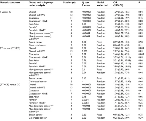
We first performed the meta-analysis on all 18 studies. The pooled ORs for allelic frequency comparison and recessive model comparison suggested that the T allele and genotype TT were significantly associated with an increased cancer risk: OR = 1.29 [95% CI (1.01, 1.65)], P = 0.04, Pheterogeneity < 0.00001, and OR = 2.18 [95% CI
3.31)], P = 0.04, Pheterogeneity < 0.0001, respectively (Table 1). The association between the genotype TT and increased cancer susceptibility was significant in Cauca-sians and in female subjects: OR = 2.40 [95% CI (1.26, 4.59)], P = 0.008, Pheterogeneity = 0.02, and OR = 3.60 [95% CI (1.17, 11.11)], P = 0.03, Pheterogeneity = 0.02 (Table 1, Figure 2, 3). A marginal significant association between the 1772 C/T polymorphism and increased cancer risk was detected in East Asians under recessive model: OR = 5.31 [95% CI (0.91, 30.83)], P = 0.06, Pheterogeneity = 0.76 (Table 1). The remaining pooled ORs from this analysis were not significant (P > 0.05) (Table 1).
Sensitivity analysis was next performed by excluding the studies with controls not in HWE. The results from the allelic frequency comparison and dominant model
com-parison showed no evidence that the 1772 C/T polymor-phism was significantly associated with an increased prostate cancer risk: OR = 1.68 [95% CI (0.94, 3.02)], P = 0.08, Pheterogeneity < 0.0001, and OR = 1.75 [95% CI (0.89, 3.47)], P = 0.11, Pheterogeneity < 0.0001, respec-tively (Table 1). The association between the genotype TT and the increased cancer risk was marginally significant in Caucasians and in female subjects: OR = 3.35 [95% CI (1.01, 11.11)], P = 0.05, Pheterogeneity = 0.01, and OR = 3.88 [95% CI (0.94, 16.01)], P = 0.06, Pheterogeneity = 0.01, respectively (Table 1, Figure 2, 3). The other results were similar to those when the studies with controls not in HWE were included (Table 1).
There was significant heterogeneity among the available studies (Table 1). To detect the source of the
heterogene-Table 1: Meta-analysis of the HIF-1α 1772 C/T polymorphism and cancer association.
Genetic contrasts Group and subgroups under analysis
Studies (n) Q test
P value
Model seclected
OR (95% CI)
P
T versus C Overall 18 <0.00001 Random 1.29 (1.01, 1.65) 0.04
Overall in HWE 13 <0.00001 Random 1.39 (1.02, 1.90) 0.04
Caucasian 11 <0.00001 Random 1.33 (0.90, 1.97) 0.15
Caucasian in HWE 7 <0.00001 Random 1.69 (0.94, 3.04) 0.08
East Asian 5 0.16 Fixed 1.05 (0.84, 1.30) 0.69
Female* 7 <0.00001 Random 1.39 (0.83, 2.35) 0.21
Female in HWE* 6 <0.00001 Random 1.48 (0.81, 2.71) 0.20
Male (prostate cancer)** 4 <0.0001 Random 1.78 (1.07, 2.94) 0.03
Male (prostate cancer) in HWE**
3 <0.0001 Random 1.68 (0.94, 3.02) 0.08
Breast cancer 3 0.12 Fixed 0.99 (0.79, 1.23) 0.90
Colorectal cancer 2 0.02 Random 0.26 (0.01, 6.38) 0.41
TT versus (CT+CC) Overall 18 0.02 Random 2.18 (1.32, 3.62) 0.003
Overall in HWE 13 0.002 Random 2.87 (1.14, 7.26) 0.03
Caucasian 11 0.02 Random 2.40 (1.26, 4.59) 0.008
Caucasian in HWE 7 0.01 Random 3.35 (1.01, 11.11) 0.05
East Asian 5 0.76 Fixed 5.31 (0.91, 30.83) 0.06
Female* 7 0.02 Random 3.60 (1.17, 11.11) 0.03
Female in HWE* 6 0.01 Random 3.88 (0.94, 16.01) 0.06
Male (prostate cancer)** 4 0.1 Fixed 1.53 (0.90, 2.60) 0.11
Male (prostate cancer) in HWE**
3 0.04 Random 1.78 (0.41, 7.74) 0.44
Breast cancer 3 0.10 Fixed 1.51 (0.55, 4.11) 0.42
Colorectal cancer 2 - Random 1.97 (0.33, 11.90) 0.46
(TT+CT) versus CC Overall 18 <0.00001 Random 1.19 (0.88, 1.59) 0.26
Overall in HWE 13 <0.00001 Random 1.34 (0.97, 1.85) 0.08
Caucasian 11 <0.00001 Random 1.15 (0.68, 1.93) 0.61
Caucasian in HWE 7 <0.00001 Random 1.70 (0.89, 3.26) 0.11
East Asian 5 0.15 Fixed 1.01 (0.80, 1.27) 0.96
Female* 7 0.0004 Random 1.28 (0.76, 2.15) 0.35
Female in HWE* 6 0.0002 Random 1.41 (0.77, 2.57) 0.26
Male (prostate cancer)** 4 <0.0001 Random 1.85 (1.04, 3.31) 0.04
Male (prostate cancer) in HWE**
3 <0.0001 Random 1.75 (0.89, 3.47) 0.11
Breast cancer 3 0.22 Fixed 0.96 (0.76, 1.21) 0.75
Colorectal cancer 2 0.02 Random 0.25 (0.01, 5.99) 0.39
ity, we performed the subgroup analyses by gender, cancer types, and ethnicity. The results showed studies in female subject subgroup and Caucasian subgroup were the main contributors of heterogeneity (Table 1).
Association of the HIF-1 1790 G/A polymorphism with cancer risk
The results on all 12 studies showed no evidence that the
HIF-1 1790 G/A polymorphism was significantly
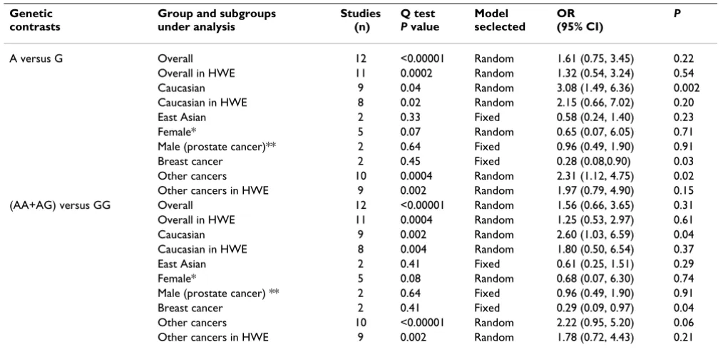
associ-ated with an increased cancer risk (P > 0.05) (Table 2, Fig-ure 4). The significant association between the A allele and the increased cancer risk was detected in other can-cers: OR = 2.31 [95% CI (1.12, 4.75)], P = 0.02, P
heterogene-ity = 0.0004 (Table IV) (Table 2). A marginal association
between the 1790 G/A polymorphism and the increased cancer risk in other cancers was also detected under dom-inant model: OR = 2.22 [95% CI (0.95, 5.20)], P = 0.06, Pheterogeneity < 0.00001 (Table 2). The pooled ORs for allelic frequency comparison and dominant model com-parison suggested the 1790 G/A polymorphism was sig-nificantly associated with an increased cancer risk in Caucasians: OR = 3.08 [95% CI (1.49, 6.36)], P = 0.002, Pheterogeneity = 0.04, and OR = 2.60 [95% CI (1.03, 6.59)], P = 0.04, Pheterogeneity = 0.002, respectively (Table 2). How-ever, reanalysis after exclusion the studies with controls not in HWE did not suggest these associations (P > 0.05) (Table 2). The pooled ORs for A versus G and (AA+AG) versus GG suggested that 1790 G/A polymorphism was significantly associated with a decreased breast cancer risk:
OR = 0.28 [95% CI (0.08, 0.90)], P = 0.03, Pheterogeneity = 0.45, and OR = 0.29 [95% CI (0.09, 0.97)], P = 0.04, P
het-erogeneity = 0.41, respectively (Table 2, Figure 4). The
remaining pooled ORs on the association of 1790 G/A polymorphism and cancer risk were not significant (P > 0.05) (Table 2).
There was significant heterogeneity for allelic frequency comparison and dominant model comparison among the available studies (Table 2). However, the heterogeneity was effectively decreased or removed in the subgroups stratified by gender, ethnicity, and cancer types (Table 2).
Publication bias
Publication bias was assayed by visual funnel plot inspec-tion and Egger's test. The funnel plots for T versus C were basically symmetric (Additional file 4A) and Egger's test did not indicate asymmetry of the plot [Intercept = 0.5092, 95% CI (-1.5454, 2.5639), P = 0.6065]. The fun-nel plots for A versus G showed some asymmetry that could suggest the existence of publication bias (Addi-tional file 4B). However, Egger's test did not show statisti-cal evidence for publication bias [Intercept = -1.82, 95% CI (-4.1611, 0.5212), P = 0.1108].
Discussion
HIF-1 plays a major role in cancer progression and metas-tasis through activation of various genes that are linked to regulation of angiogenesis, cell survival, and energy
Forest plot of the HIF-1 1772 C/T polymorphism and cancer risk [T versue C and TT versus (CT+CC)]
Figure 1
metabolism [5,6]. The HIF-1 gene was previously found to be implicated in the development and progression of cancer [5,6]. The polymorphisms analyzed in the present study consist of C to T and G to A nucleotide substitutions at positions 1772 and 1790 of the exon 12 of the HIF-1
gene [5,6]. Because a study by Tanimoto et al [6] showed that both of the substitutions displayed an increased transactivation capacity of HIF-1α in vitro, the presence of the variant alleles might be associated with increased can-cer susceptibility. However, studies focusing on the asso-ciation of the HIF-1 gene polymorphism with cancer susceptibility had controversial conclusions [5,6,8-22]. The lack of concordance across many of these studies reflects limitation in the studies, such as small sample
sizes, ethnic difference and research methodology. Meta-analysis is a powerful tool for summarizing the results from different studies by producing a single estimate of the major effect with enhanced precision. It can overcome the problem of small sample size and inadequate statisti-cal power of genetic studies of complex traits, and provide more reliable results than a single case-control study [27]. In this meta-analysis, we investigated the association between the HIF-1 1772 C/T and 1790 G/A polymor-phism and cancer risk. The subgroup analyses stratified by cancer types, ethnicity, and gender were also performed. For the HIF-1 1772 C/T polymorphism, our meta-analy-sis on the available studies showed that the T allele and genotype TT were significantly associated with increased cancer risk. These associations were very robust, which did not vary materially when the sensitivity analyses (exclu-sion the study with controls not in HWE) were performed.
Forest plot the HIF-1 1772 C/T polymorphism and cancer risk in Caucasians [TT versus (CT+CC)]
Figure 2
Forest plot the HIF-1 1772 C/T polymorphism and cancer risk in Caucasians [TT versus (CT+CC)]. A. Results from the analysis on all studies of Caucasians. B. Results from the sensitivity analysis (exclusion of the studies with controls not in Hardy-Weinberg equilibrium).
Forest plot the HIF-1 1772 C/T polymorphism and cancer risk in female subjects [TT versus (CT+CC)]
Figure 3
The effect of the genotype TT on cancer especially exists in Caucasians and female subjects. Only female specific can-cers were included in female subgroup in our meta-analy-sis, which indicates that the genotype TT is significantly associated with an increased risk for female specific can-cers. The molecular basis of gender specific effect of the
HIF-1 1772 C/T polymorphism on cancers is unclear.
Studies have shown that estrogen can induce the expres-sion of HIF-1 [28,29]. The substitution of C to T at posi-tions 1772 of the exon 12 of the HIF-1 gene further increase the transactivation capacity of the HIF-1 gene and thus promote the development of female specific can-cers. We also observed a marginally significant association between the genotype TT and increased cancer risk in East Asians. However, subjects with mutant homozygotes were only detected in two studies of East Asians. The CI for this subgroup was very wide, and the association could have been caused by chance. More studies based on larger pop-ulation should be conducted to further examine this asso-ciation.
For the HIF-1 1790 G/A polymorphism, the meta-analy-sis on all studies showed no evidence that the HIF-1 1790 G/A polymorphism was significantly associated with increased cancer risk. We also performed the stratification analyses by gender, ethnicity, and cancer types. The pooled ORs for allelic frequency comparison and domi-nant model comparison suggested the 1790 G/A poly-morphism was significantly associated with an increased
cancer risk in Caucasians. However, the sensitivity analy-sis did not suggest this association. Because the results from the sensitivity analysis were more valid, our meta-analysis does not strongly suggest the association between
the HIF-1 1790 G/A polymorphism and cancer risk in
Caucasians [23]. The pooled effects for allelic frequency comparison and dominant model comparison suggested a significant association between the HIF-1 1790 G/A polymorphism and a decreased breast cancer risk. Because the conclusion is inconsistent with the general under-standing that the 1790 A alleles enhances HIF-1 transcrip-tional activity and the presence of the variant allele might be associated with increased cancer susceptibility, we fur-ther performed the meta-analysis for the ofur-ther cancers to detect the specific effects of cancer type [6]. The results suggested a significant association between the A allele and increased cancer risk in other cancers. A marginal association between the 1790 G/A polymorphism and increased cancer risk in other cancers was also detected under dominant model. However, the reanalysis after exclusion the studies with controls not in HWE did not suggest these associations. Our meta-analysis does not strongly support the association between the HIF-1 1790 G/A polymorphism and the cancer risk in other cancers. The exact mechanism for the inverse association between
the HIF-1 1790 G/A polymorphism and breast cancer was
not clear. However, there were two factors that must be considered. First, the frequency of the HIF-1 1790 A allele was very low and only two studies were included in the
Table 2: Meta-analysis of the HIF-1 1790 G/A polymorphism and cancer association.
Genetic contrasts
Group and subgroups under analysis
Studies (n)
Q test
P value
Model seclected
OR (95% CI)
P
A versus G Overall 12 <0.00001 Random 1.61 (0.75, 3.45) 0.22
Overall in HWE 11 0.0002 Random 1.32 (0.54, 3.24) 0.54
Caucasian 9 0.04 Random 3.08 (1.49, 6.36) 0.002
Caucasian in HWE 8 0.02 Random 2.15 (0.66, 7.02) 0.20
East Asian 2 0.33 Fixed 0.58 (0.24, 1.40) 0.23
Female* 5 0.07 Random 0.65 (0.07, 6.05) 0.71
Male (prostate cancer)** 2 0.64 Fixed 0.96 (0.49, 1.90) 0.91
Breast cancer 2 0.45 Fixed 0.28 (0.08,0.90) 0.03
Other cancers 10 0.0004 Random 2.31 (1.12, 4.75) 0.02
Other cancers in HWE 9 0.002 Random 1.97 (0.79, 4.90) 0.15
(AA+AG) versus GG Overall 12 <0.00001 Random 1.56 (0.66, 3.65) 0.31
Overall in HWE 11 0.0004 Random 1.25 (0.53, 2.97) 0.61
Caucasian 9 0.002 Random 2.60 (1.03, 6.59) 0.04
Caucasian in HWE 8 0.004 Random 1.80 (0.50, 6.54) 0.37
East Asian 2 0.41 Fixed 0.61 (0.25, 1.51) 0.29
Female* 5 0.08 Random 0.68 (0.07, 6.30) 0.74
Male (prostate cancer) ** 2 0.64 Fixed 0.96 (0.49, 1.90) 0.91
Breast cancer 2 0.41 Fixed 0.29 (0.09, 0.97) 0.04
Other cancers 10 <0.00001 Random 2.22 (0.95, 5.20) 0.06
Other cancers in HWE 9 0.002 Random 1.78 (0.72, 4.43) 0.21
breast cancer subgroup. So, the association could be due to chance. Second, our meta-analysis suggests that carci-nogenic mechanism may differ in different cancers and
HIF-1 1790 G/A polymorphism may exert varying effect.
More studies will be required to further examine the asso-ciation.
The current meta-analysis has several limitations which should be noted. First, the meta-analysis was based on the aggregation of published case-control studies. 8 studies did not clearly state the use of a matching design for cases during the selection process of controls. The meta-analysis was based on unadjusted estimates. A more precise analy-sis should be conducted if more detailed individual data were available, which would allow for an adjusted esti-mate. Second, because of data limitation, we did not per-form the stratification analyses by age, smoking, or other variables. Third, several genotyping methods were used in the eligible studies. The quality control of genotyping was not well documented in some studies. Undoubtedly, the limitations mentioned should affect our final conclu-sions.
Conclusions
Our meta-analysis suggests that the HIF-1 1772 C/T pol-ymorphism is significantly associated with higher cancer risk, and the 1790 G/A polymorphism is significantly associated with decreased breast cancer risk. The effect of the 1772 C/T polymorphism on cancer especially exists in Caucasians and female subjects. Only female specific can-cers were included in female subgroup, which indicates that the 1772 C/T polymorphism is significantly associ-ated with an increased risk for female specific cancers. The association between the 1790 G/A polymorphism and lower breast cancer risk could be due to chance.
Abbreviations
CI: confidence interval; HWE: Hardy-Weinberg equilib-rium; HIF-1: hypoxia- inducible factor -1; HIF-1α: hypoxia- inducible factor -1α; OR: odds ratio; SNP: single nucleotide polymorphism.
Competing interests
The authors declare that they have no competing interests.
Forest plot the HIF-1 1790 G/A polymorphism and cancer risk [A versue G and (AA+AG) versus GG]
Figure 4
Authors' contributions
TFZ participated in the design, data acquisition, manu-script writing, and have given final approval of the version to be published. JPZ performed data analysis, data inter-pretation. JL participated in the design, data acquisition. MN participated in data analysis and drafting the script. All authors read and approved the final manu-script.
Additional material
Acknowledgements
This work was supported by National Natural Science foundation of China (Grant No: 30671007) and Natural Science foundation of Zhejiang Prov-ince, China (Grant No: Y2081111).
References
1. Jemal A, Siegel R, Ward E, Hao Y, Xu J, Thun MJ: Cancer statistics, 2009. CA Cancer J Clin 2009, 59(4):225-249.
2. Wright ME, Peters U, Gunter MJ, Moore SC, Lawson KA, Yeager M, Weinstein SJ, Snyder K, Virtamo J, Albanes D: Association of vari-ants in two vitamin e transport genes with circulating vita-min e concentrations and prostate cancer risk. Cancer Res
2009, 69(4):1429-1438.
3. Cheung WY, Liu G: Genetic variations in esophageal cancer risk and prognosis. Gastroenterol Clin North Am 2009, 38(1):75-91. 4. Hill RP, Marie-Egyptienne DT, Hedley DW: Cancer stem cells, hypoxia and metastasis. Semin Radiat Oncol 2009, 19(2):106-111. 5. Smaldone MC, Maranchie JK: Clinical implications of hypoxia inducible factor in renal cell carcinoma. Urol Oncol 2009,
27(3):238-245.
6. Tanimoto K, Yoshiga K, Eguchi H, Kaneyasu M, Ukon K, Kumazaki T, Oue N, Yasui W, Imai K, Nakachi K, Poellinger L, Nishiyama M:
Hypoxia-inducible factor-1alpha polymorphisms associated with enhanced transactivation capacity, implying clinical sig-nificance. Carcinogenesis 2003, 24:1779-1783.
7. Zhong H, De Marzo AM, Laughner E, Lim M, Hilton DA, Zagzag D:
Overexpression of hypoxia-inducible factor 1 alpha in com-mon human cancers and their metastases. Cancer Res 1999,
59:5830-5835.
8. Munoz-Guerra MF, Fernandez-Contreras ME, Moreno AL, Martin ID, Herraez B, Gamallo C: Polymorphisms in the hypoxia inducible factor 1-alpha and the impact on the prognosis of early stages of oral cancer. Ann Surg Oncol 2009, 16(8):2351-2358. 9. Foley R, Marignol L, Thomas AZ, Cullen IM, Perry AS, Tewari P,
O'Grady A, Kay E, Dunne B, Loftus B, Watson WR, Fitzpatrick JM, Woodson K, Lehman T, Hollywood D, Lynch TH, Lawler M: The HIF-1α C1772T polymorphism may be associated with sus-ceptibility to clinically localised prostate cancer but not with elevated expression of hypoxic biomarkers. Cancer Biol Ther
2009, 8(2):118-124.
10. Li H, Bubley GJ, Balk SP, Gaziano JM, Pollak M, Stampfer MJ, Ma J:
Hypoxia-inducible factor-1alpha (HIF-1alpha) gene polymor-phisms, circulating insulin-like growth factor binding protein (IGFBP)-3 levels and prostate cancer. Prostate 2007,
67(12):1354-1361.
11. Orr-Urtreger A, Bar-Shira A, Matzkin H, Mabjeesh NJ: The homozygous P582S mutation in the oxygen-dependent deg-radation domain of HIF-1 alpha is associated with increased risk for prostate cancer. Prostate 2007, 67(1):8-13.
12. Chau CH, Permenter MG, Steinberg SM, Retter AS, Dahut WL, Price DK, Figg WD: Polymorphism in the hypoxia-inducible factor 1 alpha gene may confer susceptibility to androgen-independ-ent prostate cancer. Cancer Biol Ther 2005, 4(11):1222-1225. 13. Lee JY, Choi JY, Lee KM, Park SK, Han SH, Noh DY, Ahn SH, Kim DH,
Hong YC, Ha E, Yoo KY, Ambrosone CB, Kang D: Rare variant of hypoxia-inducible factor-1alpha (HIF-1A) and breast cancer risk in Korean women. Clin Chim Acta 2008, 389(1-2):167-170. 14. Apaydin I, Konac E, Onen HI, Akbaba M, Tekin E, Ekmekci A: Single
nucleotide polymorphisms in the hypoxia-inducible factor-1alpha (HIF-factor-1alpha) gene in human sporadic breast cancer.
Arch Med Res 2008, 39(3):338-345.
15. Kim HO, Jo YH, Lee J, Lee SS, Yoon KS: The C1772T genetic pol-ymorphism in human HIF-1alpha gene associates with expression of HIF-1alpha protein in breast cancer. Oncol Rep
2008, 20(5):1181-1187.
16. Horree N, Groot AJ, Van Hattem WA, Heintz AP, Vooijs M, Van Diest PJ: HIF-1A gene mutations associated with higher microvessel density in endometrial carcinomas. Histopathology
2008, 52(5):637-639.
17. Konac E, Onen HI, Metindir J, Alp E, Biri AA, Ekmekci A: An inves-tigation of relationships between hypoxia-inducible factor-1 alpha gene polymorphisms and ovarian, cervical and endometrial cancers. Cancer Detect Prev 2007, 31(2):102-109. 18. Fransen K, Fenech M, Fredrikson M, Dabrosin C, Soderkvist P:
Asso-ciation between ulcerative growth and hypoxia inducible fac-tor-1 alpha polymorphisms in colorectal cancer patients. Mol Carcinog 2006, 45(11):833-840.
19. Kuwai T, Kitadai Y, Tanaka S, Kuroda T, Ochiumi T, Matsumura S, Oue N, Yasui W, Kaneyasu M, Tanimoto K, Nishiyama M, Chayama K: Single nucleotide polymorphism in the hypoxia-inducible factor-1alpha gene in colorectal carcinoma. Oncol Rep 2004,
12(5):1033-1037.
20. Ollerenshaw M, Page T, Hammonds J, Demaine A: Polymorphisms in the hypoxia inducible factor-1alpha gene (HIF1A) are associated with the renal cell carcinoma phenotype. Cancer Genet Cytogenet 2004, 153(2):122-126.
21. Clifford SC, Astuti D, Hooper L, Maxwell PH, Ratcliffe PJ, Maher ER:
The pVHL-associated SCF ubiquitin ligase complex: Molecu-lar genetic analysis of elongin B and C, Rbx1 and HIF-1α in renal cell carcinoma. Oncogene 2001, 20:5067-5074.
22. Ling TS, Shi RH, Zhang GX, Zhu H, Yu LZ, Ding XF: Common sin-gle nucleotide polymorphism of hypoxia-inducible factor-1 alpha and its impact on the clinicopathological features of esophageal squamous cell carcinoma. Chin J Dig Dis 2005,
6(4):155-158.
Additional file 1
The flow diagram of included/excluded studies.
Click here for file
[http://www.biomedcentral.com/content/supplementary/1756-9966-28-159-S1.JPEG]
Additional file 2
Characteristics of individual studies included in the meta-analysis.
Click here for file
[http://www.biomedcentral.com/content/supplementary/1756-9966-28-159-S2.DOC]
Additional file 3
Genotype and allele distribution of hypoxia- inducible factor -1α 1772 C/T and 1790 G/A polymorphisms of individual studies included in the meta-analysis.
Click here for file
[http://www.biomedcentral.com/content/supplementary/1756-9966-28-159-S3.DOC]
Additional file 4
Funnel plots for publication bias test. A. HIF-1α 1772 C/T: T versus C. B. HIF-1α 1790 G/A: A versus G. Each point represents a separate study for the indicated association. SE(SMD), standard error of the logarithm of the odd ratio.
Click here for file
Publish with BioMed Central and every scientist can read your work free of charge "BioMed Central will be the most significant development for disseminating the results of biomedical researc h in our lifetime."
Sir Paul Nurse, Cancer Research UK
Your research papers will be:
available free of charge to the entire biomedical community
peer reviewed and published immediately upon acceptance
cited in PubMed and archived on PubMed Central
yours — you keep the copyright
Submit your manuscript here:
http://www.biomedcentral.com/info/publishing_adv.asp
BioMedcentral
23. Thakkinstian A, McElduff P, D'Este C, Duffy D, Attia J: A method for meta-analysis of molecular association studies. Stat Med 2005,
24(9):1291-1306.
24. Egger M, Davey Smith G, Schneider M, Minder C: Bias in meta-analysis detected by a simples, graphical test. BMJ 1997,
315:629-634.
25. Bax L, Yu LM, Ikeda N, Tsuruta H, Moons KGM: Development and validation of MIX: comprehensive free software for meta-analysis of causal research data. BMC Med Res Methodol 2006,
6:50.
26. Bax L, Yu LM, Ikeda N, Tsuruta H, Moons KGM: MIX: comprehen-sive free software for meta-analysis of causal research data. Version 1.7. 2008 [http://mix-for-meta-analysis.info].
27. Yong D, QingQing W, Hua L: Association of uteroglobin G38A polymorphism with IgA nephropathy: a meta-analysis. Am J Kidney Dis 2006, 48(1):1-7.
28. Kazi AA, Jones JM, Koos RD: Chromatin immunoprecipitation analysis of gene expression in the rat uterus in vivo: estro-gen-induced recruitment of both estrogen receptor alpha and hypoxia-inducible factor 1 to the vascular endothelial growth factor promoter. Mol Endocrinol 2005, 19(8):2006-2019. 29. Hua K, Din J, Cao Q, Feng W, Zhang Y, Yao L, Huang Y, Zhao Y, Feng
![Figure 1Forest plot of the HIF-1 1772 C/T polymorphism and cancer risk [T versue C and TT versus (CT+CC)]Forest plot of the HIF-1 1772 C/T polymorphism and cancer risk [T versue C and TT versus (CT+CC)]](https://thumb-us.123doks.com/thumbv2/123dok_us/9024838.1895305/5.612.140.470.86.321/figure-forest-polymorphism-cancer-versue-forest-polymorphism-cancer.webp)
![Figure 3risk in female subjects [TT versus (CT+CC)]Forest plot the HIF-1 1772 C/T polymorphism and cancer Forest plot the HIF-1 1772 C/T polymorphism and cancer risk in female subjects [TT versus (CT+CC)]](https://thumb-us.123doks.com/thumbv2/123dok_us/9024838.1895305/6.612.56.308.92.471/figure-female-subjects-forest-polymorphism-forest-polymorphism-subjects.webp)