The Effects of High Intensity Interval Training on HNF-4
α
Gene
Expression in Liver Tissue of Type 2 Diabetic Male Wistar Rats
Elham Yadegari
1, Abdol Ali Banaeifar
2*, Mohamad Ali Azarbayjani
3, Sajad Arshadi
2Introduction
isruption of insulin function or
insufficient insulin secretion leads to
type 2 diabetes (T2D) (1). Increased
production of glucose in the liver is
responsible for increasing fasting blood
glucose (FBG) and an important part of
glucose uptake after eating a meal in diabetic
people (2).
Insulin controls the production of glucose in
the liver by controlling the expression of two
glucose-6-phosphatase
enzymes
and
phosphonalcarboxy kinase pyruvate, involved
in the gluconeogenesis process (2). These
enzymes are regulated by some genes
including
Foxo1 (Forkhead box O1), HNF-4
α
(hepatocyte
nuclear
factor-4),
PGC-1
α
(peroxisome proliferator- activated receptor
gamma coactivator- 1
α
), Sirt 1 (sirtuin 1) and
STAT3 (signal transducer and activator of
transcription 3) (3-5).
The study of genes
D
1. PhD Student, Department of Exercise Physiology, South Tehran Branch, Islamic Azad University, Tehran, Iran.
2. PhD, Department of Exercise Physiology, South Tehran Branch, Islamic Azad University, Tehran, Iran.
3. PhD, Department of Exercise Physiology, Central Tehran Branch, Islamic Azad University, Tehran, Iran.
*Correspondence:
Abdol Ali Banaeifar, PhD, Department of Physical Education and Sport Science, South Tehran Branch, Islamic Azad University, Tehran, Iran.
Tel: (98) 912 225 1779 Email:[email protected]
Received: 10 December 2018
Accepted: 24 February 2018
Published in May 2019
Abstract
Objective:
This study examined the effect of high intensity interval training (HIIT) on HNF-4α gene expression, glucose and insulin in liver tissue of type 2 diabetic male Wistar rats.Materials and Methods: In this study, 20 male Wistar rats
weighing 220 (±20) grams were selected. After type 2 diabetes (T2D) induction, the samples were divided into two groups of HIIT and control. The training program was 12 weeks and 5 times a week. Fasting glucose, serum insulin and HNF-4α gene expression in the liver tissue were measured in both groups after the last exercise session by using independent T-test.Results: The results showed that HIIT decreased significantly the fasting glucose (P-value: 0.001) and increased serum insulin (P -value: 0.001). Also, the expression of HNF-4α after HIIT significantly decreased in comparison to the control group (P-value: 0.003).
Conclusion:
Based on the findings, HIIT resulted in a significant decrease in fasting glucose and a significant increase in insulin that is likely to reduce the liver HNF-4α gene expression of T2D male Wistar rats by increasing serum insulin.Keywords
:
High intensity intermittent exercise, HNF-4α gene expression, Type 2 diabetic, Wistar ratexpression in various diseases, especially T2D,
is a new method in recent years. Therefore, in
this study, the gene expression of HNF-4
α
is
investigated.
HNF-4
α
belongs to the steroid hormone
receptor, and is also a transcription factor that
was first recognized by the interaction of
specific gene promoters for the regulatory
system of hepatic cysts (6). HNF-4
α
is a
transcription factor expressed in the liver,
intestine, pancreas and kidneys, also plays an
important role in regulating the transcription
of the liver and pancreas (6). One study found
that one of the mechanisms that inhibits the
effect of insulin on gluconeogenesis depends
on the ability to reduce the expression of
HNF-4a and Foxo1 in the liver (7). Indeed, in
insulin resistant models, both Foxo1 and
HNF-4
α
protein levels increased in steady state and
after insulin stimulation (8). It has been
suggested that HNF-4
α
modulates glucose
metabolism in the liver by controlling the
expression of glucose 6-phosphate and
phosphonalcarboxy kinase pyruvate genes (7).
On the other hand, exercise is generally
recommended because of the beneficial effects
on glucose control in the treatment of T2D (9).
Exercise improves insulin function and has a
significant
effect
on
insulin
signaling
pathways (10). It also improves glucose
hemostasis and insulin sensitivity. After acute
exercise, insulin sensitivity increases in tissues
such as skeletal muscle, fat, liver and
hypothalamus (11). One study investigated the
effect of acute exercise on reducing liver
glucose production through HNF-4a pathway
in insulin resistant mice, which resulted in a
reduction in HNF-4a pathway and, as a result
of
acute
exercise,
improved
glucose
hemostasis (12). Despite limited studies on the
effect of exercise on HNF-4a expression, as
well as the lack of a study that directly
measures the effect of exercise training on
HNF-4a expression in the liver tissue and its
interaction with insulin and glucose, The
present study aimed to determine the effect of
HIIT on HNF-4a expression in liver, insulin
and FBG in T2D rats.
Materials and Methods
The present research was done on male Wistar
rats (produced at Pasteur Institute of Iran). The
sample consisted of 20 rats in a 10-week-old
age range of 220 (± 20) grams, selected
randomly. All procedures of this study were
approved by the Ethic Committee of south
Tehran
Branch
Azad
University
(14121407951010). The rats were kept in a
laboratory environment at 22 (± 3) ºC,
humidity in the range of 30-60, 12 hours of
light and 12 hours of darkness. Every two rats
were kept in a rat cage for free access to
standard water and food. (13). After a fasting
night, for induction of T2D, a nicotine amide
solution at a dose of 110 mg per kg of rat
weight was injected in peritoneal. After 15
minutes, the freshly prepared STZ solution
was injected intraperitoneally with a dose of
65 mg / kg in citrate buffer (PH = 4.5) (14). A
week after injection, to ensure diabetes
induction in rats, blood drops were collected
from the venous vein and the glucose blood
level was measured by the glucometer (15). In
the next step, diabetic rats were randomly
divided into two groups of exercise (12 weeks
HIIT) and 10 controls. The HIIT program was
conducted for 12 weeks each week, 5 sessions
per running treadmill with 30 minute time in
accordance with Table 1 (13).
After 12 hours of fasting and 48 hours after the
last training session, intraperitoneal injection
of 10% ketamine (50 mg / kg) and xylazine
2% (10 mg / kg) were used to anesthetizing the
rats. The blood samples were taken directly
from the animal's heart. The rat liver tissue
was also sampled and washed in a physiologic
serum in 1.8
µ
m microelements containing
RNA later fluid with a 20% ratio for genetic
testing. FBG were measured using glucose
oxidase enzyme method and with glucose Kit
(Pars test compani). The coefficient of internal
and external test changes was 1.74 and 1.19%,
respectively, and the sensitivity was 5 mg/dL.
The serum insulin was measured by ELISA
and in accordance with the instructions for the
commercial kit (Demeditec Diagnostic Insulin
made in Germany). The coefficient of internal
and external test changes was 2.6 and 2.88,
respectively, and the sensitivity was 1.76.
RNA extraction was performed using the
RNeasy mini commercial kits (QIAGEN
Company). Determination of TCF mRNA by
RT-Real time PCR using the Rotator 6000
system using the One Step Single Step SYBR
TAKARA kit from Takara Company in
accordance with the company's instructions.
RNA polymerase II was used as control gene
to determine the expression of HNF-4a. The
sequence pattern of primers is given in Table
2. Reactions CTs were extracted and recorded
using Real Time PCR software. A CT
∆∆
comparison was used to quantitatively express
HNFMRNA.
After confirming the normal distribution of
data with Kolmogorov-Smirnov test and
homogeneity of data with Levene’s test,
independent T-test was used to examine the
effect of training on dependent variables. The
whole operation was performed by SPSS
software (Version 20) and the significance
level of the tests was considered to be 0.05.
Results
Body weight was measured in both groups
before and after the training period. The values
of rat weight in both HIIT and control groups
are presented in Table 3. There was no
significant difference in the weight of rats
between the two groups in the pre-exercise
program (
P
-value: 0.523).
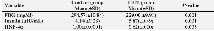
The results of independent t-test showed that
FBG level decreased significantly between the
two groups (
P
-value: 0.001). The findings also
showed a significant increase in serum insulin
levels (
P
-value: 0.001). Also, the relative
expression of HNF-4a gene in the liver tissue
in the HIIT group significantly decreased (
P
-value: 0.003) (Table 4).
Discussion
In this study, the effect of 12 weeks of HIIT on
HNF-4
α
in liver, glucose and insulin tissues in
T2D rats was studied. The results of this study
showed that HIIT led to a significant decrease
in HNF-4
α
and glucose and a significant
increase in serum insulin level in T2D rats.
Despite the fact that genetic studies refer to the
interconnection of insulin signaling pathways
with HNF-4
α
expression in T2D, excessive
production of glucose is one of the main
mechanisms for increasing FBG, which is due
to increased glycogenolysis, reduced glycogen
synthesis
and
gluconeogenesis.
Insulin
resistance with insulin deficiency in inhibiting
phosphoanol-pyruvate carboxy kinase and
glucose 6-phosphatase enzymes play an
important role in this process (16). Possibly,
there are many factors affecting the ability of
insulin
on
the
expression
of
Phosphoenolpyruvate carboxylase kinase and
glucose 6-phosphatase enzymes, one of which
is HNF-4
α
(3,7). Therefore, inhibition of
gluconeogenesis and production of glucose in
the liver is an attractive treatment in diabetic
patients, so that metformin may act by
inhibiting liver glucose production in hepatic
patients (17).
Table 1. HIIT Exercise Program
Active rest (Speed: m/min) One-minute repetitions
(Speed: m/min) Activity time (week)
10 16
1
10 20
2-3
12 25
4-5
12 30
6-7
14 33
8-9
14 36
10-12
Table 2. Primers pattern used in research
Gene Bank Tm
Product size Primer sequence
Genes
NM_001191052.1 60
159bp For: GCAGAGATGAGCCGTGTGTC
Rev: TTGATCTTGCCTGGGTCACTC HNF4
NM_001191052.1 60
159bp For: ACTTTGATGACGTGGAGGAGGAC
Rev: GTTGGCCTGCGGTCGTTC RNA Polymerase ΙΙ
Sport activity is one of the effective methods
known to control blood glucose levels in
diabetics by various mechanisms such as
reduction in liver glucose production as an
effective factor in hemostasis in glucose
(18,19). A study investigated the effect of
acute exercise on reducing liver glucose
production through FOXO1 and HNF-4
α
pathways in insulin resistant mice, which
resulted in a decrease in FOXO1 and HNF-4
α
pathways and therefore acute exercise would
improve glucose hemostasis (12). Also, the
results of the study showed that the association
between HNF-4
α
polymorphisms with glucose
and insulin is moderated by physical activity.
Therefore, the results of this study showed that
the effect of HNF-4
α
polymorphisms on the
risk of T2D is affected by physical activity
(20). The results of these studies are consistent
with the results of the present study, which
reported a reduction in HNF-4
α
expression.
Also, in several studies, the effect of exercise
activity on insulin and glucose in diabetic
animal samples has been investigated, some of
which are consistent with the present study
and some others are inconsistent. For example,
in an 8 weeks study, moderate and high
endurance training did not affect insulin levels
in diabetic rats (21). Other studies also found
no effect of exercise on insulin levels in
diabetic rats (22-25). In contrast, in a 7 weeks
treadmill study, moderate treadmill increased
the insulin level in diabetic rats (26), which is
in line with the results of this study. The
reasons of contradiction between the findings
could be due to study situation.
On the other hand, the results of this study
indicate a decrease in blood glucose levels,
which has been shown to significantly reduce
blood glucose following long-term training
(27,28). Aerobic exercise seems to control
liver
glucose
production
by
several
mechanisms. One of these mechanisms is the
protein 1C binding to the sterol responsive
element, which is a physiological inhibitor of
liver glucose production (29), which done the
expression
of
phosphoanol
pyruvate
carboxykinase by controlling the expression of
HNF-4
α
(30). Other studies also showed that
HNF-4
α
activity depends on the transmission
of insulin signal via the Foxo1 insulin pathway
(3). In the absence of insulin, HNF-4
α
, along
with Foxo1, is likely to activate the glucose
6-phosphatase gene and phosphoanol pyruvate
carboxy kinase via PGC-1
α
, which is
associated with activation of gluconeogenesis,
but in the presence of phosphorylated Foxo1
insulin it is removed from the nucleus,
resulting in HNF-4
α
being isolated, which,
with its separation, activates HNF-4
α
and the
glucokinase gene and inhibits the genes
involved in gluconeogenesis. (3,31).
In this study, there was a relationship between
the increase in insulin levels and the reduction
of HNF-4
α
expression, which is probably due
to the reduction of the expression of
phosphoanol pyruvate carboxy kinase and
glucose 6-phosphate, and ultimately the
reduction of gluconeogenesis and liver glucose
production. It has also been shown that
HNF-4
α
is involved in expression glucose
metabolism and insulin secretion genes in
Table 3. Body weight changes (grams) before and after training in the HIIT and control groups
Group Before HIIT
Mean (±SD)
After HIIT
Mean (±SD) P-value
HIIT 218.42±1.13 271.42±6.65 0.001
Control 219.28±3.25 253.14±5.61 0.001
SD: standard deviation
Table 4. Glucose and insulin levels after exercise in the HIIT and control group
Variable Control group
Mean(±SD)
HIIT group
Mean(±SD) P-value
FBG (mg/dl) 294.57(±10.84) 229.00(±9.91) 0.001
Insulin (µIU/mL) 4.14(±0.28) 5.87(±0.49) 0.001
HNF-4α 1.00(±0.0001) 0.62(±0.20) 0.003
SD: standard deviation
pancreatic beta cells (32,33). This increase in
insulin in the present study may also be
associated with expression of HNF-4
α
in Beta
cells.
Conclusions
The findings of this study showed a decrease
in the expression of HNF-4
α
, glucose uptake
and increased serum insulin levels in T2D rats
in response to HIIT. The increased liver
glucose production and gluconeogenesis as an
important pathologic factor in diabetic
patients, as well as the role of HNF-4
α
on liver
gluconeogenesis, HIIT activity may lead to
increased levels of insulin and phosphorylation
of the Foxo1 / HNF-4
α
pathway. Reducing the
expression of phosphoanol pyruvate carboxy
kinase
and
glucose
6-phosphatase
and
ultimately reducing gluconeogenesis and
glucose production. However, more studies are
needed in this area.
Acknowledgments
This research is based on a PhD thesis at the
Faculty of Physical Education of south Tehran
Branch Azad University.
Conflict of Interest
We declare that there was no conflict of
interest.
References
1. Association AD. Standards of medical care in diabetes-2013. Diabetes care 2013; 36(1):11-66. 2. Sharabi K, Tavares CD, Rines AK, Puigserver P.
Molecular pathophysiology of hepatic glucose production. Molecular aspects of medicine. 2015;46:21-33.
3. Hirota K, Sakamaki J-i, Ishida J, Shimamoto Y, Nishihara S, Kodama N, et al. A combination of HNF-4 and Foxo1 is required for reciprocal transcriptional regulation of glucokinase and glucose-6-phosphatase genes in response to fasting and feeding. Journal of Biological Chemistry. 2008;283(47):32432-41.
4. Rodgers JT, Lerin C, Gerhart-Hines Z, Puigserver P. Metabolic adaptations through the PGC1α and SIRT1 pathways. FEBS letters. 2008;582(1):46-53.
5. Nie Y, Erion DM, Yuan Z, Dietrich M, Shulman GI, Horvath TL, et al. STAT3 inhibition of gluconeogenesis is downregulated by SirT1. Nature cell biology. 2009;11(4):492.
6. Babeu JP, Boudreau F. Hepatocyte nuclear factor 4-alpha involvement in liver and intestinal inflammatory networks. World journal of gastroenterology: WJG. 2014;20(1):22.
7. Hirota K, Daitoku H, Matsuzaki H, Araya N, Yamagata K, Asada S, et al. Hepatocyte nuclear factor-4 is a novel downstream target of insulin via FKHR as a signal-regulated transcriptional inhibitor. Journal of Biological Chemistry. 2003;278(15):13056-60.
8. Ganjam GK, Dimova EY, Unterman TG, Kietzmann T. FoxO1 and HNF-4 are involved in regulation of hepatic glucokinase gene
expression by resveratrol. Journal of Biological Chemistry. 2009;284(45):30783-97.
9. Yousefipoor P, Tadibi V, Behpoor N, Parnow A, Delbari M, Rashidi S. Effects of aerobic exercise on glucose control and cardiovascular risk factor in type 2 diabetes patients. medical journal of mashhad university of medical sciences, 2015;57(9):976-84.
10. Enteshary M, Esfarjani F, Reisi J. The Comparison of 8 week combined training with two different intensity on level of serum Irisin, and glycemic indices of type 2 diabetic women. medical journal of mashhad university of medical sciences, 2018;61(2):971-84.
11. Riyahi F, Riyahi S, Yaribeygi H. Diabetes and Role of Exercise on its Control; A Systematic. hrjbaq. 2016;1(2):113-21.
12. De Souza CT, Frederico MJ, Da Luz G, Cintra DE, Ropelle ER, Pauli JR, et al. Acute exercise reduces hepatic glucose production through inhibition of the Foxo1/HNF4α pathway in insulin resistant mice. The Journal of physiology. 2010;588(12):2239-53.
13. Eizadi M, Soory R, Ravasi A, Baesy K, Choobineh S. Relationship between TCF7L2 Relative Expression in Pancreas Tissue with Changes in Insulin by High Intensity Interval Training (HIIT) in Type 2 Diabetes Rats. The Journal of Shahid Sadoughi University of Medical Sciences. 2017;24(12):981-93.
14. Shirwaikar A, Rajendran K, Kumar CD, Bodla R. Antidiabetic activity of aqueous leaf extract of Annona squamosa in streptozotocin–
nicotinamide type 2 diabetic rats. Journal of ethnopharmacology. 2004;91(1):171-5.
15. Kazemi F, Ebrahim K, Asl SZ. Effect of regular exercise-induced apelin on dyslipidemia of type 2 diabetic rats. Research in Medicine. 2016;39(4):163-8.
16. Barthel A, Schmoll D. Novel concepts in insulin regulation of hepatic gluconeogenesis. American Journal of Physiology-Endocrinology And Metabolism. 2003;285(4):685-92.
17. Wiernsperger NF, Bailey CJ. The antihyperglycaemic effect of metformin. Drugs. 1999;58(1):31-9.
18. Hoene M, Lehmann R, Hennige AM, Pohl AK, Häring HU, Schleicher ED, et al. Acute regulation of metabolic genes and insulin receptor substrates in the liver of mice by one single bout of treadmill exercise. The Journal of physiology. 2009;587(1):241-52.
19. Ropelle ER, Pauli JR, Cintra DE, Frederico MJ, De Pinho RA, Velloso LA, et al. Acute exercise modulates the Foxo1/PGC1α pathway in the liver of dietinduced obesity rats. The Journal of physiology. 2009;587(9):2069-76.
20. Stephanie-May R, John WS, Tuomo R, Claude B, Marie-Claude V, Louis P. Interaction between HNF4A polymorphisms and physical activity in relation to type 2 diabetes-related traits: results from the Quebec Family Study. diabetes research and clinical practice. 2009;84(3):211-8.
21. Salehi OR, Hosseini SA. The Effects of Endurance Trainings on Serum BDNF and Insulin Levels in Streptozotocin-Induced Diabetic Rats. Shefaye Khatam 2017;5(2):52-61. 22. Crespilho DM, de Almeida Leme JAC, de Mello
MAR, Luciano E. Effects of physical training on the immune system in diabetic rats. International journal of diabetes in developing countries. 2010;30(1):33.
23. Gomes RJ, de Oliveira CAM, Ribeiro C, Mota CSdA, Moura LP, Tognoli LMMC, et al. Effects of exercise training on hippocampus concentrations of insulin and IGF1 in diabetic rats. Hippocampus. 2009;19(10):981-7.
24. Hosseini S, Nikbakht H, Azarbayjani M. The Effect of Resistance Training on Glycemic Indexes of Streptozotocin Induced Diabetic Rats. Physical Education and Sport Science Quarterly (PESSQ). 2011;2(2):42-8.
25. Leme JACdA, Gomes RJ, Mello MARd, Luciano E. Moderate physical training increases brain insulin concentrations in experimental diabetic rats. Indian J Exp Bio. 2008;46(6):443-6. 26. Rawal S, Huang H, Novikova L, Hamedi T,
Smirnova I, Stehno Bittel L. Effects of exercise on pancreatic islets in zucker diabetic fatty rats. J Diabetes Metab S. 2013;10.
27. Madsen SM, Thorup AC, Overgaard K, Jeppesen PB. High intensity interval training improves glycaemic control and pancreatic β cell function of type 2 diabetes patients. PLoS One. 2015;10(8):0133286.
28. Malin SK, Solomon TP, Blaszczak A, Finnegan S, Filion J, Kirwan JP. Pancreatic β-cell function increases in a linear dose-response manner following exercise training in adults with prediabetes. American Journal of Physiology-Endocrinology and Metabolism. 2013;305(10):1248-54.
29. Chakravarty K, Wu S-Y, Chiang C-M, Samols D, Hanson RW. SREBP-1c and Sp1 interact to regulate transcription of the gene for phosphoenolpyruvate carboxykinase (GTP) in the liver. Journal of Biological Chemistry. 2004;279(15):15385-95.
30. Yamamoto T, Shimano H, Nakagawa Y, Ide T, Yahagi N, Matsuzaka T, et al. SREBP-1 interacts with hepatocyte nuclear factor-4α and interferes with PGC-1 recruitment to suppress hepatic gluconeogenic genes. Journal of Biological Chemistry. 2004;279(13):12027-35.
31. Rhee J, Inoue Y, Yoon JC, Puigserver P, Fan M, Gonzalez FJ, et al. Regulation of hepatic fasting response by PPARγ coactivator-1α (PGC-1): requirement for hepatocyte nuclear factor 4α in gluconeogenesis. Proceedings of the national academy of sciences. 2003;100(7):4012-7. 32. Stoffel M, Duncan SA. The maturity-onset
diabetes of the young (MODY1) transcription factor HNF4α regulates expression of genes required for glucose transport and metabolism. Proceedings of the National Academy of Sciences. 1997;94(24):13209-14.
33. Wang H, Maechler P, Antinozzi PA, Hagenfeldt KA, Wollheim CB. Hepatocyte nuclear factor 4α regulates the expression of pancreatic β-cell genes implicated in glucose metabolism and nutrient-induced insulin secretion. Journal of Biological Chemistry. 2000;275(46):35953-9