The interactive effects of multiple stressors on physiological stress
responses and club cell investment in fathead minnows
Aditya K. Manek
a,⁎
, Maud C.O. Ferrari
b, Som Niyogi
a, Douglas P. Chivers
a aDepartment of Biology, University of Saskatchewan, Saskatoon, S7N 5E2 SK, Canada
bDepartment of Biomedical Sciences, WCVM, University of Saskatchewan, Saskatoon, SK S7N 5B4, Canada
H I G H L I G H T S
•UV radiation caused a physiological stress response (cortisol release) infish. •Cd reduced cortisol levels substantially forfish exposed to UV.
•Fish exposed to UV, with or without Cd, showed decreases in club cell investment. •There was no difference in alarm cues potency from UV and Cd exposedfish. •Our work highlights the difficulty of untangling effects of multiple stressors.
a b s t r a c t
a r t i c l e i n f o
Article history: Received 15 August 2013
Received in revised form 9 December 2013 Accepted 9 December 2013
Available online 21 January 2014
Keywords: UV radiation Cadmium Cortisol
Epidermal club cells Alarm cue
Anthropogenic activities have dramatically increased over the past decades, with the consequence that many or-ganisms are simultaneously exposed to multiple stressors. Understanding how oror-ganisms respond to these stressors is a key focus for scientists from many disciplines. Here we investigated the interactive effects of two stressors, UV radiation (UVR) and cadmium (Cd) exposure on a common freshwaterfish, fathead minnow (Pimephales promelas). UVR is known to influence the density of epidermal club cells (ECCs), which are not only a key component of the innate immune system offishes, but are also the source of chemical alarm cues that serve to warn otherfishes of nearby predators. In contrast, Cd impairs the physiological stress response and ability offish to respond to alarm cues. We used an integrative approach to examine physiological stress re-sponse as well as investment in ECCs. Fish exposed to UVR had higher levels of cortisol than non-exposed con-trols, but Cd reduced cortisol levels substantially forfish exposed to UVR. Fish exposed to UVR, either in the presence or absence of Cd, showed consistent decreases in ECC investment compared to non-exposed controls. Despite differences in ECC number, there was no difference in the potency of alarm cues prepared from the skin of UVR and Cd exposed or non-exposedfish indicating that UVR and Cd exposure combined may have little influence on chemically-mediated predator–prey interactions.
© 2014 Elsevier B.V. All rights reserved.
1. Introduction
There is a rapid loss in global biodiversity as a result of anthropogen-ic changes occurring at local and global scales (Vitousek, 1994; Pereira et al., 2012). Identifying the reasons for such changes requires us to con-sider how different environmental stressors interact to affect the phys-iology, behaviour and ecology of individuals and how these changes subsequently affect species interactions. It is rare that animals are exposed to single stressors and consequently research aimed at identi-fying the interactive effects of multiple stressors is needed (Boone et al., 2007).
For much of the past century, we have witnessed increases in levels of ultraviolet radiation (hereafter UVR) hitting the surface of the earth as a result of reductions in stratospheric ozone (Newman et al., 2006). The implementation of the Montreal Protocol has ameliorated much of the ozone depletion, but it is difficult to ascertain how consistently ozone re-covery would occur due to factors such as changes in cloud cover, air pol-lutants and aerosols, all of which influence climate change (McKenzie et al., 2011). Increased levels of UVR have profound consequences for aquatic organisms. UVR damages DNA and impairs an organism's ability to reproduce by impairing egg production (Blaustein et al., 1994). It also impairs survival and growth in freshwater and marine aquatic systems (Blaustein et al., 1997), along with the ability of aquatic organisms to sense their environment, and resist disease (Williamson and Rose, 2010). Infishes, UVR causes cataracts and skin lesions (Mayer, 1992). It impairs immune function (Salo et al., 2000) and induces physiological
⁎ Corresponding author.
E-mail address:[email protected](A.K. Manek).
0048-9697/$–see front matter © 2014 Elsevier B.V. All rights reserved. http://dx.doi.org/10.1016/j.scitotenv.2013.12.042
Contents lists available atScienceDirect
Science of the Total Environment
stress responses (Manek et al., 2012). Much of the detrimental effects of solar radiation in aquatic ecosystems are negated by chromophoric dis-solved organic matter (CDOM) which blocks UVR (Scully and Lean, 1994; Williamson and Zagarese, 1994).
Cadmium (Cd) is a heavy metal that is considered to be a priority pollutant in aquatic systems because of its toxicity at very low concen-trations (Campbell, 2006). It enters the aquatic environment mainly from atmospheric deposition and effluents from smelting, metal-mining and refining (CCME, 1996). Based on the criteria of the United States Environmental Protection Agency (U.S. EPA), at a hardness of 120 mg/L, the Cd concentration that is believed to protect 95% of exam-ined freshwater species in a 24-h exposure is 2.5μg/L (U.S. EPA, 2001). Cadmium has a myriad of effects on physiology and behaviour offish. Cadmium is known to be a calcium antagonist and causes toxicity by in-ducing disruption of calcium homeostasis, particularly during acute ex-posure (Niyogi and Wood, 2004). It also acts as an immunosuppressant (Sanchez-Dardon et al., 1999) and an endocrine disruptor.Lacroix and Hontela (2004)reported that Cd inhibits adrenocorticotrophic hormone (ACTH) stimulating higher cortisol secretion from interrenal cells in rainbow trout (Oncorhynchus mykiss)—indicating that cadmium may disrupt normal stress response infish. Cadmium exposure also alters shoaling behaviour in fathead minnows and renders minnows more vulnerable to predation by largemouth bass (Micropterus salmoides) (Sullivan et al., 1978). This result may be explained by the deleterious effects of Cd on lateral line and olfactory perception. Cadmium exposure causes severe epithelial necrosis throughout the olfactory epithelium in fathead minnows (Stromberg et al., 1983) and accumulates in olfactory sensory neurons, the olfactory nerve and the anterior part of the olfacto-ry bulb (Tjälve and Gottofrey, 1986; Gottofrey and Tjalve, 1991; Scott et al., 2003). Chronic exposure to environmentally relevant levels of waterborne cadmium (2μg/L), has been shown to reduce responses to alarm cues in embryonic and larval stages of zebrafish (Danio rerio) (Blechinger et al., 2007; Kusch et al., 2007) and rainbow trout (O. mykiss) (Scott et al., 2003).
The goal of our work here was to examine the interactive effects of UVR and Cd exposures on stress physiology and epidermal club cell (hereafter ECC) investment in fathead minnows (Pimephales promelas). ECCs are ubiquitous among members of the Superorder Ostariophysi and also occur in some other groups of fishes including percids (e.g. yellow perch, darters) (Ferrari et al., 2010). Evolutionary ecologists have long been interested in understanding the evolution of these cells. They are likely the primary site for production of chemical alarm cues. These chemical cues have shown to elicit typical anti-predator responses when released through damage to the skin, and detected by nearby conspecifics (Ferrari et al., 2010). Early work concentrated on predation-centered hypotheses (e.g. kin selection, Chivers et al., 2012), given that the contents of the cells serve to warn nearby shoal mates of danger.Chivers et al. (2007)provided an alternative hypothe-sis, arguing that ECCs provide afirst line of defense against skin-penetrating pathogens. Exposure of ostariophysanfish such as fathead minnows to pathogenic water molds and larval trematodes causes an increase in ECC investment highlighting that the cells are part of the in-nate immune system.Halbgewachs et al. (2009)showed that an intra-peritoneal injection of cortisol resulted in reduced numbers of ECCs in fathead minnows. There was also a significant reduction in respiratory burst activity of kidney phagocytes indicating that there was suppres-sion of the innate immune system. Similarly, minnows exposed to Cd had a reduced ability to increase ECCs upon exposure to pathogens (Chivers et al., 2007). Furthermore,Manek et al. (2012)showed that minnows exposed to UVR exhibited a rise in cortisol production and a corresponding reduction in ECC numbers. Based on thefinding of the studies described above, we infer that reduced ECC investment is linked with immunosuppression.
Understanding the combined effects of UVR and Cd exposures on ECC investment is fascinating because of the potential causal link with corti-sol. Cadmium is not only an immunosuppressant (Sanchez-Dardon et al.,
1999), but it also reduces the physiological stress response infish. For ex-ample,Scott et al. (2003)showed that rainbow trout exposed to Cd had a reduced ability to increase cortisol when they were exposed to risk. UVR appears to suppress the immune system but it increases cortisol produc-tion infish (Salo et al., 2000; Manek et al., 2012). However, it is not known how the physiological stress response (i.e. cortisol production) infish is influenced by both UVR and Cd exposures, and whether the in-teractions of these two factors affect ECC investment. Similarly, it is also important to investigate whether changes in ECC investment impact chemically-mediated predator–prey interactions infish. Specifically, if there is a reduction in ECC number, does that reduce the effectiveness of damagedfish skin to act as a cue that mediates predator–prey interac-tions? The main objectives of this experiment were three-fold: (i) to de-termine the effects of Cd exposure on physiological stress response and ECC investment in minnows, (ii) to determine if UVR in the presence and absence of Cd influences the physiological stress response and ECC investment of minnows and; (iii) to examine the effect of UVR and Cd on the potency of alarm cues prepared from the skin of minnows ex-posed to UVR and/or Cd. We use the term“potency”in this study to de-scribe the capacity of alarm cues to elicit an antipredator response. We hypothesize that physiological stress and ECC investment will vary in minnows depending on their exposure to Cd and/or UVR and that the potency of alarm cues will vary with an alteration in ECC investment. Specifically, we predict that Cd and/or UVR exposure will result in an el-evation in cortisol. However, Cd will result in endocrine disruption and lower the characteristic elevation in cortisol typically observed upon ex-posure to UVR, as found in our previous study (Manek et al., 2012). We also predict that the elevated cortisol production in response to Cd and/or UVR exposure will result in lowered ECC investment in minnows exposed to UVR only compared to Cd and/or UVR exposed minnows. Fi-nally we predict that Cd and/or UVR exposure will lower the level of anti-predator response (potency of alarm cues) prepared from the skin of Cd and/or UVR exposed minnows.
2. Methods
2.1. Experimentalfish
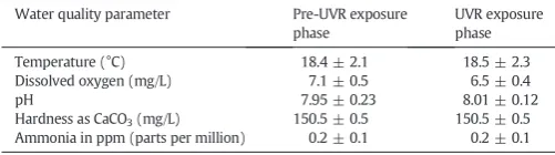
Approximately 300 adult fathead minnows (standard length ± SD = 5.27 ± 0.38 cm, weight ± SD = 2.05 ± 0.51 g) were collected between April and May 2010, from a pond located on the University of Saskatchewan campus, using Gee's improved minnow traps. Male minnows in the reproductive phase have suppressed ECC numbers due to high testosterone levels (Smith, 1973). To ensure that minnows were in the non-reproductive phase, they were acclimated in the labo-ratory for at least one month prior to the experimental procedure. Main-taining thefish in the laboratory for an extended period can also reduce variation in baseline ECC production between minnows collected from the same site (Manek et al., 2013). Fish were housed in a 73-L aquaria containing dechlorinated tap water. The water was maintained at 18.4 ± 2.3 °C and the photoperiod was set to 14:10 h light:dark cycle. The water used for the experiments originated from the Saskatoon, SK, Canada municipal water supply and was tested on alternate days for water chemistry parameters (temperature, dissolved oxygen, pH, total alkalinity, nitrite, nitrate, chlorine, ammonia, dissolved oxygen and total hardness—Table 1) during the acclimation and experimental phase.
2.2. Cadmium and UVR exposure
were fed ad libitum daily with commercialflake food. Minnows in each beaker were not independent, so we considered the‘beaker’, not the in-dividual minnows, as our replicate unit in all analysis where we obtain-ed more than one data point per beaker. For phase two which involvobtain-ed exposing minnows to UVR and/or Cd, we could expose only 2 beakers at a time to UVR in the presence and absence of a UVR blockingfilter. Thus, following phase one, two beakers housing fourfish each were sacrificed for cortisol analysis and ECC analysis (see below) and the remaining two beakers housing fourfish each were used in the second phase of the ex-periment. We had 8–10 replicates in each treatment.
The second phase (hereafter referred as the UVR exposure phase) started immediately after phase one (15–18 days after initiation of the experiment). In this phase, minnows from both the Cd and no Cd groups were exposed to solar radiation for 8 h a day for four days in an Atlas SUNTEST XLS + Solar Simulator with Xeon lamp with a Suprax Day-light Glass Filter—290 nm cut off (Atlas Material Testing Technology LLC, Chicago, USA) following the methods described byManek et al. (2012). All of the minnows were exposed to UVR, half in the presence and half in the absence of a UV blockingfilter. In the UV-filtered group, the beakers had their top and sides covered with a 2 mm thick Lexan polycarbonate sheet. The polycarbonate sheet removed around 76% of the UVB and UVA radiation. This gave us a 2 × 2 design for the UVR exposure phase with the presence and absence of Cd (0 vs. 5.4μg/L) crossed with exposure to low or high levels of UVR. Eight groups with 4 minnows per group in each treatment were held in the solar simulator in quartz beakers (diameter 13.8 cm, height 16.8 cm, QSI Quartz Scientific, USA) with a complete water change being per-formed in each beaker each day.
At the end of the UVR exposure phase,fish were euthanized. Two of four minnows from each beaker were sacrificed to prepare alarm cues for the behavioral trials. Thesefish were euthanized with a blow to the head, in accordance with UCACS Animal Care protocol # 2009091. They could not be anesthetized prior to being sacrificed because the an-esthetic could contaminate the alarm cue solution and compromise the behavioral assay. The remaining twofish were used for histological analysis and were preserved in 10% neutral buffered formalin after they were euthanized with an overdose of MS222 or Aquacalm (Syndel Laboratories, Canada). The blood from all four minnows from each bea-ker was used in the cortisol analysis. We conducted between 8 and 10 replicates per treatment.
2.3. Cadmium analysis
Water samples (1 mL) from all treatment groups (pre-UVR expo-sure phase and UVR expoexpo-sure phase) were collected in 1.5 mL polyeth-ylene microfuge tubes every day during the experimental phase, and stored at 4 °C until Cd analysis. To ensure that Cd remains in dissolved form, all water samples were acidified to afinal strength of 0.2% nitric acid (trace metal grade, VWR, Canada), as suggested by the employed analytical protocol (Perkin Elmer, USA). Cadmium concentrations were estimated using a Graphite Furnace Atomic Absorption Spectrom-eter (AAnalyst 800, Perkin Elmer, USA). The mean concentration of Cd in the Cd spiked water samples was 5.40 ± 0.45μg/L (mean ± SD, N = 3
per day/per treatment). Cadmium levels in the remaining groups were below the detectable levels (detection limit = 0.1μg/L).
2.4. Experimental protocol for blood extraction
Blood extraction for cortisol analysis followed the method described byHalbgewachs et al. (2009). Blood samples (25–50μL) were extracted from the caudal vein near the analfin region of euthanized minnows. In order to obtain enough blood for the analysis, we pooled blood from fourfish from the same beaker. This blood was placed on ice and allowed to clot for at least 1 h. Serum was extracted from the blood after centrifugation and then frozen at−20 °C until it was used. The cortisol level in the extracted serum was measured by the Endocrine Laboratory at Prairie Diagnostic Service (University of Saskatchewan) in a Coat-A-Count radioimmunoassay (Immulite-1000 Cortisol, Diag-nostic Products Corporation, USA), which is designed for quantitative measurement of cortisol in serum.
2.5. Histological analysis of the skin
Tissue preparation for the analysis of the minnow epidermis follow-ed the method describfollow-ed byHugie (1990)with modifications (details can be found inManek et al., 2012). The entirefish was initiallyfixed in 10% neutral buffered formalin until tissue processing could be per-formed. Epidermal samples were taken from the dorso/lateral surface just behind the operculum to the dorsalfin and placed in histocassettes and stored between two biopsy pads in formalin. An automatic tissue processor (MUP1, Modular Vacuum Processor) was used to dehydrate thefixed skin tissue in a series of ethanol grades and perfused with par-affin wax. Tissues were then manually embedded in paraffin wax and sectioned using a rotary microtome (HM330, Heidelberg) at 5μm thick-ness. Following sectioning, 3–5 sections were placed on a pre-cleaned suprafrost slide (VWR microslides). After the slides were dried on a slide warmer, they were deparaffinized, rehydrated and then stained with periodic acid Schiff's reagent with Harris' hematoxylin (PAS-H) to darken the mucous cells and the basement membrane (PAS) and the nucleus (hematoxylin), rendering ECCs colourless and easily recog-nizable. Images of each epidermal cross section were captured with a Zeiss Axioplan Fluorescence Microscope with an AxioCamICc1 (Color, 1.4 MP) digital camera at 10× magnification. For each slide, we record-ed the following parameters: epidermal thickness, number of ECCs per mm of skin, ECC density and ECC area, which were all quantified using Image J 1.32, an image processing and analysis program (available on the National Institute of Health's web pagehttp://rsb.info.nih.gov/ij/). The main difference between number of ECCs per mm of skin and ECC density is that ECC density takes into account epidermal width. Two
fish with the same number of ECCs per mm length of skin could have very different ECC densities, given the differences in epidermal thick-ness (Manek et al., 2013). Thus we measured both parameters to dissect
fine differences and select the stronger parameter, based on epidermal thickness results. Analyses by the observer were conducted blind to the treatment.
2.6. Behavioural assay on potency of skin extract
The skin extract for the behavioural assay was produced fromfish following the UV exposure phase [Cd + high UVR (N = 15, mean ± S.E. standard length: 5.15 ± 0.31 cm), Cd + low UVR (N = 15, 4.95 ± 0.22 cm), no Cd + high UVR (N = 15, 4.79 ± 0.51 cm) and no Cd + low UVR (N = 15, 5.35 ± 0.14 cm)]. The skin from the lateral epidermal layer on either side of the body was removed and placed in 40 mL of chilled distilled water. The skinfillet was homogenized with a Polytron homogenizer andfiltered throughfilterfloss to remove large particles. Serial dilutions were used to obtain afinal concentration of 1 cm2 of skin per 40-L, a concentration known to elicit overt
Table 1
Mean (± S.E.) water quality parameters for the pre-UVR exposure and UVR exposure phases. T-tests indicate no significant difference between treatments at alpha = 0.05.
Water quality parameter Pre-UVR exposure
phase
UVR exposure phase
Temperature (°C) 18.4 ± 2.1 18.5 ± 2.3
Dissolved oxygen (mg/L) 7.1 ± 0.5 6.5 ± 0.4
pH 7.95 ± 0.23 8.01 ± 0.12
Hardness as CaCO3(mg/L) 150.5 ± 0.5 150.5 ± 0.5
antipredator response in fathead minnows (Ferrari et al., 2005, 2006). The alarm substance was frozen at−20 °C in 20-mL aliquots until used. The behavioural bioassay was carried out to evaluate the difference in the potency of alarm cues prepared from the skin of the above four groups and dechlorinated water as a control, on the antipredator re-sponse of minnows. None of the test minnows were exposed to UVR or Cd. The assay was performed in a total of seventyfive, 37-L aquaria (60 × 30 × 40 cm) which were wrapped in black plastic on three sides so thatfish in adjacent aquaria were not visible to each other. Each aquarium wasfilled with dechlorinated water and equipped with a single air stone. Each tank had a 3 × 3-grid pattern drawn on the side. Three randomly selected minnows from the stock tank were transferred for acclimation in each testing aquarium for at least 24 h prior to the assay. On the day of the trial, 1 h prior to initiation of the trial, minnows were fed commercialflake food ad libitum to ensure con-sistency with the methodology applied byManek et al. (2012). On aver-age, 10 replicates were tested each day. Each day, each of the four alarm cues were randomly assigned to onefifth of the aquaria, while the re-maining onefifth of the aquaria received a no cue control (water). We tested a total of 15 groups offish in each of thefive treatment groups. The experiment was divided into three phases: an eight-minute pre-stimulus phase, a one-minute pre-stimulus injection phase followed by an eight-minute post-stimulus phase (Pollock and Chivers, 2004). Prior to the pre-stimulus phase, 60 mL of water from each tank was withdrawn and discarded using injection tubes (to remove any stagnant water). Following the pre-stimulus phase, we injected 10 mL of the alarm sub-stance or water. We used a well-established protocol for measuring anti-predator responses of minnows (Ferrari et al., 2005). This included recording an index of shoaling and an estimate of activity level as mea-sured by line crossing. The shoaling index of threefish was measured every 15 s by evaluating the distance between the 3fish per aquarium every 15 s during the pre- and post-stimulus periods (VWR Scientific) (1: nofish within a body length of another; 2: twofish within a body length of each other; 3: all thefish within a body length of each other). As a measure of activity, the number of line crosses was also re-corded for one of the 3 minnows (focalfish) during the pre-stimulus phase. The same focalfish was selected and observed until the end of the post-stimulus phase. An increase in shoaling index and a decrease in activity level are two typical antipredator responses observed in min-nows (Chivers and Smith, 1998).
2.7. Statistical analysis
All statistical analyses were performed using SPSS (ver. 19, SPSS Inc., USA).
2.7.1. Physiological response and histological data
Dušan et al. (2006)showed that minnows euthanized with an over-dose of Aquacalm (metomidate hydrochloride) had a lower cortisol elevation from the baseline than those euthanized with MS222 (tricaine methanesulfonate). For this reason we included both euthanasia methods (MS222 vs. Aquacalm) in our analysis. For the pre-UVR expo-sure phase, we performed 2 × 2 ANOVAs looking at the effect of Cd (Cd vs. no Cd) and euthanasia method (MS222 vs. Aquacalm) on cortisol levels and histological parameters. Similarly for the UVR exposure phase, we performed three-way ANOVAs, investigating the effect of Cd (Cd vs. no Cd), UVR (low vs. high) and euthanasia method (MS222 vs. Aquacalm). Cortisol data were log-transformed to meet homosce-dasticity assumptions. All other raw data met parametric assumptions.
2.7.2. Behavioural data
For the behavioural responses, we used the differences in shoaling index and line crossing from the pre-stimulus baseline as our raw data. The effects of cues on the behavioural response of control min-nows were tested using one-way ANOVAs followed by post hoc Tukey tests.
3. Results
3.1. Evaluation of mortality
During the 14 day pre-UVR exposure phase, no mortality was recorded in either the control group (no Cd) or the Cd exposed group. In the UVR exposure phase, 10% of minnows in the Cd + high UVR group (4 of 40) died while 5% of minnows in the no Cd + high UVR group died (2 of 40). In the Cd + low UVR group, 7.5% of minnows died (3 of 40) while nofish died in the no Cd + low UVR group. Min-nows that did not survive until the end of the exposure were excluded from further analysis.
3.2. Cortisol levels
3.2.1. Pre-UVR exposure phase
The 2 × 2 ANOVA revealed a significant effect of Cd (F1,11= 30.9,
Pb0.001) and a significant effect of euthanasia method (F1,11= 18.3,
P = 0.001), but no interaction between the two (F1,11= 1.7,
P = 0.21) on the mean cortisol levels of minnows. We found thatfish euthanized via Aquacalm had a 5-fold lower concentration of cortisol in the no Cd group and a 2-fold lower concentration of cortisol in the Cd exposed group than those euthanized with MS222 (Fig. 1). In addi-tion,fish exposed to Cd, had a significantly lower level of cortisol than those not exposed to Cd.
3.2.2. UVR exposure phase
The 3-way ANOVA revealed a significant effect of Cd (F1,22= 5.8,
P = 0.025), a significant effect of UVR (F1,22= 37.6, Pb0.001), and a
significant effect of euthanasia agent (F1,22= 24.5, Pb0.001).
How-ever, none of the 2- or 3-way interactions were significant (all PsN0.35). Similar to the pre-UVR exposure phase results, our results indicate thatfish euthanized with Aquacalm had a lower level of cortisol than those euthanized with MS222 andfish exposed to Cd had lower mean cortisol levels than those not exposed to Cd. In addition, we found that minnows exposed to UVR had a greater level of mean cortisol production than those not exposed to UVR (Fig. 2). Cortisol levels in our current study are consistent with our previous work, where we showed that exposure to UVR resulted in a significant increase in cortisol pro-duction infish (Manek et al., 2012). However, the results of our 2 × 2 analysis in the pre-UVR exposure phase and 3-way analyses in the UVR exposure phase indicate that Cd played a significant role in disrupting elevation of cortisol production. This was irrespective of the type of euthanizing agent used or combined exposure to UVR and Cd.
3.3. Histological parameters
3.3.1. Epidermal thickness
3.3.1.1. Pre-UVR exposure phase.The 2 × 2 ANOVA revealed no signifi -cant effect of Cd (F1,16= 0.16.9, P = 0.69) or euthanasia method
(F1,16= 0.77, P = 0.392) and did not reveal any interaction between
the two (F1,16= 0.03, P = 0.85) on mean epidermal thickness of
minnows (no Cd + MS222 group mean epidermal thickness ± SD = 37.6 ± 6.6μm; no Cd + Aquacalm group: 36.1 ± 3.6μm; Cd + MS222: 38.8 ± 6.6μm; Cd + Aquacalm group: 36.6 ± 2.1μm).
3.3.1.2. UVR exposure phase.The 3-way ANOVA revealed a significant effect of Cd (F1,25= 14.14, P = 0.001), but there was no effect of UVR
(F1,25= 0.35, P = 0.55), or euthanasia agent (F1,25= 0.08, P =
0.775) or any significant 2- or 3-way interactions (all PsN0.32) on epidermal thickness. Fish exposed to Cd had lower mean epidermal thickness than those not exposed to Cd (no Cd + high UVR mean epidermal thickness ± SD = 29.03 ± 6.1μm; no Cd + low UVR: 30.70 ± 5.1μm; Cd + low UVR: 35.01 ± 2.2μm; Cd + high UVR: 38.90 ± 6.0μm). These results indicate that the Cd + high UVR group had an almost 25% thicker epidermis as compared to most of the other groups. Given that Cd influenced epidermal thickness it is im-perative that this be accounted for in any analysis of ECC investment. Twofish with the same number of ECCs per mm length of skin could have very different ECC densities, given differences in thickness. Conse-quently, in our experiment we analyzed ECC density rather than num-ber of ECCs per mm of skin (data not shown).
3.3.2. ECC density
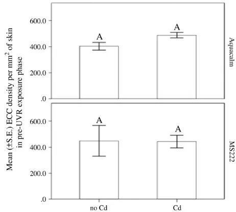
3.3.2.1. Pre-UVR exposure phase.The 2 × 2 ANOVA revealed no signifi -cant effect of Cd (F1,16= 0.35, P = 0.56) or euthanasia method
(F1,16= 0.23, P = 1.0) on mean ECC density. Also, there was no
inter-action between the two (F1,16= 0.46, P = 0.5) on the mean ECC
densi-ty of minnows (Fig. 3).
3.3.2.2. UVR exposure phase.The 3-way ANOVA revealed no significant effect of Cd (F1,25= 0.32, P = 0.571), or euthanasia agent (F1,25=
3.8, P = 0.063), but a significant effect of UVR (F1,25= 27.94,
Pb0.001). However, none of the 2- or 3-way interactions were signifi -cant (all PsN0.26). Similar to the pre-UVR exposure group results, our results indicate thatfish euthanized with Aquacalm did not differ in mean ECC density than those euthanized with MS222. However, we
found that minnows exposed to UVR had a 2-fold lower ECC density than those not exposed to UVR (Fig. 4).
3.3.3. ECC area
3.3.3.1. Pre-UVR exposure phase.The 2 × 2 ANOVA revealed no signifi -cant effect of Cd (F1,16= 0.14, P = 0.713), or euthanasia method
(F1,16= 0.31, P = 0.58) on mean ECC area. Also it revealed no
interac-tion between the two (F1,16= 0.20, P = 0.65) on the mean ECC area of
minnows (data not shown).
3.3.3.2. UVR exposure phase.The 3-way ANOVA revealed no significant effect of Cd (F1,27= 3.94, P = 0.057), or UV (F1,27= 0.97,
P = 0.333), or euthanasia agent (F1,27= 0.77, P = 0.388) on mean
ECC area. Also, none of the 2- or 3-way interactions were significant (all PsN0.51) (data not shown).
3.4. Behavioural assay
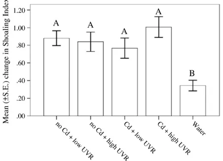
The one-way ANOVAs revealed a significant effect of cue on the be-havioural responses of minnows for both shoaling index (F4,70= 6.3,
Fig. 2.Mean ± S.E. serum cortisol levels from blood of minnows exposed to UVR (high UVR vs. low UVR) and Cd (no Cd vs. Cd) in the UVR exposure phase [N = 4–5/euthanizing agent (MS222 vs. Aquacalm)]. Different letters indicate significant differences.
Fig. 3.Mean ± S.E. ECC density per mm2
of skin of minnows exposed to Cd (no Cd vs. Cd) in the pre-UVR exposure phase. [N = 4–5/euthanizing agent (MS222 vs. Aquacalm)]. Dif-ferent letters indicate significant differences.
Fig. 4.Mean ± S.E. ECC density per mm2
Pb0.001,Fig. 5) and line crosses (F4,70= 4.1, P = 0.005,Fig. 6). For
both behavioural measures, post-hoc Tukey tests revealed a stronger antipredator response displayed by minnows exposed to alarm cues than minnows exposed to water (all Psb0.001). However, post hoc tests revealed no difference among treatments for minnows exposed to alarm cues (PN0.05).
4. Discussion
Understanding the effects of multiple stressors on organisms is an emerging discipline in stress ecology (Altshuler et al., 2011). It is par-ticularly interesting to determine the combined effects of different stressors when we predict a priori that different stressors could lead to different outcomes. Our previous studies on ECC investment infishes provide such a case. While trying to address the evolutionary role of ECCs (immune-function hypothesis),Chivers et al. (2007), showed that minnows exposed to 5.64μg/L of waterborne cadmium for 14 days were no longer able to increase production of ECCs in response to pathogenic challenge. Similarly, our previous work indicated that minnows exposed to 30μg/L of waterborne cadmium for 14 days in the absence of pathogenic zoospores did not reduce ECC density relative to that observed in controlfish (Kusch et al., unpublished data). It was also observed that a 14-day exposure to 5.64 and 30.00μg/L of water-borne Cd resulted in immunosuppression, measured as a significant re-duction in respiratory burst activity. Supporting the immune-function hypothesis,Halbgewachs et al. (2009)found an inverse relationship between cortisol and ECC investment in fathead minnows. They showed that intra-peritoneal injections of cortisol, and the associated suppres-sion of the immune system as measured by a respiratory burst assay, re-sulted in lower numbers of ECCs in fathead minnows. Likewise,Manek et al. (2012)found that exposure to UVR led to an increase in cortisol production and lower ECC production in minnows.
Exposure to Cd likely has the opposite effect on cortisol production than exposure to UVR.Lacroix and Hontela (2004)have shown using an in vitro approach that short-term acute Cd exposure to adrenocorti-cal cells offish can cause endocrine disruption by lowering the charac-teristic elevation in cortisol production in response to stress. Similarly,
Scott et al. (2003)found that in vivo exposure to waterborne Cd at a concentration of 2μg/L for 7 days resulted in a decrease of the charac-teristic elevation of cortisol observed in rainbow trout exposed to alarm cues. The impaired elevation in cortisol production is likely due to the disruption of the hypothalamic-pituitary-interrenal axis, which is the primary pathway for cortisol production infish (Wendelaar Bonga, 1997). Based on the studies described above we can suggest that there may be a fascinating link between exposure to UVR, and
immunosuppressants like Cd, which could be driven by changes in cortisol levels (Chivers et al., 2007; Manek et al., 2012). An elevation in cortisol production is associated with a reduction in ECC investment. However, it was not known what happens to cortisol production when
fish are exposed to both Cd and UVR, and subsequently how that infl u-ences ECC investment and the potency of alarm cues produced by the
fish. Normally expression of an elevated cortisol response under physi-ologically stressful conditions is necessary for an organism to maintain normal homeostasis (Sapolsky et al., 2000). If aquatic organisms that are stressed by exposure to elevated UVR are unable to express an elevated cortisol response due to factors like Cd exposure (endocrine disruption), their physiological state could be further deteriorated ulti-mately making it detrimental for the basic survival of that organism.
One of the key goals of our current study was to better understand the immune-function hypothesis with relation to cortisol production and ECC investment using multiple environmental stressors. Thus, we selected 5μg/L waterborne Cd exposures for the current study based on the observations of the previous studies mentioned above. Moreover, it is also important to note that our chosen Cd exposure concentration is environmentally relevant since similar Cd concentrations have been reported in many contaminated natural aquatic ecosystems (CCME, 1996). Similarly, the experimental setup and duration of UVR exposure in our current study is almost identical to the one described byManek et al. (2012). The total level of UVR emitted by the solar simulator in the present study was 250 W/m2which is a cumulative dose that
in-cludes UVR and PAR (photochemically active radiation). If we dissect out the actual UVR exposure only, it was around 45 W/m2, which is
comparable to natural levels of UVR in mid-summer in Saskatchewan (43 W/m2). The levels of UVR that fathead minnows are exposed to across their geographical range can vary 2 fold depending on their lati-tude (Goncalves et al., 2010). The level of UVR exposure in our current study was a cumulative dose of exposure which is comparable to levels of UVR in mid-summer in Saskatchewan (Sereda et al., unpublished results).
Results of the histological parameters in our current study revealed an interesting link between ECC investment and cortisol. We found that minnows exposed to UVR had lowered ECC density irrespective of the type of euthanizing agent used. Despite the reduction in cortisol production due to Cd exposure in the Cd + high UVR group, we did notfind a significant difference in mean ECC density or ECC area be-tween the Cd + high UVR group and no Cd + high UVR group. This in-dicates that the reduction in cortisol production due to Cd was not enough to change ECC investment. An elevation in cortisol production may trigger the reduction in ECC investment, but the increased levels
Fig. 5.Mean ± S.E. change in shoaling index for fathead minnows exposed to alarm cues prepared from the skin of minnows exposed to UVR in the presence and absence of a UVR blockingfilter (high UVR vs. low UVR) crossed with the presence and absence of cadmium (Cd vs. no Cd). N = 15/treatment. Different letters indicate significant differences.
of cortisol in our current study was probably not sufficient enough to lead to greater reductions in ECC investment in fathead minnows rela-tive to controls.
We did notfind any significant difference in antipredator response to alarm cues from the skin of UVR and Cd exposed minnows. This be-havioural observation is consistent with our previous observations (Manek et al., 2012). Previous research has shown thatfish often exhibit a graded response to chemical alarm cues, such that a high concentra-tion of alarm cues can elicit a high level of antipredator response infi sh-es as compared to medium and low concentrations (Ferrari et al., 2005). For our behavioural experiment, we used a high concentration of alarm cues (1 cm2in 40 L of water, as inFerrari et al., 2005) to test antipred-ator responses (line crosses and shoaling index) in control minnows (not exposed to either UVR or Cd). It is possible that minnows were responding at their highest intensity to these cues, the result of which was that we could not detect anyfine differences in antipredator response. One more factor complementing this observation could be that an almost 2-fold reduction in ECC production is not enough to alter alarm cue potency. Future studies where minnows are exposed to a medium and low concentration of alarm cues could help reveal if there exists any modest difference in response to alarm cues from the skin of minnows exposed to UVR or Cd. A lack of significant difference in behavioural response to alarm cues despite a 2-fold reduction in ECC production also contributes to a growing body of evidence that sug-gests that alarm cues may not be exclusively produced in ECCs but else-where in the skin of cyprinidfish skin (Carreau-Green et al., 2008).
Overall, the keyfindings of our current study support most of our predictions. At the waterborne Cd exposure concentration used in our study, we found that: (i) Cd can influence UVR driven increases in cortisol elevation with no downregulation of ECC investment, (iia) the duration of UVR exposure in our study in the absence of Cd results in characteristic elevation of cortisol levels and downregulates ECC invest-ment, (iib) the duration of UVR exposure in our study in the presence of Cd, disrupts characteristic elevation of cortisol production and downregulates ECC investment and; (iii) UVR and Cd did not elicit dif-ferent behaviours at the concentration of alarm cue prepared from UVR and/or Cd exposed minnows despite reduction in ECC investment. Our work points to the fact that it is difficult to predict the physiological, behavioural and ecological effects of exposure to multiple stressors. Fu-ture studies could build upon this framework to identify the relative im-portance of different stressors under different conditions. For example, water chemistry can play a significant role in affecting bioavailability of heavy metals and levels of UVR exposure. Specifically, water hardness has been demonstrated to be the most significant modulator of water-borne Cd bioavailability tofish (Niyogi et al., 2008). It is well character-ized that hardness cations, particularly Ca2+ions, compete with free Cd
ions (Cd2+) for binding sites on thefish gill, and thereby decrease Cd
bioavailability and toxicity (Niyogi et al., 2008). In our current experi-mental setup, we used dechlorinated Saskatoon city water, which has a moderate level of hardness (150–160 mg/L as CaCO3). This could
have resulted in lowered bioavailability of Cd2+ions to thefish in our study, which might have resulted in lack of effect on ECC investment and alarm cue production. Thus, it would be interesting to examine the effects of UVR and Cd infish during exposure at a relatively lower hardness. The DOC concentration in dechlorinated Saskatoon city water varies between 2.6 and 3.2 mg/L (Manek et al. unpublished data). Likewise, it would be interesting to examine the effects of UVR in the presence of higher levels of DOC, which is known to attenuate UVR (Williamson et al., 2001). The DOC levels across lakes and wetlands in Saskatchewan range from 4.1 to 156.2 mg/L (Arts et al., 2000), so there is considerable scope for empirical testing. In addition to identify-ing the relative importance of multiple stressors, we need to understand time lag effects. There was no effect of Cd on skin thickness pre-UVR, but an effect after the UVR phase. Could this be due to duration of Cd expo-sure instead of UVR expoexpo-sure? In our study minnows were exposed to Cd for 18 days prior to being euthanized. We know that Cd accumulates
in various target tissues like the liver, kidney, gill and ovary/testes infish due to its long half-life and low rate of excretion (Norey et al., 1990; Sorensen, 1991; Hollis et al., 2000). Perhaps the disrupted cortisol pro-duction would be much greater in magnitude with a longer exposure time. A depuration period following Cd exposure, prior to any exposure to UVR, could help understand if the disrupted cortisol production as a result of endocrine disruption can be reversed in thesefish.
With ever increasing disturbance to aquatic and terrestrial ecosys-tems, we are seeing organisms exposed to multiple stressors. Our results highlight that identifying the effects of simultaneous exposure to these stressors will remain a challenge for environmental scientists.
Acknowledgments
Funding was provided by NSERC Discovery Grants to DPC and SN, and the University of Saskatchewan. We would like to thank Zach Hoover for helping withfish collection.
References
Altshuler I, Demiri B, Xu S, Constantin A, Yan ND, Cristescu ME.An integrated multi-disciplinary approach for studying multiple stressors in freshwater ecosystems: Daphniaas a model organism. Integr Comp Biol 2011;51(4):623–33.
Arts MT, Robarts RD, Kasai F, Waiser MJ, Tumber VP, Plante AJ, et al.The attenuation of ultraviolet radiation in high dissolved organic carbon waters of wetlands and lakes on the northern Great Plains. Limnol Oceanogr 2000;45(2):292–9.
Blaustein AB, Hoffman PD, Hokit G, Kiesecker JM, Walls SC, Hays JB.UV repair and resistance to solar UV-B in amphibian eggs: a link to population declines? Proc Natl Acad Sci U S A 1994;91:1791–5.
Blaustein AB, Kiesecker JM, Chivers DP, Anthony RG.Ambient UV-B radiation causes deformities in amphibian embryos. Proc Natl Acad Sci U S A 1997;94(25):13735–7. Blechinger SR, Kusch RC, Haugo K, Matz C, Chivers DP, Krone PH.Brief embryonic
cadmium exposure induces a stress response and cell death in the developing olfactory system followed by long-term olfactory deficits in juvenile zebrafish. Toxicol Appl Pharmacol 2007;224:72–80.
Boone MD, Semlitsch RD, Little EE, Doyle MC.Multiple stressors in amphibian communities: effects of chemical contamination, bullfrogs, andfish. Ecol Appl 2007;17(1):291–301. Campbell PGC.Cadmium—a priority pollutant. Environ Chem 2006;3:387–8. Carreau-Green DN, Mirza SR, Martinez LM, Pyle GG.The ontogeny of chemically mediated
antipredator responses of fathead minnowsPimephales promelas. J Fish Biol 2008;73: 2390–401.
CCME.Canadian water quality guidelines. Canadian water quality guidelines. Canadian Council of Resource and Environment Ministers; 1996.
Chivers DP, Smith RJF.Chemical alarm signalling in aquatic predator–prey systems: a review and prospectus. Ecoscience 1998;5:338–52.
Chivers DP, Wisenden BD, Hindman CJ, Michalak TA, Kusch RC, Kaminskyj SG, et al. Epi-dermal‘alarm substance’cells offishes maintained by non-alarm functions: possible defence against pathogens, parasites and UVB radiation. Proc R Soc Lond B Biol Sci 2007;274:2611–9.
Chivers DP, Brown GE, Ferrari MCO.The evolution of alarm substances and disturbance cues in aquatic animals. In: Hansson, Bronmark, editors. Chemical ecology in aquatic systems. Oxford, UK: Oxford University Press; 2012.
Dušan P, Herolt DM, Andreasen CB, Menzel BW, Roth J.Anesthetic efficacy of tricaine methanesulfonate, metomidate and eugenol: effects on plasma cortisol concentration and neutrophil function in fathead minnows (Pimephales promelasRafinesque, 1820). Aquaculture 2006;254:675–85.
Ferrari MCO, Trowell J, Brown G, Chivers DP.The role of leaning in the development of threat-sensitive predator avoidance in fathead minnows. Anim Behav 2005;270: 777–84.
Ferrari MCO, Kapitania-Kwok T, Chivers DP.The role of learning in the development of threat-sensitive predator avoidance: the use of predator cue concentration by fathead minnows. Behav Ecol Sociobiol 2006;60:522–7.
Ferrari MCO, Wisenden BD, Chivers DP.Chemical ecology of predator–prey interactions in aquatic ecosystems: a review and prospectus. Can J Zool 2010;88:698–724. Goncalves RJ, Soula MS, Aigo J, Modonutti B, Balseiro E, Villafane VE, et al.Response of
plankton andfish to temperate zones to UVR and temperature in a context of global change. Ecol Austral 2010;20:129–53.
Gottofrey J, Tjälve H.Axonal transport of cadmium in the olfactory nerve of the pike. Pharmacol Toxicol 1991;69:242–52.
Halbgewachs CF, Marchant TA, Kusch RC, Chivers DP.Epidermal club cells and the innate immune system of minnows. Biol J Linn Soc Lond 2009;98:891–7.
Hollis L, McGeer JC, McDonalds DG, Wood CM.Effects of long term sublethal Cd exposure in rainbow trout during soft water exposure: implications for biotic ligand modelling. Aquat Toxicol 2000;51:93–105.
Hugie DM.An intraspecific approach to the evolution of chemical alarm signalling in the Ostariophysi. (MSc Thesis)University of Saskatchewan; 1990.
Lacroix A, Hontela A.A comparative assessment of the adrenotoxic effects of cadmium in two teleost species, rainbow troutOncorhynchus mykiss, and yellow perchPerca flavescens. Aquat Toxicol 2004;67:13–21.
Manek AK, Ferrari MCO, Sereda J, Niyogi S, Chivers DP.The effects of ultraviolet radiation on a freshwater preyfish: physiological stress response, club cell investment and alarm cue production. Biol J Linn Soc Lond 2012;105:832–41.
Manek AK, Ferrari MCO, Pollock RJ, Vicente D, Weber LP, Chivers DP.Within and between population variation in epidermal club cell investment in a freshwater preyfish: a cautionary tale for evolutionary ecologists. PLoS One 2013;8.
Mayer SJ.Stratospheric ozone depletion and animal health. Vet Rec 1992;131:120. McKenzie RL, Aucamp PJ, Bais AF, Bj¨orn LO, Ilyas M, Madronich S.Ozone depletion and
climate change: impacts on UV radiation. Photochem Photobiol Sci 2011;10:182. Newman PA, Nash ER, Kawa R, Montzka SA, Schauffler SM.When will the Antarctic ozone
hole recover? Geophys Res Lett 2006;33:LI2814.
Niyogi S, Wood CM.The biotic ligand model,flexible tool for developing site-specific water quality guidelines for metals. Environ Sci Technol 2004;38:6177–92. Niyogi S, Kent R, Wood CM.Effects of water chemistry variables on gill binding and acute
toxicity of cadmium in rainbow trout (Oncorhynchus mykiss): a biotic ligand model (BLM) approach. Comp Biochem Physiol C 2008;148:305–14.
Norey CG, Cryer A, Kay J.A comparison of cadmium induced metallothionein gene expression and Me2+distribution in the tissue of cadmium sensitive (rainbow trout; Salmo gairdneri) and tolerant (stone loach;Noemacheilus barbatulus) species of freshwaterfish. Comp Biochem Physiol 1990;97C:221–5.
Pereira HM, Navarro LM, Martins IS.Global biodiversity change: the bad, the good, and the unknown. Annu Rev Environ Resour 2012;37:25–50.
Pollock MS, Chivers DP.The effects of density on learned recognition of heterospecific alarm cues. Ethology 2004;110:341–9.
Salo H, Jokienen E, Markkula S, Aaltonen T, Penttila H.Comparative effects of UVA and UVB irradiation on the immune system offish. Photochem Photobiol B Biol 2000;56:154–62.
Sanchez-Dardon J, Voccia I, Hontela A, Chilmonczyk S, Dunier M, Boermans H, et al. Immunomodulation by heavy metals tested individually or in mixtures in
rainbow trout (Oncorhynchus mykiss) exposedin vivo. Environ Toxicol Chem 1999;18: 1492–7.
Sapolsky RM, Romero LM, Munck AU.How do glucocorticoids influence stress responses? Integrating permissive, supportive, stimulatory, and preparative actions. Endocr Rev 2000;21:55–89.
Scott GR, Sloman KA, Rouleau C, Wood CM.Cadmium disrupts behavioural and physiological response to alarm substance in juvenile rainbow trout (Oncorhynchus mykiss). J Exp Biol 2003;206:1779–90.
Scully NM, Lean DR.The attenuation of ultraviolet radiation in temperate lakes. Arch Hydrobiol 1994;43:135–43.
Smith RJF.Testosterone eliminates alarm substance in male fathead minnows. Can J Zool 1973;51:875–6.
Sorensen EM.Metal poisoning infish. Boca Raton FL: CRC Press; 1991175–234. Stromberg PC, Ferrante JE, Carter S.Pathology of lethal and sublethal exposure of fathead
minnows,Pimephales promelas, to cadmium: a model for aquatic toxicity assessment. J Toxicol Environ Health Sci 1983;11:2.
Sullivan JF, Aitchison GJ, Kolar DJ, McIntosh AW.Changes in the predator–prey behavior of fathead minnows (Pimephales promelas) and largemouth bass (Micropterus salmoides) caused by cadmium. J Fish Res Board Can 1978;35:446–51.
Tjälve H, Gottofrey J.Tissue disposition of 109 Cd2+in the brown trout (Salmo trutta)
stud-ied by autoradiography and impulse counting. Toxicol Environ Chem 1986;12:31–45. United States Environmental Protection Agency (U.S. EPA).2001 update of ambient water quality criteria for cadmium (EPA/822/R-01/001). Washington, USA: Author; 2001. Vitousek PM.Beyond global warming: ecology and global change. Ecology 1994;75(7):
1861–76.
Wendelaar Bonga SE.The stress response infish. Physiol Rev 1997;77:591–625. Williamson CE, Rose KC.When UV meets fresh water. Science 2010;329:637. Williamson CE, Zagarese HE.The impact of UVB radiation on pelagic freshwater ecosystems.
Arch Hydrobiol 1994;43:9–11.
![Fig. 1. Mean ± S.E. serum cortisol levels from blood of minnows exposed to Cd (no CdAquacalm)]](https://thumb-us.123doks.com/thumbv2/123dok_us/267350.2023025/4.595.323.548.556.711/mean-serum-cortisol-levels-blood-minnows-exposed-cdaquacalm.webp)