1
Sumana Nagapattinam
*2
Vinod Babu. K
3
Sai Kumar. N
4
Ayyappan. V. R
CORRESPONDING AUTHOR
*2Vinod Babu. K., MPT, MD (A.M)
Associate Professor,
K.T.G. College of Physiotherapy and KTG Multi Specialty Hospital, Bangalore-560 091, India.
Int J Physiother. Vol 2(5), 840-849, October (2015) ISSN: 2348 - 8336
ABSTRACT
Background: The principal target of any stroke rehabilitation is the motor impairments. Many studies have been advocated on the effect of Functional electrical stimulation and Task specific mirror therapy. Hence, the purpose of the study is to find the combined effect of task specific Mirror therapy with Functional Electrical Stimulation on upper limb function for subjects with sub-acute hemiplegia. Methods: An experimental study design, 60 subjects with sub-acute Hemiplegia randomised into 3 groups, functional electrical stimulation group (n=20), task specific mirror therapy group (n=20), and combined group (n=20). Each group received the corresponding regimen of treatment for 30 minutes with rest period for total 12 sessions over 2 weeks along with conventional physiotherapy. The outcome measure such as action research arm test was measured before and after two weeks of intervention. Result: When means of action research arm test were analyzed within the groups, there was a significant difference within all the three groups. When means were compared between three groups there is no statistically significant difference in pre- intervention and post intervention means.
Conclusion: It is concluded that a combination therapy of task specific mirror therapy with functional electrical stimulation for two weeks duration, is shown to be effective for recovery of upper limb function in subjects with sub-acute hemiplegia. However, the combination of task specific mirror therapy and functional electrical stimulation is shown to have similar improvements as only task specific mirror therapy and functional electrical stimulation.
Key words: Sub-acute hemiplegia, upper limb function, mirror therapy, task specific mirror therapy, functional electrical stimulation, action research arm test
DOI: 10.15621/ijphy/2015/v2i5/78243
1Physiotherapist,
3Professor & Principal.
4Associate Professor,
K.T.G. College of Physiotherapy and KTG Multi Specialty Hospital. Bangalore. India. Affiliated to Rajiv Gandhi University of Health Sciences, Karnataka. India.
Received 19th September 2015, revised 04th October 2015, accepted 06th October 2015
INTRODUCTION
Stroke can be referred to as a cerebrovascular disease due to vascular pathology depriving the
brain tissue of its blood supply.1 The prevalence of
stroke in India is 44–843/100,000, and the Indian Council of Medical Research (ICMR) estimates that by 2015, India will report 1.6 million cases of stroke
per year.2,3 The classical sign of cerebrovascular
disease is hemiplegia with other symptoms.1 It has
been reported that about 55% to 75% of them have impairment of function relative to upper limb paralysis which forms the integral part of
rehabilitation.4
The principal target of any stroke rehabilitation is the motor impairments with the introduction of many therapeutic techniques in the recent decade, among these techniques, large interest is devoted to treatment approaches aimed to improve motor functions, includes constraint- induced movement therapy, mental practice, mirror therapy, virtual
reality, robotics, and brain stimulation techniques.5
Mirror therapy is a technique using a parasagittal mirror or a modified mirror device in rehabilitation. The concept of MT is that the mirror provides a “virtual feedback” that the paretic limb is moving with a normal pattern of movement as the mirror reflection of unaffected limb movement gets superimposed on the affected extremity creating an illusion in a similar way as action
observation or motor imagery.4-6 It has been
observed that in recent literature, emphasis has been on non-task related MT. A recent study by Young-Rim Paiket et., al showed that Task specific mirror therapy (TSMT) shown improvement in upper limb functioning in stroke patients by
comparing simple and task oriented MT.7
Functional electrical stimulation (FES) shown to have significant effect in upper limb functional
recovery after stroke.8,9 It has been suggested by
Ziling Lin., et,.al that FES being able to produce repetitive exercise and meaningful movement of limbs may activate cortical synapses similar to
repetitive task training.10 Furthermore, it has been
observed by Gergely., et., al that a combination of voluntary effort and FES has greater potential to induce motor cortex plasticity than with FES or voluntary training alone owing to excitability of corresponding motor areas through afferent input
from electrical stimulation.11
Many studies have proven the effect of Functional Electrical Stimulation (FES) and Mirror Therapy (MT) on functional improvement of arm in
hemiplegia, improving gait ability.12 Sneha S.
Khandare et al., found that Task Specific Mirror Therapy (TSMT) was more effective than MT or
task specific exercises alone.13 Since no study has
been found on combination of TSMT with FES on upper limb functioning in sub-acute hemiplegia. Therefore, the Study with research question, Does task specific Mirror therapy with Functional Electrical Stimulation have an effect on upper limb function for sub-acute hemiplegia? Hence, the purpose of this study is to find the combined effect of TSMT with FES on upper limb function for sub-acute hemiplegia. It was null hypothesized that there will be no significant effect of task specific
Mirror therapy with Functional Electrical
Stimulation on upper limb function for sub-acute hemiplegic subjects.
METHODOLOGY
An experimental study design with three groups, Group A: Task specific Mirror Therapy Group (TSMT Group), Group B: Functional Electrical Stimulation Group (FES Group), Group C: Study group - Combination of Task specific Mirror Therapy (TSMT) With Functional Electrical Stimulation (FES). As this study involved human subjects the Ethical Clearance was obtained from the Ethical Committee of KTG College of Physiotherapy and K.T.G. Hospital, Bangalore as per the ethical guidelines of Bio-medical research on human subjects. This study was registered under Rajiv Gandhi University of Health Sciences for subject for registration for dissertation with registration number 09_T031_47181. Subjects included in the study were unilateral hemiplegic
stroke right or left, 14,15 subjects between 6 weeks
and 6 months post stroke, 15 Ischemic stroke,13 age
group between 18-60 years,15 both male and female
subjects, Brunstrom stage of motor recovery of 2 to
5,14 Modified Ashworth scale score ≥1,16 Voluntary
extension of wrist and fingers of at least 10 degrees
from the resting position.16 Subjects were excluded
with above 60 years of age, Brunstrom stage 1 or 6, Wrist and/or finger contracture, presence of Cardiac pacemaker or other metal implants,
significant visual, auditory and cognitive
duration of thirty minutes. The subject was positioned on a height adjustable table comfortably while easily accommodating the mirror between the affected and unaffected limbs. The affected limb was placed behind the mirror and unaffected limb in front of the mirror. The mirror was positioned in front of the patient’s midline, so that the affected limb was fully covered by the mirror and the reflection of the unaffected limb was completely visible. The subject was first asked to perceive the limb differently to how it actually is. They were then instructed to observe the mirror reflection for one to two minutes, trying to visualize the mirror image as the affected limb. Once the subject got engaged with the mirrored limb they were asked to perform slow, easy to achieve bilateral movements (perceived bilateral movements) while continuing to look at the reflected image. The exercises that were performed by unaffected limb were active wrist extension and fingers extension in mid-prone and pronated forearm, task specific grasping and releasing of a half-litre bottle. Rest period was given for every 15 repetitions. The same set of exercises was given to both the limbs simultaneously. All movements were executed very slowly, as this facilitates the intensity of the mirror illusion. The gaze direction of the patient was regularly checked in the mirror and feedback of performance was given.
Procedure for intervention for Group B: 10, 20 In Group B, subjects were treated with Functional electrical stimulation simultaneously with two tasks, for a total duration of thirty minutes, for six days over a period of two weeks. The subject was positioned on a height adjustable table with the mirror placed in front of the midline. The positive electrode and negative electrode of the muscle stimulator (Johari Digitals AP 439) were placed over the muscle belly of the wrist extensors on forearm over the motor point of extensor digitorum communis/ extensor carpi radialis brevis/ extensor carpi radialis longus (between one-third and half-way from the proximal end of the dorsal forearm) of the affected upper limb. The subject was instructed to look into the opaque side of the mirror while the stimulation was given and was asked to perform the following exercises synchronously with the duty cycle of the stimulation such as active wrist extension and fingers extension in mid-prone and pronated forearm, task specific grasping and releasing of a half-litre bottle. Rest period was given for every 15 repetitions.The parameters of the stimulation included a frequency of 35 Hz, pulse width of 250 μs, symmetrical biphasic waveform, duty cycle of
5 s on and 5 s off, and the amplitude of the current was adjusted to the maximal tolerance of the patient, in a range up to 90 mA. The total number of cycles was 180 in a single treatment session. Procedure for intervention for Group C: 17, 10, 18, 20
In Group C, subjects were treated with a combination therapy of task specific mirror therapy and functional electrical stimulation, consisting of two tasks, for a total duration of thirty minutes. The subject was positioned on a height adjustable table with the mirror placed in front of the midline same as explained in Group A. The positive electrode and negative electrode of the muscle stimulator were placed as explained in Group B. The subject was then instructed to observe the mirror reflection for one to two minutes, trying to visualize the mirror image as the affected limb. Once the subject got engaged with the mirrored limb they were asked to perform slow, easy to achieve simultaneous bilateral movements (perceived bilateral movements) while continuing to look at the reflected image, with the affected limb performing synchronously with the duty cycle of electrical stimulation. The exercises that performed were active wrist extension and fingers extension in mid-prone and pronated forearm, task specific grasping and releasing of a half-litre bottle.
Figure-1: Group-A Task specific mirror therapy
Figure-3: Group- C Combination therapy of Task specific mirror therapy and functional electrical
stimulation Conventional exercises:21
All the three groups were prescribed with the same set of conventional exercise in an individualized format. The exercises included Range of motion exercises and stretches to the shoulder, elbow, wrist, fingers, hip, ankle, and trunk for ten repetitions in two sets progressing from passive to active exercises with manual and verbal cuing. Twenty minutes of upper limb training related to functional use were practiced such as opening drawers, putting away dishes, folding towels, closing blinds and counting change with emphasis on coordination for activities of daily living. This was followed by sets of balance exercises including step ups such as, repeated stepping anteriorly and laterally onto a step with affected lower limb progressing to higher step and decreasing upper limb support; repeated rising from a sitting position, progressing from using arms to not using arms and from high surface to lower; wall exercises such as standing against a wall at a distance and falling backwards while touching wall and coming back to neutral, progressing from short to large distances from the wall; repeated marching in place, progressing from upper limb support to no support were given. Also repeated rising up on toes, progressing from using upper limb support to no support; kicking a ball with either foot followed by abrupt stops and turns while walking were given. The number of repetitions prescribed was ten in two sets with sufficient rest periods.
Outcome Measurements:
The pre and post intervention measurements of upper limb function was measured using Action Research Arm Test.
Action Research Arm Test: This test was used to assess the upper extremity motor function and status in a standardized format using 19 tests of upper extremity function across 4 subsets: grasp, pinch, grip and gross movement, both distally and
proximally.58The test was administrated using
non-standardised equipment (various sized blocks of wood, cricket ball, stone, jug, glass, tube, washer and bolt, ball bearing and marble). The subject was seated in a chair in front of the table with the box
of accessories placed at reachable distance. After counting up to three and stating “yes” to cue the subject, the subtest was begun till the subject placed his hand back on the table. The baseline to complete the task was 60 seconds taking a total of approximately 10 minutes to complete the full
test.59 ARAT scale shown to have intra-class
correlation coefficient (ICQ) of 0.98 indicating very high inter-rater reliability and supported the value of using this scale for measuring recovery of upper
limb function in hemiplegia.22
Statistical Methods
Descriptive statistical analysis was carried out in the present study. Out Come measurements
analyzed are presented as mean SD. Significance
is assessed at 5 % level of significance with p value was set at 0.05 less than this is considered as statistically significant difference. Paired ‘t’ test as a parametric and Wilcoxon signed rank test as a non-parametric test have been used to analysis the variables pre-intervention to post-intervention with calculation of percentage of change. One-way ANOVA, Tukey HD post-hoc test, post-hoc test Homogeneous Subsets and Kruskal-Wallis Test have been used to compare the means of variables between the three groups and multiple pair-wise comparisons with calculation of percentage of difference between the means. The Statistical software namely SPSS 16.0, Stata 8.0, MedCalc 9.0.1 and Systat 11.0 were used for the analysis of the data and Microsoft word and Excel have been used to generate graphs, tables etc.
RESULTS
The study carried on total 60 subjects ( Table-1) in Group A there were 20 subjects with mean age of 44.65 years, in Group B there were 20 subjects with mean age of 44.15 years.
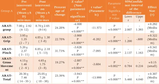
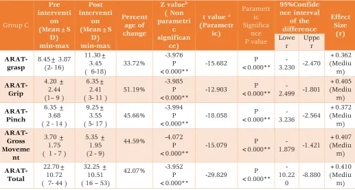
When means were analyzed within the groups (Table-2, 3, 4) shows that in Group A, B and C there is a statistically significant change in means of Action Research Arm Test – grasp, grip, pinch, Gross Movement, total when means were analyzed from pre intervention to post intervention within the groups with p<0.000 with positive percentage of change showing there is increase in post means. In Group A and C there is moderate clinical significant improvement with medium effect size, In Group B there is weak clinical significant improvement with small effect size.
When pre intervention means and post
Table 1: Basic Characteristics of the subjects studied
Basic Characteristics of the subjects studied
Group A (Task specific Mirror Therapy Group) Group B (Functional Electrical Stimulation Group) Group C (Combination group) a. Between the groups Significance
Total number of subjects
studied (n) 20 20 20 --
Age in years (Mean± SD)
44.65 ± 11.23 (25-60)
44.15± 8.64 (27-60)
45.75± 8.93
(25-58) p= 0.818(NS)
Gender Males n=12 60% n=14 70% n=14 70% --
Females n=8 40% n=6 30% n=6 30%
Duration of stroke(Months) 4.17± 1.15 (2.0-6.0) 4.10± 1.16 (2.5-6.0) 4.42± 1.09
(2.0-6.0) p= 0.656 (NS)
Brunnstrom's stage 3.75± 0.78
(3-5)
3.70± 0.86 (2-5)
4.15± 0.74
(3-5) p= 0.158 (NS)
Modified Ashworth's scale (MAS)
1 n=4 20% n=5 25% n=7 35% --
1+ n=9 45% n=9 45% n=11 55% --
2 n=7 35% n=6 30% n=2 10% --
a. Kruskal Wallis Test
Table 2: Analysis of Action Research Arm Test within Group A (Pre to post test analysis)
Group A Pre interventi on (Mean±S D) min-max Post interventi on (Mean±S D) min-max Percent age of change
Z valueb
(Non parametri
c significan
ce)
t valuea
(Parametr ic) Parametr ic Significa nce P value 95%Confide nce interval of the difference Effect Size (r) Lowe r Uppe r ARAT-grasp 7.00± 2.92 (4- 12) 8.70±2.65
(6-14) 24.28%
-4.008 P <0.000** -11.573 P <0.000** -2.007 -1.393 +0.292 (Mediu m) ARAT-Grip 3.95± 1.35 (2 – 7 )
4.85± 1.30
( 3 -8) 22.78%
-3.900 P <0.000** -6.282 P <0.000**
-1.200 -.600
+0.322 (Mediu m) ARAT-Pinch 5.20± 2.37 ( 3 - 11)
6.85± 2.18
( 5 - 13) 31.73%
-3.626 P <0.000** -7.095 P <0.000** -2.137 -1.163 +0.341 (Mediu m) ARAT-Gross Moveme nt 4.15± 1.46 ( 2 - 6 )
4.65± 1.75 (3 - 8)
19.27% -2.887
P =0.004** -3.684 P =0.002** -0.784 -0.216 +0.15 (small) ARAT-Total 20.30± 7.46 ( 14 - 35 )
25.05± 7.26 ( 18 - 38)
23.39% -3.943
P =0.001** -13.999 P <0.000** -5.460 -4.040 +0.307 (Mediu m)
Table 3: Analysis of Action Research Arm Test within Group B (Pre to post test analysis) Group B Pre interventi on (Mean±S D) min-max Post interventi on (Mean±S D) min-max Percenta ge of change
Z valueb
( Non parametri
c significan
ce)
t value a (Parametr ic) Parametr ic Significan ce P value 95%Confide nce interval of the difference Effec t Size (r) Lowe r Uppe r ARAT-grasp 7.55 ± 3.53 (2- 12) 9.45 ± 2.89 (5-13)
25.16% -3.869
P <0.000** -7.933
P <0.000** -2.401 -1.399 +0.2 82 (Smal l) ARAT-Grip 4.75 ± 2.46 (1– 8 )
5.65 ± 2.34 ( 2-9)
18.94% -3.307
P <0.001** -4.723
P <0.000** -1.299 -.501 +0.1 84 (Smal l) ARAT-Pinch 6.55 ± 3.39 (2 - 12)
8.00 ± 3.02 (4- 14)
22.13% -3.477
P <0.001** -6.175
P <0.000** -1.941 -.959 +0.2 20 (Smal l) ARAT- Gross Moveme nt 4.35 ± 1.84 ( 1 - 6 )
5.25 ± 1.71 (2 - 7)
20.68% -3.448
P <0.001** -5.604
P <0.000** -1.236 -.564 +0.2 46 (Smal l) ARAT-Total 23.20± 10.53 ( 8 - 37 )
28.35± 9.14 ( 16 – 41)
22.19% -3.931
P <0.000** -10.791
P <0.000** -6.149 -4.151 +0.2 53 (Smal l)
** Statistically Significant difference p<0.05; NS- Not significant; a. Pared t test. b. Wilcoxon Signed Ranks Test
Table 4: Analysis of Action Research Arm Test within Group C(Pre to post test analysis)
Group C Pre interventi on (Mean±S D) min-max Post interventi on (Mean±S D) min-max Percent age of change
Z valueb
( Non parametri
c significan
ce)
t value a (Parametr ic) Parametr ic Significa nce P value 95%Confide nce interval of the difference Effect Size (r) Lowe r Uppe r ARAT-grasp 8.45± 3.87 (2- 16) 11.30± 3.45 ( 6-18)
33.72% -3.976 P <0.000** -15.682 P <0.000** -3.230 -2.470 +0.362 (Mediu m) ARAT-Grip 4.20 ± 2.44 (1– 9 )
6.35± 2.41 ( 3- 11 )
51.19% -3.985 P <0.000** -12.903 P <0.000** -2.499 -1.801 +0.405 (Mediu m) ARAT-Pinch 6.35 ± 3.68 ( 2 - 14 )
9.25± 3.55 ( 5- 17 )
45.66% -3.994 P <0.000** -18.058 P <0.000** -3.236 -2.564 +0.372 (Mediu m) ARAT- Gross Moveme nt 3.70 ± 1.75 ( 1 - 7 )
5.35 ± 1.95 (2 - 9)
44.59% -4.072
P <0.000** -15.079 P <0.000** -1.879 -1.421 +0.407 (Mediu m) ARAT-Total 22.70± 10.72 ( 7- 44 )
32.25 ± 10.51 ( 16 – 53)
42.07% -3.952
P <0.000** -29.829 P <0.000** -10.22 0 -8.880 +0.410 (Mediu m)
Table 5: Comparison of means of Action Research Arm Test between GroupA, Group B and Groups C (PRE AND POST INTERVENTION COMPARISION)
Group A (Mean±SD)
min-max
Group B (Mean±SD)
min-max
Group C (Mean±SD)
min-max
Kruskal-Wallis
Chi-Square
a. Kruskal-Wallis Test Significance
(P value)
b. Tukey HSD Significance
(P value)
PRE INTERVENTION
ARAT-grasp 7.00±2.92
(4-12)
7.55 ± 3.53 (2- 12)
8.45± 3.87
(2- 16) 1.019 p=0.601 (NS) p=0.388 (NS)
ARAT-Grip 3.95± 1.35
(2 -7 )
4.75 ± 2.46 (1– 8 )
4.20 ± 2.44
(1– 9 ) 0.774 p =0.679 (NS) p=0.472 (NS)
ARAT-Pinch 5.20± 2.37
( 3 - 11)
6.55 ± 3.39 (2 - 12)
6.35 ± 3.68
( 2 - 14 ) 1.290 p=0.525 (NS) p=0.383 (NS)
ARAT- Gross Movement
4.15± 1.46 (2 - 6 )
4.35 ± 1.84 ( 1 - 6 )
3.70 ± 1.75
(1 - 7 ) 1.542 p =0.462 (NS) p=0.450 NS)
ARAT-Total 20.30±7.46
( 14 - 35 )
23.20± 10.53 ( 8 - 37)
22.70± 10.72
(7- 44 ) 0.771 p=0.680 (NS) p=0.613 (NS)
POST INTERVENTION
ARAT-grasp 8.70±2.65
(6-14)
9.45 ± 2.89 (5-13)
11.30± 3.45
(6-18) 6.095 p=0.047* p=0.137 (NS)
ARAT-Grip 4.85± 1.30
(3 -8)
5.65 ± 2.34 (2-9)
6.35± 2.41
( 3- 11 ) 3.692 P =0.158 (NS) p=0.067 (NS)
ARAT-Pinch 6.85± 2.18
(5 - 13)
8.00 ± 3.02 (4- 14)
9.25± 3.55
(5- 17 ) 5.905 p=0.052 (NS) p=0.445 (NS)
ARAT- Gross Movement
4.65± 1.75 (3 - 8)
5.25 ± 1.71 (2 - 7)
5.35 ± 1.95
(2 - 9) 2.166 P =0.339 (NS) p=0.445 (NS)
ARAT-Total 25.05± 7.26
(18 - 38)
28.35± 9.14 (16 – 41)
32.25 ± 10.51
(16 – 53) 6.245 P =0.044* p=0.488 (NS)
** Statistically Significant difference p<0.05; NS- Not significant. a. Kruskal-Wallis Test; b. One way ANOVA Post Hoc Tests -Tukey HSD (Homogeneous Subsets)
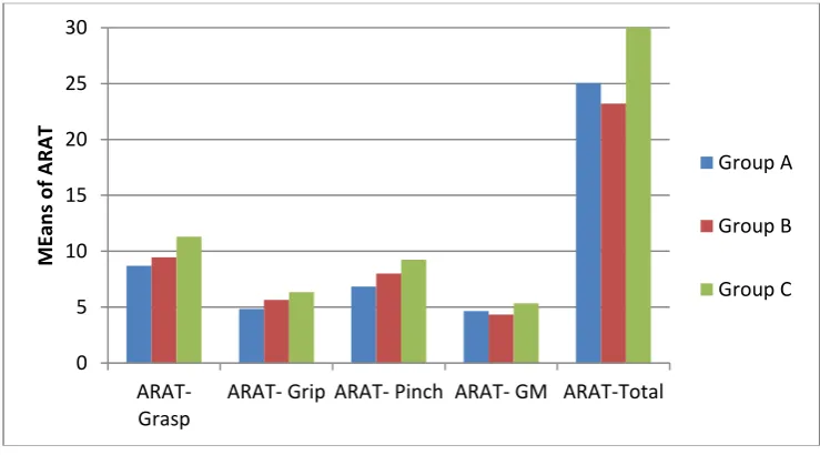
Graph - 1: Comparison of post intervention means of Action Research Arm Test between Group A , Group B and Group C (Post-intervention comparative analysis)
The above graph shows that there is no statistically significant change in means of Action Research Arm Test – grasp, grip, pinch, Gross Movement, total when post intervention means were compared between three groups ( p<0.000).
DISCUSSION
In this study, it was found from the analysis that there is no statistically significant difference in improvement in hand function in means of Action reaction arm test in the combination group when compared to the TSMT and FES group, however, all
0 5 10 15 20 25 30
ARAT-Grasp
ARAT- Grip ARAT- Pinch ARAT- GM ARAT-Total
M
Ean
s o
f A
R
A
T
Group A
Group B
the three groups showed statistically and clinically significant improvements from pre to post intervention means after 2 weeks.
In the Task specific mirror therapy group, there
was a significant improvement in post
interventions scores of ARAT. This could be because of the effect of the therapy that might have influenced the activation of the mirror neuron system that involved in action perception and are generally activated when an individual either performs a given motor act or observe a similar one which is functionally related. This coupling of observation and execution of congruent tasks
facilitates formation of motor memories.23,24 Mirror
therapy is one such multisensory stimulation technique using a mirror image of the normal limb superimposed on the affected limb. It has been suggested that on viewing the reflected image of one’s moving hand on the mirror, neuronal excitability occurs in the ipsilateral primary cortex
than when direct viewing of inactive hand.25, 26
Marian E. Michielsen et al., in their study investigated the neuronal basis of MT in stroke patients by using fMRI and showed that mirror illusion caused an increased activity in the precuneus and the posterior cingulate cortex, areas associated with awareness of the self and spatial attention during bimanual movement. This effect can be attributed to increase in spatial attention towards affected limb, thereby the mirror illusion
has aided in overcoming non-use.27 Therefore, MT
can be said to activate visual memory systems, thereby providing a recall of performance of affected limb when it was intact leading to
improved sensory feedback and motor output.17
Hence, it seems important to consider the use of task specific movements in respect to a
learning-dependent model of neural plasticity.13
In this present study, the FES group showed significant difference in pre and post intervention scores in ARAT measure. The mechanism underlying recovery of function following FES has been assumed to be based on neurophysiological models. In case of flexor spasticity, such as in hemiplegia, improvement through FES of extensor muscle can be thought to provide sufficient strength to overcome flexor spasticity or through reciprocal inhibition to the flexors group at the spinal cord level. Furthermore, FES can also evoke repetitive movements thereby facilitating motor
recovery through neural plasticity.16 Dilek Karakuş
et.,al, in their study specified that FES could be effective in reorganization and neurological healing and aid in improving functional gains especially in upper-limb in acute and sub-acute
phases of hemiplegia.15 Afferent input from
electrical stimulation of the periphery is said to excite their corresponding motor areas and the level of cortical excitability can be considered as an indicator of neuroplasticity and motor re-learning. In the combination group, there was a significant difference in findings between pre and post interventions scores of ARAT related to upper limb function. This could be due to neuroplasticity that could have occurred from two different forms of sensory motor stimulation techniques that were applied synergistically causing a recovery of upper limb function. Christian Dohle et al., in their study proposed that MT after stroke leads to a referral of sensation to the other hand, thus modulating motor and somatosensory representations of cortex and causing functionally relevant improvements in
motor, sensory and attention domains.28
Ramachandran et al., suggested that paralysis resulting in a restraint of movement could be due to a learnt factor or learned nonuse which can be reversed by using a synergic effect of MT and FES thereby increasing the self-consciousness and spatial attention to the affected limb. Further, MT involving bimanual movements can stimulate the neural networks in bilateral cerebral hemispheres
aiding in recovery of motor skills.19 Whereas FES, a
process of combining electrical stimulation with a functional task such as grasping objects, causes muscle contractions and improves functional abilities, it can be considered that the simultaneous application of mirror therapy shall aid in
revitalizing the muscles through active
participation. Gergely I. Barsi t et al., in their study suggested that the combination of voluntary effort and FES has greater potential to induce cortical plasticity than application of FES or voluntary
training alone in stroke rehabilitation.11
Gi Jeong Yun et al., in their study, applied electrical stimulation and MT with active movements simultaneously on sub-acute stroke
patients and found significant effects.20 But in our
present study first of its kind where the use of task specific mirror therapy has been applied along with FES for evaluating its effect on upper limb function. While Jackie Boscht et al., in their systemic review embarked on the effect of
task-oriented practice,29 Sneha S. Khandare et al.,
provided promising results regarding, adding task
specific exercises to MT.13
performance which was not applied appropriately in the present study as more practice is said to
produce better recovery.29 In the present study,
the total duration of MT or FES was only 30 minutes with rest periods, for twelve sessions over two weeks and this short training may only lead to a motor learning effect and improvement of motor control of the paretic arm would require much
longer training.4 Also the number of tasks was only
two.
Since FES and TSMT are considered to be effective as adjuncts in rehabilitation of upper limb functioning, the protocol of conventional exercises is an important aspect of any physiotherapy program. In this study emphasis was given to upper limb functional exercises related to coordination in daily activities as a conventional program to all the three groups, which could have masked the effectiveness of the combination
therapy.21
Limitations of the study
The study was conducted only on sub-acute hemiplegia between 6 weeks and 6 months post stroke. The duration of treatment was only for 30 minutes for 12 sessions over a period of 2 weeks. Only ARAT measure score was used for pre and post intervention analysis in this study. The effect of combination therapy on spasticity was not evaluated. The number of tasks selected for the study was less.
Recommendation for future research
Further studies on large population may prove beneficial in interpreting the actual effects of simultaneous TSMT and FES applications. Further studies on effects of TSMT and FES on spasticity and ROM may be required. Further studies using more number of tasks and higher dosage of NMES for a longer duration may be evaluated. Further studies evaluating long term effects of combination of TSMT and FES need to be considered.
CONCLUSION
The present study concludes that a combination therapy of task specific mirror therapy with functional electrical stimulation is shown to be effective for recovery of upper limb function in subjects with sub-acute hemiplegia. In two weeks duration, the combination of task specific mirror therapy and functional electrical stimulation is shown to have similar improvements as only task specific mirror therapy and functional electrical stimulation. Further long term studies are needed to find the effectiveness of this combination therapy. It is clinically recommended that incorporating more tasks, more number of practice trials and longer duration, may be more effective
while considering the use of combination therapy of task specific mirror therapy and functional
electrical stimulation in a comprehensive
rehabilitation program. Acknowledgement
Authors were expressing their sense of gratitude’s to the people who helped and encouraged them for the guidance and completion of this study.
Conflicts of interest: None REFERENCES
1. Allan H Ropper, Robert H Brown. Adams and
Victor’s principles of Neurology. 8th edi;2005.
2. Kameshwar Prasad, Deepti Vibha and
Meenakshi. Cerebrovascular disease in South Asia. Part I: A burning problem J R Soc Med Cardiovasc Dis. 2012;1:20.
3. Tenneti V.D. Sasisekhar, Madhavi Kodali, Sai
Kiran. Post Stroke Functional, Cognitive and Psychological outcomes and Mortality: Data from a Tertiary centre in South India. Int J Health Rehabil Sci. 2013; 2(1): 1-7.
4. Ruud W. Selles, Marian E. Michielsen,
Johannes B. J. Bussmann, Henk J. Stam, Henri L. Hurkmans, Iris Heijnen, Danielle de Groot, Gerard M. Ribbers. Effects of a Mirror-Induced Visual Illusion on a Reaching Task in Stroke Patients: Implications for Mirror Therapy Training. Neurorehabil Neural Repair. 2014 Sep;28(7):652-9.
5. Sjoerd de Vries, Theo Mulder. Motor imagery
and stroke rehabilitation: A critical discussion. J Rehabil Med. 2007;39(1):5-13.
6. Andreas Stefan Rothgangela, Susy M. Brauna,
Anna J. Beurskensa, Rudiger J. Seitzg, Derick T. Wade. The clinical aspects of mirror therapy in rehabilitation: A systematic review of the literature. Int J Rehabil Res. 2011 Mar;34(1):1-13.
7. Young-Rim Paik, Su-Kyoung Kim,Jae-Shin Lee,
Byoung-Jin Jeon. Simple and Task-oriented Mirror Therapy for Upper Extremity Function in Stroke Patients: A Pilot Study. Hong Kong Journal of Occupational Therapy.2014; 24(1): 6-12.
8. Barbara M. Doucet, Amy Lamb, Lisa Griffin.
Neuromuscular Electrical Stimulation for Skeletal Muscle Function. The Yale journal of biology and medicine. 2012; 85(2):201-215.
9. Shu-Shyuan Hsu, Ming-Hsia Hu, Jer-Junn Luh,
10.Ziling Lin, MD and Tiebin Yan. Long-Term Effectiveness of Neuromuscular Electrical Stimulation for Promoting Motor Recovery of the Upper Extremity after Stroke. J Rehabil Med. 2011; 43(6): 506–510.
11.Gergely I. Barsi, Dejan B. Popovic, Ina M.
Tarkka, Thomas Sinkjær, Michael J. Grey. Cortical excitability changes following grasping exercise augmented with electrical stimulation. Exp Brain Res. 2008; 191(1):57–66.
12.Sang-Goo Ji, Hyun-Gyu Cha, Myoung-Kwon
Kim,Chang-Ryeol Lee. The Effect of Mirror
Therapy Integrating FunctionalElectrical
Stimulation on the Gait of Stroke Patients. J Phys Ther Sci.2014; 26(4): 497–499.
13.Sneha S. Khandare, R. M. Singaravelan,
Subhash M. Khatri. Comparison of Task Specific Exercises and Mirror Therapy to Improve Upper Limb Function in Subacute Stroke Patients. IOSR-JDMS. 2013; 7(1): 05-14.
14.Yavuzer G, Selles R, Sezer N, Sütbeyaz S,
Bussmann JB, Köseog˘lu F, Atay MB, Stam HJ. Mirror therapy improves hand function in sub-acute stroke: a randomized controlled trial. Arch Phys Med Rehabil. 2008; 89(3):393-8.
15.Dilek Karakuş, Murat Ersöz, Gönül Ko yuncu,
Dilek Türk, Fatma Münevver Şaşmaz, Müfit
Akyüz. Effects of Functional Electrical Stimulation on Wrist Function and Spasticity in Stroke: A Randomized Controlled Study. Turk J Phys Med Rehab. 2013;59:97-102.
16.De Kroon JR, IJzerman MJ, Lankhorst GJ,
Zilvold G. Electrical stimulation of the upper limb in stroke: Stimulation of the extensors of the hand vs. alternate stimulation of flexors and extensors. Am J Phys Med Rehabil. 2004; 83(8):592–600.
17.Candy McCabe. Mirror Visual Feedback
Therapy. A Practical Approach. J Hand Ther. 2011 Apr-Jun;24(2):170-8.
18.Rothgangel AS, Braun SM. Mirror therapy:
Practical protocol for stroke rehabilitation 2013.
19.HyunJin Kim, GyuChang Lee, ChangHo Song.
Effect of Functional Electrical Stimulation with Mirror Therapy on Upper Extremity Motor Function in Poststroke Patients. Journal of Stroke and Cerebrovascular Diseases.2014;23 (4): 655-661.
20.Gi Jeong Yun, Min Ho Chun, Ji Young Park,
and Bo Ryun Kim. The Synergic Effects of
Mirror Therapy and Neuromuscular Electrical Stimulation for Hand Function in Stroke Patients. Ann Rehabil Med. 2011 June; 35(3): 316–321.
21.Pamela Duncan, Stephanie Studenski, Lorie
Richards, Steven Gollub, Sue Min Lai, Dean Reker, Subashan Perera, Joni Yates, Victoria Koch, Sally Rigler, Dallas Johnson. Randomized clinical Trial of Therapeutic Exercise in Sub-acute Stroke. Stroke. 2003; 34: 2173-2180.
22.60Ching-Ljn Hsieh, I-Ping Hsueh, Fu-Mei
Chiang, Po-Hsin Ljn. Inter-rater reliability and validity of the Action Research arm test in stroke patients. Age and Ageing. 1998; 27(2): 107-113.
23.Giacomo Rizzolatti, Maddalena Fabbri-Destro,
Luigi Cattaneo. Mirror neurons and their clinical relevance. Nature clinical practice neurology. 2009; 5(1)24-34.
24.Heyes, C. M. Where do mirror neurons come
from? Neuroscience and Biobehavioural Reviews. 2010; 34(4):575-58.
25.Alessio Faralli, Matteo Bigoni, Alessandro
Mauro, Ferdinando Rossi, Daniela Carulli. Noninvasive Strategies to Promote Functional Recovery after Stroke.Review Article. Neural Plast. 2013;2013:854597.
26.Suzanne S. Kuys, Tara Edwards, Norman R.
Morris. Effects and adherence of Mirror therapy in people with chronic upper limb Hemiparesis: A Preliminary study. ISRN 2012. Article ID 926784, 9 pages.
27.Marian E. Michielsen, Marion Smits, Gerard M.
Ribbers, Henk J.Stam, Jos N. van der Geest, Johannes B.J. Bussmann, Ruud W. Selles. The neuronal correlates of mirror therapy: an fMRI study on mirror induced visual illusions in
stroke patients. J Neurol Neurosurg
Psychiatry.2011 Apr;82(4):393-8.
28.Christian Dohle, Judith Püllen, Antje Nakaten,
Jutta Küst, Christian Rietz, and Hans Karbe. Mirror Therapy Promotes Recovery From Severe Hemiparesis: A Randomized Controlled Trial. Neurorehabil Neural Repair. 2009 Mar-Apr;23(3):209-17.
29.Jackie Bosch, Martin J. O’Donnell, Susan
Barreca, Lehana Thabane and LaurieWishart. Does Task-Oriented Practice Improve Upper Extremity Motor Recovery after Stroke? A Systematic Review. ISRN Stroke 2014. Article ID 504910, 10 pages.
Citation
Sumana Nagapattinam, Vinod Babu. K, Sai Kumar. N, & Ayyappan. V. R. (2015). EFFECT OF TASK SPECIFIC MIRROR THERAPY WITH FUNCTIONAL ELECTRICAL STIMULATION ON UPPER LIMB