COMPARATIVE EVALUATION OF AGE ESTIMATION BY NOLLA’S METHOD AND CHRONOLOGICAL
AGE OF 6-13 YEARS OLD CHILDREN
*
Shreyal Deshmukh, Dr. Darshana Shah, Dr. Rasika Pawar, Dr. Archana Gupta,
Dr. Sangeeta Palaskar and
Sinhgad Denatal College and Hospital, India
ARTICLE INFO ABSTRACT
Background:
an individual. Developing teeth are used to estimate age in number of disciplines using various techniques of age estimation.
Aim: To compare chronological age with a Materials and Methods:
visiting Sinhgad Dental College and Hospital, Pune in the age group of 6
Calcification stages of mandibular left and right canine were assessed and graded according Nolla’s calcification stages for determination of dental age and compared with the chronological age.Paired ‘t’ test and Pearson’s correlation test was used for statistical analysis.
Result:
chronological age on left as well as right mandibular canine. Conclusion:
should be carried out on larger sample size and variable age grou
Copyright©2017, Shreyal Deshmukh et al. This is an open access article distributed under the Creative Commons Att use, distribution, and reproduction in any medium, provided the original work is properly cited.
INTRODUCTION
Age is one of the essential factors, which play
role in every aspect of life. Age estimation is carried out for various reasons in forensic odontology such as criminal cases, mass disasters, rape, kidnapping, illegal immigration, employment, marriage, premature births, adoption etc (Panchbhai, 2011; Cameriere, 2007). Dental radiograph plays an important role in human age estimation. One among the various radiographic methods is Nolla’s method introduced by Nolla in 1960 for age estimation in children
Body development is not completely associated with biological and chronological age. In many cases, chronological age may not be same as biological age, due to the developmental variations. Dental development is more reliable as an indicator of biological maturity in children. Dental maturity is more relevant as it is less affected by nutritional and endocrine status. Also dental tissues are resistant to mechanical, chemical and thermal changes (McKenna, 2002).
provide a two dimensional view of the dental tissues it much helpful in forensic dentistry. Nolla’s method was applied on various populations and noticed that there was correlation between dental age and chronological age (Sachan
*Corresponding author: Shreyal Deshmukh,
Sinhgad Denatal College and Hospital, India.
ISSN: 0975-833X
Article History:
Received 14th June, 2017 Received in revised form 17th July, 2017
Accepted 28th August, 2017
Published online 29th September, 2017
Citation: Shreyal Deshmukh, Dr. Darshana Shah, Dr. Rasika Pawar
chronological age of 6-13 years old children – A cross sectional analytical study
Key words: Dental age, Mandibular Canine, Calcification Stages, Forensic Odontology.
RESEARCH ARTICLE
COMPARATIVE EVALUATION OF AGE ESTIMATION BY NOLLA’S METHOD AND CHRONOLOGICAL
13 YEARS OLD CHILDREN – A CROSS SECTIONAL ANALYTICAL STUDY
Shreyal Deshmukh, Dr. Darshana Shah, Dr. Rasika Pawar, Dr. Archana Gupta,
Dr. Sangeeta Palaskar and Dr. Rupal Punse
Sinhgad Denatal College and Hospital, India
ABSTRACT
Background: In recent years forensic odontology has gained a lot of importance in age estimation of an individual. Developing teeth are used to estimate age in number of disciplines using various techniques of age estimation.
To compare chronological age with age estimated by Nolla’s method in children
Materials and Methods: Digital orthopantomogram of 100 children (50 males and 50 females) visiting Sinhgad Dental College and Hospital, Pune in the age group of 6
Calcification stages of mandibular left and right canine were assessed and graded according Nolla’s calcification stages for determination of dental age and compared with the chronological age.Paired ‘t’ test and Pearson’s correlation test was used for statistical analysis.
Result: There was astrong positive correlation found between age chronological age on left as well as right mandibular canine.
Conclusion: Nolla’s method is simple and reliable method in estimating dental age. should be carried out on larger sample size and variable age groups to generalize the result.
is an open access article distributed under the Creative Commons Attribution License, which use, distribution, and reproduction in any medium, provided the original work is properly cited.
Age is one of the essential factors, which plays an important role in every aspect of life. Age estimation is carried out for various reasons in forensic odontology such as criminal cases, mass disasters, rape, kidnapping, illegal immigration, employment, marriage, premature births, adoption etc Dental radiograph plays an important role in human age estimation. One among the various radiographic methods is Nolla’s method introduced by Nolla in 1960 for age estimation in children (Freny, 2009). ompletely associated with biological and chronological age. In many cases, chronological age may not be same as biological age, due to the developmental variations. Dental development is more reliable as an indicator tal maturity is more relevant as it is less affected by nutritional and endocrine status. Also dental tissues are resistant to mechanical, chemical . As radiographs provide a two dimensional view of the dental tissues it is very much helpful in forensic dentistry. Nolla’s method was applied on various populations and noticed that there was correlation
Sachan, 2013).
With this background the present study was aimed to compare chronological age with age estimated by Nolla’s method in children residing in Pune, Maharashtra.
MATERIALS AND METHODOLOGY
The present study was conducted on randomly
healthy children (50males and 50 females) visiting Sinhgad Dental College and Hospital, Pune in the age group of 6 yrs. The reporting of this study follows the Strengthening the Reporting of Observational Studies in Epidemiology (STROBE) guide.
Criteriafor for case selection
Subjects who are undergoing or
treatment were excluded from the study.
All the subjects without any history of deformities,
major illness in the past or history of trauma or surgery in the dentofacial region, congenital abnormalities, muscular dystrophy etc.
The subjects with muscular dystrophy, congenital
abnormalities affecting growth or development of jaws and teeth were excluded.
International Journal of Current Research
Vol. 9, Issue, 09, pp.57159-57163, September, 2017
Shreyal Deshmukh, Dr. Darshana Shah, Dr. Rasika Pawar et al. 2017. “Comparative evaluation of age estimation by nolla’s method and A cross sectional analytical study”, International Journal of Current Research
COMPARATIVE EVALUATION OF AGE ESTIMATION BY NOLLA’S METHOD AND CHRONOLOGICAL
A CROSS SECTIONAL ANALYTICAL STUDY
Shreyal Deshmukh, Dr. Darshana Shah, Dr. Rasika Pawar, Dr. Archana Gupta,
In recent years forensic odontology has gained a lot of importance in age estimation of an individual. Developing teeth are used to estimate age in number of disciplines using various
ge estimated by Nolla’s method in children of 6-13 years. Digital orthopantomogram of 100 children (50 males and 50 females) visiting Sinhgad Dental College and Hospital, Pune in the age group of 6-13 years were selected. Calcification stages of mandibular left and right canine were assessed and graded according to Nolla’s calcification stages for determination of dental age and compared with the chronological age.Paired ‘t’ test and Pearson’s correlation test was used for statistical analysis.
strong positive correlation found between age estimation by Nolla’s method and
Nolla’s method is simple and reliable method in estimating dental age. Such studies ps to generalize the result.
ribution License, which permits unrestricted
With this background the present study was aimed to compare chronological age with age estimated by Nolla’s method in children residing in Pune, Maharashtra.
MATERIALS AND METHODOLOGY
The present study was conducted on randomly selected 100 healthy children (50males and 50 females) visiting Sinhgad Dental College and Hospital, Pune in the age group of 6-13 yrs. The reporting of this study follows the Strengthening the Reporting of Observational Studies in Epidemiology
case selection
Subjects who are undergoing or completed orthodontic treatment were excluded from the study.
All the subjects without any history of deformities, major illness in the past or history of trauma or surgery acial region, congenital abnormalities,
The subjects with muscular dystrophy, congenital abnormalities affecting growth or development of jaws and teeth were excluded.
INTERNATIONAL JOURNAL OF CURRENT RESEARCH
omparative evaluation of age estimation by nolla’s method and
METHODS
This is a cross sectional study in which digital orthopantomogram of 100 children (50 Males and 50 Females) were taken from the dental records. Calcification stages of mandibular left and right canine were assessed and graded according to Nolla’s calcification stages for determination of dental age.
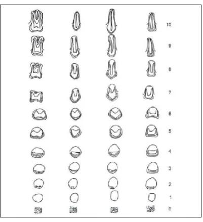
Nollas’s Developmental Stages : (Figure 1)
Stage 10: apical end of root completed Stage 9: root almost complete, apex open Stage 8: two-third of the root completed Stage 7: one-third of the root completed Stage 6: crown completed
Stage 5: crown almost completed Stage 4: two-third of crown completed Stage 3: one-third of crown completed Stage 2: Initial calcification
Stage 1: presence of crypt Stage 0: absence of crown
According to above mentioned stages, Mandibular left and right canine were graded. When the radiographic reading lay between two grades this appraisal was indicated as the value of 0.5. When the radiograph showed a reading that was slightly greater than illustrated grade, but not as much as half way between that stage and the next, the value of 0.2 was added. If development of the tooth were slightly less than the grade indicated the value 0.7 was added.
Analysis
Statistical analysis was done using SPSS 21.0v. Descriptive analysis was done to estimate the mean age. Paired t- test was done to compare between age estimation by Nolla’s method and chronological age. Pearson’s correlation test was used to assess the relation between the two methods. The level of statistical significance was kept at p< 0.05.
RESULTS
The mean chronological age and age estimated by Nolla’s method in overall population for left canine was found to be
10.06±1.91 and 9.31±1.94 respectively. The mean
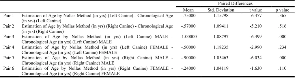
chronological age and age estimation by Nolla’s method in overall population for right canine was found to be 10.06±1.91 and 9.49±1.99. The mean chronological age of left and right mandibular canine in males is 10.12±1.78 and in females is 10.00±2.04. The mean age estimated by Nolla’s method in Mandibular left canine in males was found to be 9.12±1.86 and in females was found to be 9.5±2.01. Similarly, the mean age estimated by Nolla’s method in Mandibular right canine in males was found to be 9.22±1.91 and in females was found to be 9.76±2.05 (Figure 2, 3& 4). When the age estimation by Nolla’s method was compared with chronological age in total study population, the mean difference was found to be 0.75 for the left side canine and 0.57 for the right side canine. These difference was statistically not significant (p=0.365 for left side and p=0.516 for right side). Among males, this difference was 1.0 for left side and 0.9 on right side which is statistically significant (p<0.05) on the left as well as right side. Among the
females, the mean difference in age estimated by Nolla’s method when compared with chronological age was found to be 0.5 for left canine and 0.24 on right side. This difference is statistically not significant (p=0.23 for left side and p=0.11 on right side). Thus Nolla’s method can be used to estimate age in females accurately, whereas in males Nolla’s method shows an underestimation of age as compared to chronological age. (Table 1) In total study population of 100, there was a strong positive correlation between age estimation by nolla’s method and chronological age on left as well as right side (r=0.819 on left side, r=0.843 on right side). This correlation was statistically significant (p<0.05). Similarly among males there was a strong positive correlation between age estimation by nolla’s method and chronological age on left as well as right side (r=0.822 on left side, r=0.830 on right side). This correlation was statistically significant (p<0.05).
Table 1. Mean Difference between chronological age and age estimation of left and right canine in overall population and in males and females and their corresponding p value
Paired Differences
Mean Std. Deviation t value p value Pair 1 Estimation of Age by Nollas Method (in yrs) (Left Canine) - Chronological Age
(in yrs) (Left Canine)
-.75000 1.15798 -6.477 .365
Pair 2 Estimation of Age by Nollas Method (in yrs) (Right Canine) - Chronological Age (in yrs) (Right Canine)
-.57000 1.09411 -5.210 .516
Pair 3 Estimation of Age by Nollas Method (in yrs) (Left Canine) MALE - Chronological Age (in yrs) (Left Canine) MALE
-1.00000 1.08797 -6.499 .000
Pair 4 Estimation of Age by Nollas Method (in yrs) (Left Canine) FEMALE - Chronological Age (in yrs) (Left Canine) FEMALE
-.50000 1.18235 -2.990 .234
Pair 5 Estimation of Age by Nollas Method (in yrs) (Right Canine) MALE - Chronological Age (in yrs) (Right Canine) MALE
-.90000 1.05463 -6.034 .000
Pair 6 Estimation of Age by Nollas Method (in yrs) (Right Canine) FEMALE - Chronological Age (in yrs) (Right Canine) FEMALE
[image:2.595.43.558.348.484.2]-.24000 1.04119 -1.630 .110
Table 2. Correlation between Chronological age and age estimation by Nolla’s Method
Correlation Sig. Pair 1 Estimation of Age by Nollas Method (in yrs) (Left Canine) & Chronological Age (in yrs) (Left Canine) .819 .000 Pair 2 Estimation of Age by Nollas Method (in yrs) (Right Canine) & Chronological Age (in yrs) (Right Canine) .843 .000 Pair 3 Estimation of Age by Nollas Method (in yrs) (Left Canine) MALE & Chronological Age (in yrs) (Left Canine) MALE .822 .000 Pair 4 Estimation of Age by Nollas Method (in yrs) (Left Canine) FEMALE & Chronological Age (in yrs) (Left Canine) FEMALE .830 .000 Pair 5 Estimation of Age by Nollas Method (in yrs) (Right Canine) MALE & Chronological Age (in yrs) (Right Canine) MALE .839 .000 Pair 6 Estimation of Age by Nollas Method (in yrs) (Right Canine) FEMALE & Chronological Age (in yrs) (Right Canine)
FEMALE
Figure 1. Nollas developmental stages
Figure 2. Mean and SD of chronological age and age estimated by Nolla’s method in overall population (left and right canine)
Figure 3. Mean and SD of chronological age and age estimated by Nolla’s method in Males
[image:3.595.136.470.457.590.2] [image:3.595.132.470.622.782.2]Among females there was a strong positive correlation between age estimation by nolla’s method and chronological age on left as well as right side (r=0.839 on left side, r=0.871 on right side). This correlation was statistically significant (p<0.05). (Table 2)
DISCUSSION
One of the important aspects in forensic science is age estimation which is helpful in human identification and determining legal age in criminal cases (Gray, 1958). It can also be carried out for various reasons such as identification of mutilated victims of mass disasters, rape, kidnapping, employment, marriage, premature births, adoption, illegal immigration, pediatric endocrinopathy, and orthodontic
malocclusion (Altunsoy et al., 2013). Age is estimated on the
basis of chronological age, bone age, dental age, mental age, and various other factors such as height, weight etc. Dental age is the most reliable indicators of chronological age and is widely applicable in forensic and legal dentistry.Tooth being the integral part of body shows less variability and other developmental features and are indestructible. They can even survive after death and also remains relatively unchanged for
thousands of years (Priyadarshini, 2015; Altunsoy et al.,
2013). Amongst the various methods available for age determination in individuals, the radiological method has more benefits over histological and biochemical methods.The study of morphological parameters on dental x-ray of teeth is more valid as it is simple, noninvasive, easily reproducible on living
and dead person and not likely to get altered by local factors
(Panchbhai, 2011; Thomas et al., 2014). Age estimation
methods applied in children and adolescent are Schour and Masseler method, Moorees, Fanning and Hunt method, Demirjian, Goldstein and Tanner method, Nolla’s method and age estimation using open apices. Among these Nolla’s method is known as the most accurate method and is based on the time of emergence of tooth in oral cavity and tooth calcification and is less influenced by environmental factors
(Sachan, 2013; Mohammed et al., 2015). One of the
advantages of Nolla’s method is that it can be applied to an
individual with or without the third molar (Priyadarshini et al.,
2015). In the present study, dental age from 6-13 years in children was calculated using Nolla’s method based on developmental stages (0-10) of right and left mandibular canine. Dental age was given according to development of right and left mandibular canine in females and males
independently. The mean difference between actual
(chronological) age and estimated (dental) age was stastically not significant in total population which means that both age advances at the same time. Nolla has given age norms for females and males separately and also excluding and including
3rd molars (Thomas et al., 2014). Comparing the chronological
and dental age calculated by Nolla method for age assessment in left and right mandibular canine; the p value was found to be statistically insignificant (p>0.05) in females whereas it was statistically significant (p<0.05) in males. Among the females, the mean difference was 0.5 on the left side and 0.24 on the right side.
This difference is statistically not significant. It indicates that both the age assessed by mandibular right and left canine by Nolla’s method and the chronological age, advanced in the same direction and is more or less similar to chronological age. Among the males, mean difference was 1.0 on the left side and 0.9 on the right side which is stastically significant on the right
as well on the left side .The difference between chronologic age and dental age shows variability which is called as individuality of growth in tooth development. For this reason lower than average difference in the canine development among male was found in our study (Nolla, 1960). Our study was supported by Anderson and his associates who found that Dental development is more strongly related to morphological development than to skeletal development in both genders
(Anderson, 1975). Another study by Green found that dental
age shows the highest degree of correlation with chronological age (r = 0.6774) and the lowest correlation with skeletal age
(r = 0.4616). (Green, 1961)In another study on Mangalorean
children Nolla’s method showed good correlation between chronological age and the estimated age and statistically no significant difference was found in males and females (Thomas
et al., 2014). In our study no statistically difference was found
in chronological age and estimated age among females whereas male group showed statistically significant difference. The mean dental ages were underestimated on both right and left sidecould be due to individuality in the development of teeth and growthin males and the sample size. The overall difference was insignificant between chronological and dental age which indicates that dental maturation in terms of development of
canine also increased with the chronological age. (Anderson et
al., 1975; Gray, 1958) Also according to our study females
were ahead in dental maturation than males in all the ages
(Castellanos et al., 1996; Koshy et al., 1998; Prabhakar et al.,
2002; Hunter et al., 1966; Muller-Bolla et al., 2003). Thus it is
possible to calculate dental age using Nolla’s method. Further study has to be conducted in larger population of different age group and geographical location to generalize the outcome. In this study we used Nolla’s method for age estimation, which would be the strength of the study. Smaller sample size can be the limitation of the study. Further studies on larger sample size are recommended for extrapolation of the result.
Conclusion
Nolla’s method is a simple and reliable method to determine dental age. It matches with chronological age in overall studied population. To validate the result such studies should be carried out on larger sample size and variable age groups.
Acknowledgements
I thank Dr. Vineet Vinay for his help in statistical analysis and other detailing.
Conflicts of Interest
There are no conflicts of interest.
REFERENCES
Altunsoy M, Nur BG, Akkemik O, Ok E, Evcil MS. 2013. Dental Age Assessment: Validity of the Nolla Method in a
Group of Western Turkish Children. Marmara Dental
Journal, 2:49-52.
Anderson DL, Thompson GW, Popovitch F.1975.
Interrelationships of dental maturity, skeletal maturity, height and weight from age 4 to 14 years, Growth. 39:453-62.
Peruvian sample by the Cameriere and Demirjian methods.
Ann Hum Biol, 34:547-556.
Castellanos J, Carmona A, Catalina-Herrera CJ, Viρuales M. 1996. Skeletal maturation of wrist and hand ossification centers in normal spanish boys and girls: A study using the Greulich Pyle method. ActaAnat (Basel);155:206-11. Freny R Karjodkar. 2009. Textbook of dental and maxillofacial
radiology. Jaypee brothers medical publishers, New Delhi,India.2:940-944
Gray. L. 1958. A study of the relationship between tooth eruption age, skeletal development age and chronological
age in sixty-one Atlanta children. Am J Orthod., 44:687-91.
Green LJ. 1961. Interrelationship among height, weight and chronological, dental and skeletal ages. Angle Orthod, 31:189-93.
Hunter CJ. 1966. The correlation of facial growth with body
height and skeletal maturation at adolescence. Angle
Orthod, 36:44-54.
Koshy S, Tandon S. 1998. Dental age assessment: The applicability of Demirjian's method in South Indian children. Forensic SciInt, 94:73-85.
McKenna CJ, James H, Taylor JA, Townsend GC. 2002.
Tooth development standards for South Australia. Aust
Dent J., 47:223-7.
Mohammed RB, Sanghvi P, Perumalla KK, Srinivasaraju
D, Srinivas J, Kalyan US, et al. 2015. Accuracy of four
dental age estimation methods in southern Indian children.
J ClinDiagn Res., 9(1):1-8.
Muller-Bolla M, Lupi-Pegurier L, Quatrechomme G, Velly AM, Bolla M. 2003. Age estimation from teeth in children
and adolescents. J Forensic Sci., 48:140-148.
Nolla CM. 1960. The development of the permanent teeth. J
Dent Child, 27:254-66.
Panchbhai AS. 2011. Dental radiographic indicators, a key to
age estimation. DentomaxillofacRadiol;40:199-212.
Prabhakar AR, Panda AK, Raju OS. 2002. Applicablity of Demirjian's method of age assessment in children of
Davangere. J Indian SocPedodPrev Den., 20:54-62.
Priyadarshini C, Puranik MP, Uma SR. 2015. Dental Age
Estimation Methods: A Review. Int J Adv Health Sci.,
1(12):19-25.
Sachan K, Sharma VP, Tandon P. 2013. Reliability of Nolla’s
dental age assessment method for Lucknow population. J
Pediatr Dent.,1(1):8-13.
Thomas D, Shenai P, Chatra L, Veena K M, Rao PK, Prabhu
R, et al. 2014. Age Assessment Using Nolla’s Method in a
Group of Mangalore Population: A Study on 25 Children. J
Contemp Med. 4(3):121-127.