Curvature-driven lipid sorting: coarse-grained dynamics
simulations of a membrane mimicking a hemifusion
intermediate
Manami Nishizawa, Kazuhisa Nishizawa*
Department of Laboratory Medicine, Teikyo University School of Medical Technology, Tokyo, Japan; *Corresponding Author:
Received 26 May 2010; revised 29 June 2010; accepted 5 July 2010.
ABSTRACT
How membrane curvature influences lipid dis-tribution is under intensive research. In this short report, after a brief review of recent stud-ies, the results of our coarse-grained (CG) mo-lecular dynamics simulations of membranes with “hemifused ribbons” geometry are dis-cussed. When membranes of a binary mixture of (dipalmitoyl-phosphatidylcholine (DPPC) / diol- eoyl-phosphatidylethanolamine (DOPE) were used, DOPE accumulated in the negatively curved region of the monolayer that formed as the proximal monolayers fused (i.e., cis leaflets). However, the enrichment was dependent on the presence of tethering molecules which kept the curvature high (the curvature radius of ~1 nm), placing the cis monolayers ~2-2.5 nm from each other. Simulations in which DOPE was replaced with dioleoyl-phosphatidylcholine (DOPC) sh- owed an insignificant degree of DOPC accumu-lation, suggesting the importance of lateral in-teraction among DOPE molecules for the cur-vature sorting. The above composition was not close to a demixing point and our radial distri-bution function analysis suggested that the DOPE accumulation was not assisted by the lipid phase separation which has been shown to promote curvature-driven lipid sorting. Rele-vance of curvature-driven lipid sorting to bio-logical membrane fusion is discussed.
Keywords: Membrane Fusion; Raft; Membrane Domain; Cholesterol; Lipid Phase Separation
1. INTRODUCTION
Inhomogeneous distribution of lipids within a lipid
After Helfrich formulated curvature energy [8], theo-retical frameworks for lateral segregation based on the lipid spontaneous curvature were proposed [9-11]. Markin [9] considered the composition dependence of spontaneous curvature and the bending stiffness of membranes. Kozlov and Helfrich [10] developed a framework for thermodynamic analyses of lipid distribu-tion, bending stiffness and curvature. Curvature-induced phase segregation was also analyzed in a phenomenol-ogical continuum theory [11]. Curvature-driven lateral segregation of lipid was also computed in more recent works such as [12,13].
Experimental studies have significantly advanced the understanding of the curvature sorting of lipids. Using giant unilamellar vesicles (GUVs) formed from a ternary mixture of sphingomyelin, dioleoylphosphatidylcholine (DOPC) and cholesterol, in which Lo (liquid-ordered) and Ld (liquid-disordered) phases coexist, Baumgart et al [14] showed a correlation between domain composition and local membrane curvature. (At temperatures where single component PC bilayers would be in the fluid Lα
phase, cholesterol gives rise to an intermediate degree of organization in PC membranes known as the Lo phase (e.g., [15]). Using microfabricated surfaces, Parthasara-thy et al [16] showed that the Lo domains formed in DOPC/DPPC/cholesterol membranes, are preferentially localized at regions of low curvature. Several recent studies adopted a well-studied approach; tube- pulling from vesicles. With this method, very thin mem-brane tubes with a diameter of several tens of nanome-ters are formed by pulling a small portion of the mem-brane from GUVs (e.g., [17]). When applied to vesicles prepared from sphingomyelin, PC and cholesterol, this method showed that the tubes are essentially composed of membranes in the Ld phase enriched in DOPC, whereas GM1, which is known to segregate into the Lo phase, was excluded from tubes [18]. Lipid sorting by membrane curvature in these studies was therefore asso-ciated with phase separation (Lo and Ld), which is a col-lective behavior of lipids.
In both experiments and simulations, however, curva-ture sorting of lipids based on spontaneous curvacurva-ture (of individual lipid molecule) has exhibited only a weak effect, not leading to strong segregation of lipids. A fluorescent-labeled lipid was used in the tube-pulling system [19], showing that spontaneous curvature is not a strong factor influencing partition of the fluorescent lipid.
monolayers of vesicles of various sizes was compared [20]. The difference in lipid density between the mono- layers was present but small, indicating that the curva-ture sorting is weak [20]. In a simulation study using a CG representation of lipid (three beads for one lipid) [21], no significant sorting by curvature was observed. Theoretical consideration [13] has also suggested the weak effect of spontaneous curvature of individual lipids on curvature sorting.
Collectively, these studies have led to the consensus that lipid segregation is not significant when lipid sorting is driven only by spontaneous curvature of individual lipids. In most of the experimental settings, not only the curvature, but also the propensity toward phase separa-tion is important for curvature sorting. That is, for cur-vature to induce strong lipid sorting, the lipid composi-tion must be close to the demixing point. Thus, the en-thalpy related to curvature fitness is not likely to be enough to overcome the entropic penalty that prefers good mixing.
One of the current models for membrane fusion pro-poses formation of a stalk (Figures 1(a) and (b)) (i.e., an hourglass-shaped structure) and subsequently hemifu-sion intermediates (Figures 1(c) and (d)), leading to fusion pore formation (Figure 1(e)) [22-24]. The hemi-fusion intermediate is a state where contacting (proximal) monolayers are fused but the distal monolayers stay in-tact and therefore there is no content mixing. The model suggests that, for membrane fusion, properties of the monolayer are also important. The intrinsic curvature of lipid monolayers as well as bilayers has been studied [25,26]. However, the potential usefulness of their data has not been fully explored possibly because it is diffi-cult to study biologically relevant “monolayers”. With interest in hemifusion, we applied molecular dynamics simulation to a system mimicking a hemifusion interme-diate. In this system, the curvature of a monolayer could be higher than physiologically relevant curvatures formed by bilayers. However, note that our system (i.e., hemi-fused ribbon or ‘’-system) has not yet been applied to membranes containing cholesterol. Therefore, more ana- lyses are necessary in order to consider the behavior of physiological membranes in fusion intermediates.
2. SIMULATION DETAILS – HEMIFUSED
MEMBRANE SYSTEM
Figure 1.The stalk-hemifusion model (a-e) and the -system (f,g). In the stalk-hemifusion model [24], two apposed bilayers (a) initially form a stalk (b) and subsequently leads to hemifusion (c), which undergoes the hemifusion rupture (d) and finally ends with the full fusion (e). The arrow from (b) to (e) indicates a direct transition from the stalk-like structure to fusion pore formation, which has been observed in the CG-simulation studies [27,28]. (f) The -system. The periodic boundary condition allows the -shaped (hemifused) bilayer ribbons to have an infinite length in the y-direction. For clarity, water is hidden. The definition of the x,y and z directions is also shown. (g) The definition of the curvature strength. Shown are the angle between the two outer leaflets and the zone boundaries. To determine them, first, C1, the circle that has the longest radius located inside the phosphorus atoms of any lipid monolayers, is found. Two circles C2 and C3, which have the same center as that of C1 are drawn (in this study, the ra-dius of C2 and C3 was set at 4 nm and 10 nm, respectively). C2 defines the curved region of the outer leaflet. The planar regions of the outer leaflet are defined as the regions located between the two circles whose sizes are 8nm and 12nm (radius) and the cen-ter is the same as C1. The com of each lipid molecule was used for reference. The points of incen-tersection between C3 and the mid-planes are used to define the angle . C4, the circle that best fits the phosphorus atoms belonging to the outer leaflet and located inside of C2, is determined. The radius of C4 represents the curvature of the outer leaflet.
taining bend(s) were carried out. We mainly used “-shaped” membranes (Figure 1(f)). This structure consists of two bilayer ribbons that are partly fused such that the cross section of the ribbon has a -like shape (Figures 1(f) and (g)). This structure has 2D-curvature; i.e., the mem-branes projected onto the xz-plane are curved whereas they are basically flat along the y-axis (Figure 1(f)). For
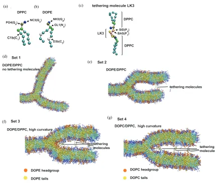
membranes, the “outer” leaflet refers to the monolayer which is highly negatively curved in Figure 1(g), mim-icking the fused cis monolayers. For the outer leaflet, the planar and the curved regions are defined as described in the legend for Figure 1. CG simulations were performed using the MARTINI forcefield (version 2.0) as in Mar-rink et al [29]. For example, for DPPC, the choline is represented as Qo particle and the phosphate group as Qa,
(d)
(e)
[image:4.595.80.508.74.439.2](g) (f)
Figure 2. The CG model of lipids and representative snapshots of the simulations. (a) The CG model of DPPC; (b) The CG model of DOPE; (c) The CG model of the tethering molecule. Shown here is LK3 along with DPPC. For (a)-(c), particle name (e.g., NC3) and type (e.g., Qo) used are indicated; (d)-(g) Representative snapshots obtained from the simulations. (d) Set 1, the simulations of DPPC/DOPE membrane without the tethering molecule; (e) Set 2, the DPPC/DOPE membrane simulated with the tethering molecules restrained at 12nm from the center of the circle C1 defined in Figure 1; (f) Set 3, the DPPC/DOPE with the tethering molecules used at three different positions; (g) Set 4, the DOPC/DPPC with the tethering molecules at the same positions as in (f). For (d)-(g) the leftmost part of the membrnae is hidden; this part consists of the DPPC molecules whose com was restrained to reduce the large drift of membranes. The representation scheme for (d)-(g): blue spheres, DPPC headgroup particles (from NC3 to GL2); light blue spheres, DPPC hydrophobic tail particles; orange spheres, DOPE (or DOPC) headgroup particles; yellow spheres, DOPE hydrophobic tail particles; gray spheres, particles representing the tethering molecules.
membrane. For the initial structure, a membrane of DPPC was prepared and, subsequently, an appropriate number of DPPC chosen randomly were replaced with DOPE. Four water molecules were modeled by one par-ticle as in [29]. For all simulations 11423 water parti-cles were used. A typical size of the simulation box was 31, 4.5, 16 (nm) in x-, y-, and z-direction respectively. The artificial tethering (crosslinking) molecules that tether the cis monolayers were CG-modeled peptides consisting of three (“LK3”) or six (“LK6”) Ser residues (Figure 2(c)). As in [30,31], the peptide backbone was
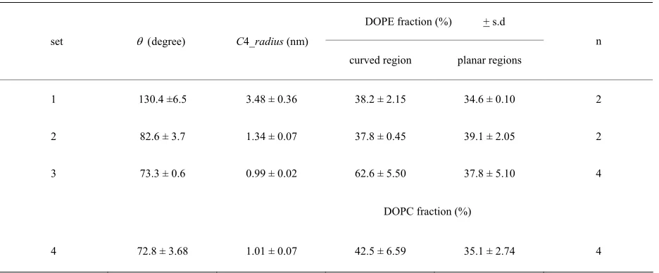
Table 1. Outer leaflet curvature and DOPE (DOPC) distribution in the curved and planar regions.
DOPE fraction (%) + s.d set degree)C4_radius (nm)
curved region planar regions
n
1 130.4 ±6.5 3.48 ± 0.36 38.2 ± 2.15 34.6 ± 0.10 2
2 82.6 ± 3.7 1.34 ± 0.07 37.8 ± 0.45 39.1 ± 2.05 2
3 73.3 ± 0.6 0.99 ± 0.02 62.6 ± 5.50 37.8 ± 5.10 4
DOPC fraction (%)
4 72.8 ± 3.68 1.01 ± 0.07 42.5 ± 6.59 35.1 ± 2.74 4
Set 1-3 are DOPE/DPPC simulations, where the overall DOPE fraction, DOPE / (DOPE+DPPC) = 34.5%. For set 4, the DOPC content was also 34.5%. s.d was calculated from the n average values, each of which was computed from the last 400ns period of each trajectory; n is the number of simulation runs (i.e., trajectories).
the C1 center. For the remaining part of the -membrane, a harmonic restraint was applied to the com of each teth-ering molecule.
Nonbonded interactions are described by a Lennard- Jones potential [29]. In addition, the charged particles interact via a Coulombic energy function with a relative dielectric constant= 15, which represents screening effect. The cut-off lengths and the switching function were set as in [29]. Gromacs (ver 3.3) was used [32] for 500 ns simu-lations performed at 323 K with the time step of 20 fs and with semi-isotropic pressure coupling at 1bar. Due to the increased diffusion rate of molecules with the CG system [29], in the following we show the time after the 4-fold multiplication. The pressure coupling time constant was 0.2 ps and the compressibility set at 3 × 10⁻⁵ bar-1.
3. RESULTS
Four sets of simulations, each consisting of two to five independent 2000ns simulations, were performed (Table 1). For sets 1-3, the DOPE/DPPC membranes were used, whereas for set 4 the DOPC/DPPC membrane was used. Table 1 shows the average (+ s.d.) of the mean values (each obtained from the final 400 ns of each trajectory) of θ, the C4 radius and the DOPE (or DOPC) density in the curved region and planar regions. For all these re-sults the s.d. was small, which was not surprising be-cause the 1600 ns pre-run allowed sufficient equilibration time. When no tethering molecule was used (set 1), the three bilayers quickly became equivalent (at ~400 ns); the angle between any two bilayers equally approached 120 degree (Figure 2(d)). The DOPE density relative to
DPPC became similar between the curved and the planar regions (Table 1, set 1). Note that the enrichment of DPPC at the edge, generally increased the DOPE frac-tion above the nominal 34.5%.
When two LK3 molecules were used for tethering at 12nm from the C1 center, and the C4 radius decreased, indicating negative curvature. However, the outer leaflet had horseshoe-like roundness (Figure 2(e)). In this case, the DOPE accumulation in the negatively curved region was not significant (Table 1).
When the number of tethering molecules was in-creased and some positioned close to the junction (as described in the above section), the higher curvature was maintained (Figure 2(f)). Then, the significant enrich-ment in DOPE was observed for the negatively curved region compared to the planar region (set 3) (p < 0.01; t-test). In contrast, when the DOPE molecules of the DOPE/DPPC membrane (set 3) were all replaced with DOPC (set 4, Figure 2(g)), the DOPC density in the negatively curved region was lower than the density of DOPE of set 3 simulations (p < 0.05).
(a) (b)
[image:6.595.127.473.95.417.2](c) (d)
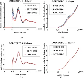
Figure 3. Radial distribution function (rdf) analyses of the CG lipids used in this study. 6000ns pilot simulations with planar bilayers composed of 512 lipid molecules were performed and the last 400ns data were used for this analysis. (a) DOPE/DPPC 1:1 membrane; (b) Same as (a) but the cumulative rdf is shown; (c) DOPC/DPPC 1:1 membrane; (d) Same as (c) but the cumulative rdf is shown. For example, the “DOPE-DOPE” curve shows the density (relative to the density over the whole system) of the DOPE NH3 particle at the indicated distance from each DOPE NH3 particle. Similarly, the DOPE-DPPC curve shows the density of DPPC NC3 around DOPE NH3, whereas DOPC-DPPC shows the DPPC NC3 density around DOPC NC3.
DPPC interaction and the DPPC-DPPC interaction (Fig- ures 3(c) and (d)). Therefore, for the CG model used, the between-headgroups interaction determines the lat-eral distribution in planar bilayers, whereas the be-tween-acyl chains interaction is not important. We also note that the DOPE-DOPE interaction is very local and there is no lipid clustering on scales greater than ~1 nm; in fact, at >2 nm from each DOPE molecule the density of DOPE is the same as that of DPPC (Figure 3(a)). Therefore, the DOPE and DPPC membrane is not demixed; only subtle enrichment in DOPE occurs in the close proximity (~0.5-1 nm) of each DOPE molecule. While we cannot rule out the possibility that further demixing of DOPE from DPPC could take place in longer simulations, there was no significant difference in the rdf results between the ~1000 ns and ~5600 ns data
(details not shown), suggesting that the DOPE/DPPC planar bilayer was equilibrated earlier than 5600 ns.
4. DISCUSSION AND PERSPECTIVES
DOPE/DPPC membrane until the end of the simulation (6000 ns).
While these results may be of physicochemical rele-vance, several questions remain in terms of membrane physiology. First, biological relevance of the tethering molecules is unclear; in the real fusion, proteins are gen-erally large, likely making the curvature of the fused monolayers lower than our setting (e.g., [33,34]; and references cited therein). However, in a cell fusion study using SNARE proteins expressed on the cell surface, it has been shown that GPI (glycerophosphatidylinosi-tol)-anchored EYFP (Enhanced Yellow Fluorescent Protein consisting of 239 amino acid residues) does not diffuse between hemifused cells whereas GPI-anchored short peptide (named GPI-AU1) does (Figure 7 of [35]). In the latter case, the negative curvature between the fused monolayers may be so high that membrane pro-teins with a large ectodomain are excluded from the be-tween-cell diffusion.
Second, results with the 2D-curvature system have to be carefully interpreted; our 2D-curvature system is ob-viously different from a small hemifusion diaphragm having a saddle-like (i.e., high Gaussian) curvature be-tween the fused monolayers [24]. It should also be re-membered that, while a large hemifusion diaphragm is observed especially with a high PE fraction, it is possible that the hemifusion diaphragm may not be a major in-termediate in the fusion pathway of physiologic mem-branes [36,37].
It still seems relevant to ask why theoretical and in vi-tro experimental studies show only a weak degree of curvature sorting of lipids, despite in vivo evidence ar-guing for the curvature sorting effects that support cel-lular activities [5,6,38]. As several authors have sug-gested, the lateral interaction between lipids may have a profound impact on the efficacy of curvature sorting. The question arises then, how such lipid-lipid interac-tions modulate the lipid partitioning into differently curved membrane portions. This question may be con-sidered based on two scenarios. First, a strong lipid-lipid interaction increases the effective area of the lipid molecule. For example, if DOPE favors interaction with DOPE, but not with DPPC, this leads to lipid sorting and can be regarded as an increase in the effective molecule area of both DOPE and DPPC. The second scenario is that the sorting can be based on the bending stiffness [19,39]. Once the curvature facilitates phase segregation in the membrane close to a demixing point, relatively soft and rigid domains may be generated. While soft domains can move into highly curved regions, rigid do-mains favor locations in the relatively flat regions.
Consider the first scenario. How does the increase in molecule area (surface area per molecule) influence curvature sorting. In the study employing tube pulling, a
model considering individual molecule curvature sorting based on spontaneous curvature has been proposed [19,39]. For a membrane consisting of lipids α and β, based on the assumption of equal chemical potentials of molecule α (and β), on tether and vesicle, the authors have derived the following relationship:
kc a (C –Cst + Csv) (C–C)
= kT ln (ov/ot) + kT ln (ot /ov), (1)
where, kc is the bending stiffness, a is the molecule area, C = 1/Rt is the tube curvature (with Rt being the tube radius), Cst and Csv are the spontaneous curvature of the outer monolayer of the tube and the vesicle, respectively, k is the Bolzmann constant, ov, for example, is the mole fraction of molecule in the outer leaflet of the vesi-cle. Eq.1 is derived via the partial differentiation of the total energy function by Not, which is the number of
molecule in the outer monolayer of the tube. The left side of Eq.1 corresponds to the potential regarding the bending energy, whereas the right side corresponds to the entropy term. The weighted average spontaneous curvature is assumed to be Cst = (CNot + CβNβot) / Not,
where C is the spontaneous curvature of molecule α.
Our λ system can be considered similarly; the curved region corresponds to the tube, while the planar regions correspond to the vesicle. Let αrepresent DOPE and β
represent DPPC. Let ov = 0.38, ot = 0.63 (set 3), C =
– 0.3 nm-1 [25]. The remaining terms may be approxi-mated as: kc = 4 × 10-20 J [25]; C = – 1.0 nm-1; Cst = – 0.19 nm-1; Csv = – 0.11 nm-1; C
= ~0 nm-1; kT = ~4 ×
10-21 J. These values lead to a = ~0.4 nm2, smaller than the real value for the CG system (0.64 nm2 and 0.66 nm2 for DPPC and DOPE respectively). This may be because Cand Cβfor the CG-model of DOPE and DPPC are not
known and also our definition of C and the curved re-gion may not be suitable.
The above relationship helps to infer the impact of molecule area. For example, if the DOPE-DOPE interac-tion and the DPPC-DPPC interacinterac-tion are greater than the DOPE-DPPC interaction so that DOPE and DPPC be-have, on average, as a particle with a 1.5-fold effective molecular area, then this would increase the fraction of DOPE in the curved region from 0.63 to ~0.75. (Here, Nov >> Not was assumed.) The reality should be far more complex, yet this notion supports the view [20] that in-dividual lipids have a small molecular area compared with transmembrane proteins or lipid clusters, and therefore, although some lipids have high spontaneous curvatures, the membrane curvature preference of these lipids is weak.
tion compared with the binary mixture lacking choles-terol [15]. In addition, different fluid phases may have different bending stiffness. The Ld region (consisting of sphingomyelin and cholesterol) has a higher bending stiffness kc value compared with the Lo phase [14]. While our analyses do not take into account the kc dif-ference between the membrane regions, this difdif-ference may be important in the case of cholesterol-containing mixtures as considered in [19,39]. Another intriguing finding regarding cholesterol segregation is that choles-terol accumulates in high curvature regions in a mem-brane composed of brominated di18:0 PC/cholesterol 2:1 [43].
Characterization of cholesterol-containing membranes is a topic of computational research. Several groups conducted atomistic simulations and showed several features such as hydrogen bond formation between cho-lesterol and phospholipids, which are consistent with experimental findings [44,45]. In general, the simula-tions supported the consensus that cholesterol interacts with phospholipid molecules and gives rise to the inter-mediate level of organization in lipid membranes, Lo [46]. (In the Lo phase, lipid hydrocarbon chains are or-dered as in a Lc gel state, but phospholipid molecules are nearly as mobile as in a Lfluid phase [47], for review
see [41]). In terms of domain formation, Vattulainen and coworkers used a coarse-grained model and showed the formation of cholesterol-rich and cholesterol-poor do-mains at intermediate cholesterol concentrations [48,49]. Recently, Risselada and Marrink [50] showed that the CG model can reproduce the experimental findings on the formation of distinct phases (Lo and Ld (liquid-dis- ordered) phase). The lateral organization of cholesterol of the raft-like structure in [50] resembles that derived from the atomistic preassembled raft-like mixtures [51]. The necessity of cholesterol for the phase separation has also been shown in [50]; in the absence of cholesterol, the DPPC/diC18:2-PC 3:1 mixture exhibited nonrandom (nonideal) mixing, yet macroscopic phase separation did not occur. From this finding, and also considering the choice of 323K in our system, it seems unlikely that the phase separation occurs in our DOPE/DPPC planar bi-layer even when the simulation is extended longer than 6000ns. Future simulation analyses could focus on the phase separation in curved regions, especially those containing the stalk or the rim of the hemifusion dia-phragm. As stated above, curvature sorting is significant
virus HA2 fusion peptide enhances the rate of stalk for-mation [52], it is yet unknown whether such fusion pep-tides associate with and stabilize a particular lipid do-main or, more positively, draw particular types of lipid, creating a domain in the proximity of the peptide, thereby modulating the fusion rate. (The fusion peptide was also studied in recent atomistic simulations of vesi-cle fusion [53]). One difficulty is the limitation in the simulation time; atomistic simulations typically cover 10-100ns and therefore very subtle lipid sorting cannot be studied with this approach.. This problem may be partially remedied by using initial structures based on the ones obtained from well equilibrated CG simulations. Phase separation simulation in [50] may provide an im-portant framework for analyses of interactions between peptides and lipid domains.
Another interesting problem in membrane fusion is the mechanism by which transmembrane (TM) peptide domains control the fusion. Findings from Langosch and other researchers ([54] and references cited therein) gen-erally argue that, while the ectodomains (hydrophilic domains located between the cis membranes) of fusion proteins are important in stalk formation and/or in the hemifusion intermediate, TM domains play a crucial role in later steps of fusion, such as, the rupture of the hemi-fusion diaphragm and/or pore formation. Considering the practical difficulties in computation (i.e., system size and simulation time), it is hoped that 2D-curved mem-brane systems such as ours will provide a useful starting point for such peptide analyses.
5. CONCLUSIONS
curvature could exert more pronounced lipid sorting than the one shown in this study.
6. ACKNOWLEDGEMENTS
This work was supported by Grant-in-Aid for Scientific Research from the Ministry of Education, Culture, Sports, Science, and Technology of Japan.
REFERENCES
[1] Holthuis, J.C. and Levine, T.P. (2005) Lipid traffic: Floppy drives and a superhighway. Nature Reviews
Mo-lecular Cell Biology, 6(3), 209-220.
[2] Van Meer, G., Voelker, D.R. and Feigenson, G.W. (2008) Membrane lipids: Where they are and how they behave.
Nature Reviews Molecular Cell Biology, 9(2), 112-124.
[3] De Matteis, M.A. and Luini, A. (2008) Exiting the Golgi complex. Nature Reviews Molecular Cell Biology, 9(4), 273-284.
[4] Mukherjee, S., Soe, T.T. and Maxfield, F.R. (1999) En-docytic sorting of lipid analogues differing solely in the chemistry of their hydrophobic tails. Journal of Cell
Bi-ology, 144(6), 1271-1284.
[5] Van Meer, G. and Lisman, Q. (2002) Sphingolipid trans- port: Rafts and translocators. Journal of Biological Che-
mistry,277(29), 25855-25858.
[6] Ostrowski, S.G., Van Bell, C.T., Winograd, N. and Ew-ing, A.G. (2004) Mass spectrometric imaging of highly curved membranes during Tetrahymena mating. Science,
305(5680), 71-73.
[7] Fratti, R.A., Jun, Y., Merz, A.J., Margolis, N. and Wick-ner, W. (2004) Interdependent assembly of specific re- gulatory lipids and membrane fusion proteins into the vertex ring domain of docked vacuoles. Journal of Cell
Biology, 167(6), 1087-1098.
[8] Helfrich, W. (1973) Elastic properties of lipid bilayers: Theory and possible experiments. Zeitschrift fur
Natur-forschung C, 28(11), 693-703.
[9] Markin, V.S. (1981) Lateral organization of membranes and cell shapes. Biophysical Journal, 36(1), 1-19. [10] Kozlov, M.M. and Helfrich, W. (1992) Effects of a
co-surfactant on the stretching and bending elasticities of a surfactant monolayer. Langmuir,8(11), 2792-2797. [11] Seifert, U. (1993) Curvature-induced lateral phase
seg-regation in two-component vesicles. Physical Review Let-ters,7(9), 1335-1338.
[12] Iglic, A., Hägerstrand, H., Veranic, P., Plemenitas, A. and Kralj-Iglic, V. (2006) Curvature-induced accumula-tion of anisotropic membrane components and raft for-mation in cylindrical membrane protrusions. Journal of
Theoretical Biology, 240(3), 368-373.
[13] Derganc, J. (2007) Curvature-driven lateral segregation of membrane constituents in Golgi cisternae. Physical
Biology, 4(4), 317-324.
[14] Baumgart, T., Hess, S.T. and Webb, W.W. (2003) Imag-ing coexistImag-ing fluid domains in biomembrane models coupling curvature and line tension. Nature, 425(6960), 821-824.
[15] Radhakrishnan, A. and McConnell, H. (2005) Condensed complexes in vesicles containing cholesterol and phos-pholipids. Proceedings of the National Academy of
Sci-ences of the United States of America,102(36), 12662-
12666.
[16] Parthasarathy, R., Yu, C.H. and Groves, J.T. (2006) Cur-vature-modulated phase separation in lipid bilayer mem-branes. Langmuir,22(11), 5095-5099.
[17] Bo, L. and Waugh, R.E. (1989) Determination of bilayer membrane bending stiffness by tether formation from giant, thin-walled vesicles. Biophysical Journal, 55(3), 509-517.
[18] Roux, A., Cuvelier, D., Nassoy, P., Prost, J., Bassereau, P. and Goud, B. (2005) Role of curvature and phase tran-sition in lipid sorting and fission of membrane tubules.
European Molecular Biology Organization Journal, 24(8),
1537-1545.
[19] Tian, A. and Baumgart, T. (2009) Sorting of lipids and proteins in membrane curvature gradients. Biophysical
Journal, 96(7), 2676-2688.
[20] Kamal, M.M., Mills, D., Grzybek, M. and Howard, J. (2009) Measurement of the membrane curvature prefer-ence of phospholipids reveals only weak coupling be-tween lipid shape and leaflet curvature. Proceedings of the National Academy of Sciences of the United States of
America,106(52), 22245-22250.
[21] Cooke, I.R. and Deserno, M. (2006) Coupling between lipid shape and membrane curvature. Biophysical Jour-nal, 91(2), 487-495.
[22] Chernomordik, L.V., Melikyan, G.B. and Chizmadzhev, Y.A. (1987) Biomembrane fusion: A new concept de-rived from model studies using two interacting planar lipid bilayers. Biochimica et Biophysica Acta, 906(3), 309- 352.
[23] Tamm, L.K., Crane, J. and Kiessling, V. (2003) Mem-brane fusion: A structural perspective on the interplay of lipids and proteins. Current Opinion in Structural Biol-ogy, 13(4), 453-466.
[24] Chernomordik, L.V. and Kozlov, M.M. (2008) Mechan-ics of membrane fusion. Nature Structural & Molecular
Biology, 15(7), 675-683.
[25] Marsh, D. (2006) Elastic curvature constants of lipid monolayers and bilayers. Chemistry and Physics of Lip-ids, 144(2), 146-159.
[26] Fuller, N. and Rand, R.P. (2001) The influence of lysol-ipids on the spontaneous curvature and bending elasticity of phospholipid membranes. Biophysical Journal, 81(1), 243-254.
[27] Marrink, S.J. and Mark, A.E. (2003) The mechanism of vesicle fusion as revealed by molecular dynamics simu-lations. Journal of American Chemical Society, 125(37), 11144-11145.
[28] Kasson, P.M., Kelley, N.W., Singhal, N., Vrljic, M., Brunger, A.T. and Pande, V.S. (2006) Ensemble molecular dy-namics yields submillisecond kinetics and intermediates of membrane fusion. Proceedings of the National
Acad-emy of Sciences of the United States of America,103(32),
11916-11921.
[29] Marrink, S.J., Risselada, H.J., Yefimov, S., Tieleman, D.P. and De Vries, A.H. (2007) The MARTINI force field: Coarse grained model for biomolecular simulations.
[32] Lindahl, E., Hess, B. and Van der Spoel, D. (2001) GROMACS 3.0: A package for molecular simulation and trajectory analysis. Journal of Molecular Modeling,7(8), 306-317.
[33] Weissenhorn, W., Hinz, A. and Gaudin, Y. (2007) Virus membrane fusion. FEBS Letters, 581(11), 2150-2155. [34] Wickner, W. and Schekman, R. (2008) Membrane fusion.
Nature Structural & Molecular Biology, 15(7), 658-664.
[35] Giraudo, C.G., Hu, C., You, D., Slovic, A.M., Mosharov, E.V., Sulzer, D., Melia, T.J. and Rothman, J.E. (2005) SNAREs can promote complete fusion and hemifusion as alternative outcomes. Journal of Cell Biology, 170(2), 249- 260.
[36] Liu, T., Wang, T., Chapman, E.R. and Weisshaar, J.C. (2008) Productive hemifusion intermediates in fast vesi-cle fusion driven by neuronal SNAREs. Biophysical Jour-nal, 94(4), 1303-1314.
[37] Kasson, P.M. and Pande, V.S. (2007) Control of mem-brane fusion mechanism by lipid composition: Predic-tions from ensemble molecular dynamics. PLoS
Compu-tational Biology, 3(11), 2228-2238.
[38] Van Meer, G. and Sprong, H. (2004) Membrane lipids and vesicular traffic. Current Opinion in Cell Biology,
16(4), 373-378.
[39] Tian, A., Capraro, B.R., Esposito, C. and Baumgart, T. (2009) Bending stiffness depends on curvature of ternary lipid mixture tubular membranes. Biophysical Journal,
97(6), 1636-1646.
[40] Edidin, M. (2003) The state of lipid rafts: From model membranes to cells. Annual Review of Biophysics and
Biomolecular Structure, 32(1), 257-283.
[41] Jacobson, K., Mouritsen, O.G. and Anderson, R.G. (2007) Lipid rafts: At a crossroad between cell biology and physics. Nature Cell Biology, 9(1), 7-14.
[42] Mannock, D.A., Lewis, R.N., McMullen, T.P. and McEl- haney, R.N. (2010) The effect of variations in phosphol-ipid and sterol structure on the nature of lphosphol-ipid-sterol in-teractions in lipid bilayer model membranes. Chemistry
and Physics of Lipids, 163(6), 403-448.
[43] Wang, W., Yang, L. and Huang, H.W. (2007) Evidence of cholesterol accumulated in high curvature regions:
lar dynamics simulations of phospholipid bilayers with cholesterol. Biophysical Journal, 84(4), 2192-2206. [46] Pandit, S.A., Khelashvili, G., Jakobsson, E., Grama, A.
and Scott, H.L. (2007) Lateral organization in lipid-cho- lesterol mixed bilayers. Biophysical Journal, 92(2), 440- 447.
[47] McMullen, T.P. and McElhaney, R.N. (1995) New as-pects of the interaction of cholesterol with dipalmitoyl-phosphatidylcholine bilayers as revealed by high-sensi- tivity differential scanning calorimetry. Biochimica et Bio-
physica Acta, 1234(1), 90-98.
[48] Murtola, T., Falck, E., Patra, M., Karttunen, M. and Vat-tulainen, I. (2004) Coarse-grained model for phospholipid/ cholesterol bilayer. Journal of Chemical Physics, 121(18), 9156-9165.
[49] Murtola, T., Karttunen, M. and Vattulainen, I. (2009) Sys-tematic coarse graining from structure using internal states: Application to phospholipid/cholesterol bilayer.
Journal of Chemical Physics, 131(5), (055101)1-15.
[50] Risselada, H.J. and Marrink, S.J. (2008) The molecular face of lipid rafts in model membranes. Proceedings of the National Academy of Sciences of the United States of
America, 105(45), 17367-17372.
[51] Niemelä, P.S., Ollila, S., Hyvönen, M.T., Karttunen, M. and Vattulainen, I. (2007) Assessing the nature of lipid raft membranes. PLoS Computational Biology, 3(2), 304- 312.
[52] Nishizawa, M. and Nishizawa, K. (2010) Molecular dy-namics simulation analyses of viral fusion peptides in membranes prone to phase transition: Effects on mem-brane curvature, phase behavior and lipid-water interface destabilization Journal of Biophysical Chemistry, 1(1), 19-32.
[53] Kasson, P.M., Lindahl, E. and Pande, V.S. (2010) Atomic- resolution simulations predict a transition state for vesi-cle fusion defined by contact of a few lipid tails. PLoS
Computational Biology, 6(6), e1000829.
[54] Langosch, D., Hofmann, M. and Ungermann, C. (2007) The role of transmembrane domain in membrane fusion.
![Figure 1. The stalk-hemifusion model (a-e) and the -system (f,g). In the stalk-hemifusion model [24], two apposed bilayers (a)](https://thumb-us.123doks.com/thumbv2/123dok_us/8982284.393944/3.595.58.538.83.381/figure-stalk-hemifusion-model-stalk-hemifusion-apposed-bilayers.webp)