Crystal structure of (
E
)-2-({[2-(1,3-dioxan-2-yl)phenyl]imino}methyl)phenol
Zhengyi Li, Song Shi, Kun Zhou, Liang Chen and Xiaoqiang Sun*
Jiangsu Key Laboratory of Advanced Catalytic Materials and Technology, School of Petrochemical Engineering, Changzhou University, Changzhou 213164, Jiangsu, People’s Republic of China. *Correspondence e-mail: [email protected]
Received 20 April 2015; accepted 23 April 2015
Edited by H. Stoeckli-Evans, University of Neuchaˆtel, Switzerland
The title compound, C17H17NO3, prepared by the
condensa-tion reaccondensa-tion of 2-(1,3-dioxan-2-yl)aniline and salicylaldehyde,
has an E conformation about the C N bond. The
six-membered O-heterocycle adopts a chair conformation, with the bond to the aromatic ring located at its equatorial position. The dihedral angle between the aromatic rings is 36.54 (9). There is an intramolecular N—H O hydrogen bond forming anS(6) ring motif. In the crystal, molecules are linked by C— H O hydrogen bonds, forming chains along the a-axis direction. Within the chains, there are C—H interactions involving adjacent molecules.
Keywords:crystal structure; acetal; Schiff base; intramolecular hydrogen bonding; N—H O hydrogen bonds.
CCDC reference:1061272
1. Related literature
For general background to acetals, see: Cismas¸et al.(2005); Sun et al. (2010). For Schiff bases of salicylaldehyde having important applications in biological and pharmacological chemistry, see: Gupta & Sutar (2008); Jime´nez-Sa´nchez et al. (2013). For further background to related Schiff base ligands and their various properties, see: Arod et al. (2005); Chat-ziefthimiouet al.(2006).
2. Experimental 2.1. Crystal data
C17H17NO3
Mr= 283.32
Orthorhombic,Pna21
a= 8.4873 (18) A˚
b= 10.821 (2) A˚
c= 16.232 (3) A˚
V= 1490.8 (5) A˚3
Z= 4
MoKradiation
= 0.09 mm1
T= 296 K
0.260.240.22 mm
2.2. Data collection
Bruker APEXII CCD area-detector diffractometer
Absorption correction: multi-scan (SADABS; Bruker, 2009)
Tmin= 0.978,Tmax= 0.981
9005 measured reflections 3123 independent reflections 2494 reflections withI> 2(I)
Rint= 0.043
2.3. Refinement
R[F2> 2(F2)] = 0.039
wR(F2) = 0.097
S= 1.00 3123 reflections 190 parameters
1 restraint
H-atom parameters constrained max= 0.15 e A˚
3 min=0.21 e A˚
3
Table 1
Hydrogen-bond geometry (A˚ ,).
Cg1 is the centroid of the C1–C6 ring.
D—H A D—H H A D A D—H A
O1—H1 N1 0.85 1.90 2.632 (2) 144
C7—H7 O2i
0.93 2.48 3.364 (2) 160
C15—H15A Cg1ii 0.97 2.77 3.694 (3) 160
Symmetry codes: (i)xþ1 2;yþ
1
2;z; (ii)x1;y;z.
Data collection: APEX2 (Bruker, 2009); cell refinement: SAINT (Bruker, 2009); data reduction:SAINT; program(s) used to solve structure:SHELXS97(Sheldrick, 2008); program(s) used to refine structure: SHELXL97 (Sheldrick, 2008); molecular graphics: SHELXTL(Sheldrick, 2008); software used to prepare material for publication:SHELXTL.
Acknowledgements
We gratefully acknowledge financial support from the NSFC (No. 21002009), the Scientific and Technological Project of Jiangsu Province (BY2014037–01), the Major Program for Natural Science Research of Jiangsu Colleges and Universities
data reports
Acta Cryst.(2015).E71, o357–o358 doi:10.1107/S2056989015008051 Liet al.
o357
(12KJA150002 and 14KJA150002), and the Qing Lan Project of Jiangsu Province.
Supporting information for this paper is available from the IUCr electronic archives (Reference: SU5122).
References
Arod, F., Gardon, M., Pattison, P. & Chapuis, G. (2005).Acta Cryst.C61, o317– o320.
Bruker (2009).APEX2,SAINTandSADABS. Bruker AXS Inc., Madison, Wisconsin, USA.
Chatziefthimiou, S.-D., Lazarou, Y.-G., Hadjoudis, E., Dziembowska, T. & Mavridis, I. M. (2006).J. Phys. Chem. B,110, 23701–23709.
Cismas¸, C., Terec, A., Mager, S. & Grosu, I. (2005).Curr. Org. Chem.9, 1287– 1314.
Gupta, K.-C. & Sutar, A.-K. (2008).Coord. Chem. Rev.252, 1420–1450. Jime´nez-Sa´nchez, A., Farfa´n, N. & Santillan, R. (2013).Tetrahedron Lett.54,
5279–5283.
Sheldrick, G. M. (2008).Acta Cryst.A64, 112–122.
supporting information
sup-1
Acta Cryst. (2015). E71, o357–o358
supporting information
Acta Cryst. (2015). E71, o357–o358 [https://doi.org/10.1107/S2056989015008051]
Crystal structure of (
E
)-2-({[2-(1,3-dioxan-2-yl)phenyl]imino}methyl)phenol
Zhengyi Li, Song Shi, Kun Zhou, Liang Chen and Xiaoqiang Sun
S1. Comment
Schiff bases of salicylaldehyde have important applications in biological chemistry and pharmacological chemistry
(Gupta et al., 2008; Sánchez et al., 2013). In addition, Schiff bases of salicylaldehyde has good optical properties with the
ability of distinctive ultraviolet absorption (Chatziefthimiou et al., 2006). Herein (E)-2-{[2-(1,3-dioxan-2-yl) phenyl)
imino]methyl} phenol was prepared by the condensation reaction of 2-(1,3-dioxan-2-yl) aniline and salicylaldehyde, and
the structure was confirmed by X-ray diffraction analysis.
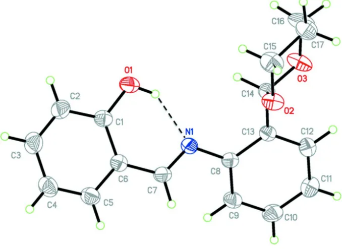
In the molecular structure of the title compound, Fig. 1, the two aromatic rings (C1—C6 and C8—C13) are linked by
the double bond C7═N1, with the dihedral angle between the two rings being 36.54 (9) °. The C7═N1 bond is coplanar
with the benzene ring (C1—C6), and atom N1 forms an intramolecular hydrogen bond, O1—H1···N1, with the hydroxyl
group on ring (C1—C6) [Fig. 1 and Table 1]. The six-membered O-heterocycle adopts a chair conformation with the (C8
—C13) ring located at its equatorial position.
In the crystal, molecules are linked by C—H···O hydrogen bonds forming chain along the a axis. Within the chains there
are C—H···π interactions involving adjacent molecules (Table 1 and Fig. 2).
S2. Experimental
A mixture of 2-(1,3-dioxan-2-yl) aniline (1.8 g, 10 mmol) and salicylaldehyde (1.32 g, 11 mmol) in 20 ml methanol,
stirred for 20 h at room temperature. After the reaction had finished, the solution was left overnight at at 273 K, and
yellow block-like crystals were obtained on slow evaporation of the solvent (yield: 82%; m.p.: 321 K).
S3. Refinement
The OH and C-bound H atoms were placed in geometrically idealized positions and constrained to ride on their parent
Figure 1
The molecular structure of the title compound, with atom labelling. Displacement ellipsoids are drawn at the 30%
probability level. The intramolecular O—H···N hydrogen bond is shown as a dashed line (see Table 1 for details).
Figure 2
A partial view of the crystal packing of the title compound, view along the c axis. Hydrogen bonds are shown as dashed
lines (see Table 1 for details).
(E)-2-({[2-(1,3-Dioxan-2-yl)phenyl]imino}methyl)phenol
Crystal data
C17H17NO3
Mr = 283.32
Orthorhombic, Pna21
Hall symbol: P 2c -2n
a = 8.4873 (18) Å
b = 10.821 (2) Å
V = 1490.8 (5) Å3
Z = 4
F(000) = 600
Dx = 1.262 Mg m−3
Mo Kα radiation, λ = 0.71073 Å
[image:4.610.136.474.378.540.2]supporting information
sup-3
Acta Cryst. (2015). E71, o357–o358
µ = 0.09 mm−1
T = 296 K
Block, yellow
0.26 × 0.24 × 0.22 mm
Data collection
Bruker APEXII CCD area-detector diffractometer
Radiation source: fine-focus sealed tube Graphite monochromator
phi and ω scans
Absorption correction: multi-scan (SADABS; Bruker, 2009)
Tmin = 0.978, Tmax = 0.981
9005 measured reflections 3123 independent reflections 2494 reflections with I > 2σ(I)
Rint = 0.043
θmax = 27.7°, θmin = 2.3°
h = −10→11
k = −13→13
l = −17→21
Refinement
Refinement on F2
Least-squares matrix: full
R[F2 > 2σ(F2)] = 0.039
wR(F2) = 0.097
S = 1.00
3123 reflections 190 parameters 1 restraint
Primary atom site location: structure-invariant direct methods
Secondary atom site location: difference Fourier map
Hydrogen site location: inferred from neighbouring sites
H-atom parameters constrained
w = 1/[σ2(F
o2) + (0.0565P)2 + 0.020P]
where P = (Fo2 + 2Fc2)/3 (Δ/σ)max < 0.001
Δρmax = 0.15 e Å−3 Δρmin = −0.21 e Å−3
Special details
Geometry. All e.s.d.'s (except the e.s.d. in the dihedral angle between two l.s. planes) are estimated using the full covariance matrix. The cell e.s.d.'s are taken into account individually in the estimation of e.s.d.'s in distances, angles and torsion angles; correlations between e.s.d.'s in cell parameters are only used when they are defined by crystal symmetry. An approximate (isotropic) treatment of cell e.s.d.'s is used for estimating e.s.d.'s involving l.s. planes.
Refinement. Refinement of F2 against ALL reflections. The weighted R-factor wR and goodness of fit S are based on F2,
conventional R-factors R are based on F, with F set to zero for negative F2. The threshold expression of F2 > σ(F2) is used
only for calculating R-factors(gt) etc. and is not relevant to the choice of reflections for refinement. R-factors based on F2
are statistically about twice as large as those based on F, and R- factors based on ALL data will be even larger.
Fractional atomic coordinates and isotropic or equivalent isotropic displacement parameters (Å2)
x y z Uiso*/Ueq
C1 0.36289 (19) 0.02306 (15) 0.41243 (12) 0.0456 (4)
C2 0.4402 (2) 0.0087 (2) 0.48668 (13) 0.0594 (5)
H2 0.4360 −0.0668 0.5140 0.071*
C3 0.5234 (3) 0.1054 (2) 0.52064 (14) 0.0666 (6)
H3 0.5749 0.0945 0.5707 0.080*
C4 0.5314 (2) 0.2185 (2) 0.48125 (14) 0.0645 (5)
H4 0.5872 0.2835 0.5047 0.077*
C5 0.4561 (2) 0.23357 (17) 0.40717 (14) 0.0567 (5)
H5 0.4616 0.3096 0.3807 0.068*
C6 0.37108 (19) 0.13744 (15) 0.37051 (12) 0.0442 (4)
C7 0.2987 (2) 0.15509 (15) 0.29062 (12) 0.0455 (4)
H7 0.3059 0.2324 0.2659 0.055*
C8 0.16825 (19) 0.09047 (14) 0.17144 (11) 0.0409 (4)
H9 0.3494 0.1962 0.1303 0.061*
C10 0.1966 (2) 0.18018 (19) 0.03623 (13) 0.0572 (5)
H10 0.2533 0.2288 −0.0005 0.069*
C11 0.0567 (2) 0.12752 (18) 0.01177 (12) 0.0549 (5)
H11 0.0185 0.1407 −0.0412 0.066*
C12 −0.0266 (2) 0.05486 (16) 0.06659 (12) 0.0482 (4)
H12 −0.1208 0.0188 0.0499 0.058*
C13 0.02793 (18) 0.03466 (13) 0.14640 (10) 0.0390 (3)
C14 −0.06859 (19) −0.03996 (13) 0.20547 (11) 0.0415 (4)
H14 0.0019 −0.0855 0.2424 0.050*
C15 −0.2564 (3) −0.0245 (2) 0.31050 (16) 0.0704 (6)
H15A −0.3223 0.0334 0.3404 0.084*
H15B −0.1891 −0.0664 0.3499 0.084*
C16 −0.3585 (2) −0.1176 (2) 0.26691 (17) 0.0710 (6)
H16A −0.4128 −0.1685 0.3071 0.085*
H16B −0.4371 −0.0752 0.2340 0.085*
C17 −0.2593 (3) −0.1966 (2) 0.21304 (19) 0.0797 (7)
H17A −0.1945 −0.2500 0.2470 0.096*
H17B −0.3267 −0.2486 0.1794 0.096*
N1 0.22509 (16) 0.06900 (12) 0.25250 (9) 0.0427 (3)
O1 0.28135 (16) −0.07360 (11) 0.38050 (9) 0.0606 (4)
H1 0.2368 −0.0545 0.3354 0.091*
O2 −0.16132 (15) 0.04108 (10) 0.25196 (9) 0.0536 (3)
O3 −0.16055 (18) −0.12428 (12) 0.16085 (10) 0.0670 (4)
Atomic displacement parameters (Å2)
U11 U22 U33 U12 U13 U23
C1 0.0412 (9) 0.0476 (8) 0.0479 (9) −0.0025 (7) 0.0094 (8) 0.0012 (7)
C2 0.0610 (11) 0.0654 (11) 0.0517 (11) −0.0032 (10) 0.0004 (9) 0.0117 (10)
C3 0.0688 (13) 0.0826 (14) 0.0484 (11) −0.0044 (11) −0.0071 (10) 0.0028 (11)
C4 0.0687 (12) 0.0662 (11) 0.0586 (13) −0.0109 (10) −0.0074 (10) −0.0122 (10)
C5 0.0586 (11) 0.0474 (9) 0.0642 (12) −0.0064 (8) −0.0016 (10) −0.0024 (9)
C6 0.0400 (8) 0.0445 (8) 0.0480 (9) 0.0015 (7) 0.0043 (8) −0.0020 (7)
C7 0.0459 (9) 0.0382 (7) 0.0525 (10) −0.0014 (7) 0.0030 (8) 0.0037 (7)
C8 0.0410 (8) 0.0364 (7) 0.0452 (9) −0.0001 (6) 0.0038 (7) 0.0011 (7)
C9 0.0460 (9) 0.0530 (9) 0.0531 (11) −0.0096 (8) 0.0056 (8) 0.0053 (8)
C10 0.0583 (11) 0.0591 (10) 0.0542 (11) −0.0029 (9) 0.0128 (9) 0.0105 (8)
C11 0.0559 (11) 0.0643 (11) 0.0446 (10) 0.0081 (9) 0.0046 (8) 0.0013 (9)
C12 0.0428 (9) 0.0521 (9) 0.0496 (10) 0.0037 (7) 0.0009 (8) −0.0063 (8)
C13 0.0376 (8) 0.0335 (7) 0.0460 (9) 0.0031 (6) 0.0042 (7) −0.0044 (6)
C14 0.0361 (8) 0.0364 (7) 0.0519 (9) 0.0016 (6) 0.0014 (7) 0.0004 (7)
C15 0.0669 (13) 0.0810 (14) 0.0632 (14) −0.0131 (12) 0.0203 (11) −0.0020 (12)
C16 0.0464 (10) 0.0842 (13) 0.0824 (16) −0.0132 (10) 0.0135 (11) 0.0118 (13)
C17 0.0778 (15) 0.0588 (11) 0.103 (2) −0.0284 (11) 0.0238 (14) −0.0027 (13)
N1 0.0381 (7) 0.0429 (6) 0.0470 (8) −0.0035 (5) 0.0011 (7) 0.0035 (6)
O1 0.0697 (8) 0.0510 (6) 0.0612 (8) −0.0186 (6) −0.0049 (7) 0.0099 (7)
supporting information
sup-5
Acta Cryst. (2015). E71, o357–o358
O3 0.0723 (9) 0.0561 (7) 0.0726 (10) −0.0275 (7) 0.0215 (8) −0.0191 (7)
Geometric parameters (Å, º)
C1—O1 1.357 (2) C10—H10 0.9300
C1—C2 1.381 (3) C11—C12 1.382 (3)
C1—C6 1.414 (2) C11—H11 0.9300
C2—C3 1.378 (3) C12—C13 1.393 (3)
C2—H2 0.9300 C12—H12 0.9300
C3—C4 1.382 (3) C13—C14 1.497 (2)
C3—H3 0.9300 C14—O2 1.399 (2)
C4—C5 1.372 (3) C14—O3 1.402 (2)
C4—H4 0.9300 C14—H14 0.9800
C5—C6 1.399 (3) C15—O2 1.435 (2)
C5—H5 0.9300 C15—C16 1.505 (3)
C6—C7 1.448 (3) C15—H15A 0.9700
C7—N1 1.281 (2) C15—H15B 0.9700
C7—H7 0.9300 C16—C17 1.485 (3)
C8—C13 1.396 (2) C16—H16A 0.9700
C8—C9 1.399 (2) C16—H16B 0.9700
C8—N1 1.420 (2) C17—O3 1.426 (3)
C9—C10 1.380 (3) C17—H17A 0.9700
C9—H9 0.9300 C17—H17B 0.9700
C10—C11 1.375 (3) O1—H1 0.8501
O1—C1—C2 119.27 (16) C11—C12—H12 119.4
O1—C1—C6 121.05 (17) C13—C12—H12 119.4
C2—C1—C6 119.67 (17) C12—C13—C8 119.10 (15)
C3—C2—C1 120.50 (19) C12—C13—C14 119.90 (14)
C3—C2—H2 119.8 C8—C13—C14 120.91 (15)
C1—C2—H2 119.8 O2—C14—O3 111.92 (14)
C2—C3—C4 120.8 (2) O2—C14—C13 108.36 (11)
C2—C3—H3 119.6 O3—C14—C13 108.94 (15)
C4—C3—H3 119.6 O2—C14—H14 109.2
C5—C4—C3 119.21 (19) O3—C14—H14 109.2
C5—C4—H4 120.4 C13—C14—H14 109.2
C3—C4—H4 120.4 O2—C15—C16 110.1 (2)
C4—C5—C6 121.66 (19) O2—C15—H15A 109.6
C4—C5—H5 119.2 C16—C15—H15A 109.6
C6—C5—H5 119.2 O2—C15—H15B 109.6
C5—C6—C1 118.13 (17) C16—C15—H15B 109.6
C5—C6—C7 120.13 (16) H15A—C15—H15B 108.2
C1—C6—C7 121.71 (15) C17—C16—C15 109.61 (17)
N1—C7—C6 122.95 (15) C17—C16—H16A 109.7
N1—C7—H7 118.5 C15—C16—H16A 109.7
C6—C7—H7 118.5 C17—C16—H16B 109.7
C13—C8—C9 119.22 (16) C15—C16—H16B 109.7
C9—C8—N1 121.46 (15) O3—C17—C16 111.56 (16)
C10—C9—C8 120.40 (18) O3—C17—H17A 109.3
C10—C9—H9 119.8 C16—C17—H17A 109.3
C8—C9—H9 119.8 O3—C17—H17B 109.3
C11—C10—C9 120.59 (18) C16—C17—H17B 109.3
C11—C10—H10 119.7 H17A—C17—H17B 108.0
C9—C10—H10 119.7 C7—N1—C8 119.61 (14)
C10—C11—C12 119.44 (19) C1—O1—H1 111.6
C10—C11—H11 120.3 C14—O2—C15 111.33 (13)
C12—C11—H11 120.3 C14—O3—C17 112.17 (17)
C11—C12—C13 121.21 (17)
O1—C1—C2—C3 −179.79 (19) C9—C8—C13—C12 −2.4 (2)
C6—C1—C2—C3 0.8 (3) N1—C8—C13—C12 −179.32 (15)
C1—C2—C3—C4 0.0 (3) C9—C8—C13—C14 −178.78 (15)
C2—C3—C4—C5 −0.4 (3) N1—C8—C13—C14 4.3 (2)
C3—C4—C5—C6 0.1 (3) C12—C13—C14—O2 −94.14 (18)
C4—C5—C6—C1 0.7 (3) C8—C13—C14—O2 82.23 (17)
C4—C5—C6—C7 −177.36 (18) C12—C13—C14—O3 27.85 (19)
O1—C1—C6—C5 179.50 (15) C8—C13—C14—O3 −155.78 (15)
C2—C1—C6—C5 −1.1 (2) O2—C15—C16—C17 52.4 (3)
O1—C1—C6—C7 −2.5 (3) C15—C16—C17—O3 −51.1 (3)
C2—C1—C6—C7 176.88 (16) C6—C7—N1—C8 −174.91 (15)
C5—C6—C7—N1 177.01 (16) C13—C8—N1—C7 −146.14 (15)
C1—C6—C7—N1 −1.0 (3) C9—C8—N1—C7 37.0 (2)
C13—C8—C9—C10 2.6 (3) O3—C14—O2—C15 60.3 (2)
N1—C8—C9—C10 179.47 (18) C13—C14—O2—C15 −179.59 (16)
C8—C9—C10—C11 −1.3 (3) C16—C15—O2—C14 −57.2 (2)
C9—C10—C11—C12 −0.3 (3) O2—C14—O3—C17 −58.6 (2)
C10—C11—C12—C13 0.5 (3) C13—C14—O3—C17 −178.37 (16)
C11—C12—C13—C8 0.9 (2) C16—C17—O3—C14 54.4 (3)
C11—C12—C13—C14 177.32 (15)
Hydrogen-bond geometry (Å, º)
Cg1 is the centroid of the C1–C6 ring.
D—H···A D—H H···A D···A D—H···A
O1—H1···N1 0.85 1.90 2.632 (2) 144
C7—H7···O2i 0.93 2.48 3.364 (2) 160
C15—H15A···Cg1ii 0.97 2.77 3.694 (3) 160