RESEARCH ARTICLE
Activity, not submergence, explains diving heart rates of captive
loggerhead sea turtles
Cassondra L. Williams1,*,‡, Katsufumi Sato2and Paul J. Ponganis1
ABSTRACT
Marine turtles spend their life at sea and can rest on the seafloor for hours. As air-breathers, the breath-hold capacity of marine turtles is a function of oxygen (O2) stores, O2 consumption during dives and hypoxia tolerance. However, some physiological adaptations to diving observed in mammals are absent in marine turtles. This study examined cardiovascular responses in loggerhead sea turtles, which have even fewer adaptations to diving than other marine turtles, but can dive for extended durations. Heart rates (fH) of eight undisturbed loggerhead turtles in shallow tanks were measured using self-contained ECG data loggers under five conditions: spontaneous dives, resting motionless on the tank bottom, resting in shallow water with their head out of water, feeding on squid, and swimming at the surface between dives. There was no significant difference between restingfHwhile resting on the bottom of the tank, diving or resting in shallow water with their head out of water.fHrose as soon as turtles began to move and was highest between dives when turtles were swimming at the surface. These results suggest cardiovascular responses in captive loggerhead turtles are driven by activity and apneicfHis not reduced by submergence under these conditions.
KEY WORDS: ECG,Caretta caretta, Cardiovascular, Diving
INTRODUCTION
Loggerhead sea turtles can remain submerged at sea for as long as 7 to 8 h (Hawkes et al., 2007; Hochscheid et al., 2007). Although not as extraordinary as the initial reports in the 1970s of marine turtles hibernating on the ocean floor for months (Carr et al., 1980; Felger et al., 1976), these are notable durations considering that, unlike freshwater turtles, marine turtles do not have significant extrapulmonary respiration capacity (Lutz and Bentley, 1985). Thus, as in marine mammals, the breath-hold capacity of marine turtles is a function of the amount of stored oxygen (O2), how fast they consume O2during dives, and how tolerant they are to low O2 levels. However, some of the key physiological adaptations observed in diving mammals are not present or are unknown in marine turtles. Loggerhead turtles in particular represent a unique model to study cardiovascular responses in a diving reptile because,
despite their long dive durations, they have even fewer adaptations to diving than other marine turtles.
First, unlike marine mammals, which cut off muscle blood flow during forced submersions (Scholander, 1940), muscle blood flow appears to occur in forcibly submerged loggerhead turtles (Lutz and Bentley, 1985). An intense peripheral vasoconstriction during forced submersion was initially described in seals (Scholander, 1940), the archetypal diving mammal, and considered a mechanism to limit O2 uptake by tissues and to defend central arterial pressure in the face of a severe reduction in heart rate (fH). Peripheral vasoconstriction was sufficient to result in the cessation of muscle perfusion, which was demonstrated by the simultaneous build-up of muscle lactate content and the lack of blood lactate accumulation during the forced submersion, and, then, the subsequent large increase in blood lactate after the seal was allowed to breathe again (Scholander, 1940). In green turtles, blood lactate remained low during the initial 30 to 60 min of a forced submersion, but then increased in the late stages of the submersion, suggesting peripheral vasoconstriction occurred initially, but could not be maintained for the entire submersion. In contrast, blood lactate accumulated throughout forced submersions of loggerhead turtles, implying continuous muscle perfusion in this species (Lutz and Bentley, 1985).
Second, although O2 stores are elevated in marine mammals and, to a lesser degree, leatherback turtles, other marine turtles do not have increased O2 stores (Lutcavage et al., 1990, 1992; Ponganis, 2015). In many homeothermic divers, myoglobin (Mb) concentrations are up to 15 times higher than in terrestrial animals, providing a significant O2store in muscle (Butler and Jones, 1997). The Mb–O2store can support maintenance of aerobic metabolism if muscle blood flow is limited during diving (Scholander, 1940; Williams et al., 2011). However, Mb concentrations in the muscle of loggerhead turtles are not elevated above those of terrestrial animals (Lutz and Bentley, 1985).
Although reduced fHis the hallmark of the dive response, few studies have examined cardiac responses to diving in marine turtles. The classic dive response includes a rapid drop infHas the dive begins and lowfHduring the dive until the ascent, whenfHincreases to near surface levels (Butler and Jones, 1997; Ponganis, 2015). The reduction in fH in diving mammals is variable: ranging from minimal in manatees and moderate in the majority of dives of many pinnipeds and dolphins to severe (equivalent to forced submersion) in long dives of some animals (Andrews et al., 1997; Gallivan et al., 1986; McDonald and Ponganis, 2014; Thompson and Fedak, 1993; Williams et al., 2017; Wright et al., 2014). The one study on marine turtles diving at sea described only mild fH reduction in diving leatherback turtles relative to surfacefH(Southwood et al., 1999).
The high cost of endothermy at least partially necessitates the extreme mechanisms required for marine mammals to remain submerged for extended durations. However, with marine turtles’ low metabolic rate and hypoxic tolerance (including of the brain) (Berkson, 1966; Lutz et al., 1980; Lutz and Bentley, 1985), cardiac Received 29 January 2019; Accepted 26 March 2019
1Center for Marine Biotechnology and Biomedicine, Scripps Institution of Oceanography, University of California San Diego, 8655 Kennel Way, La Jolla, CA 92037, USA.2Atmosphere and Ocean Research Institute, The University of Tokyo, 5-1-5 Kashiwanoha, Kashiwa, Chiba 277-8564, Japan.
*Present address: National Marine Mammal Foundation, 2240 Shelter Island Drive, Suite 200, San Diego, CA 92106, USA.
‡Author for correspondence ([email protected])
C.L.W., 3977-2900; K.S., 7557-4784; P.J.P., 0000-0002-1556-770X
Journal
of
Experimental
adjustments to reduce O2consumption during dives may not be as critical to their aquatic lifestyle. Studies on freshwater turtles suggest fH changes are related to activity level and not whether turtles are submerged or on land (e.g. Krosniunas and Hicks, 2003). With few studies onfHin marine turtles, the effect of activity level and submergence onfHis unknown.
During summer, sub-adult and adult-sized loggerhead turtles forage in the nearshore areas of the Sanriku Coast of Japan (Narazaki et al., 2015). On occasion, they are incidentally caught in set nets by local fishermen. We took advantage of this seasonal incidental capture of loggerhead turtles to investigate their cardiac responses to voluntary dives. Our goals were to describefHpatterns and investigate three questions aboutfHof loggerhead turtles. First, isfHduring dives reduced relative to (1) surfacefHand (2)fHwhen resting in shallow water with their head out of water? We predicted that fH at the surface would be higher owing to the costs of ventilation and increased activity during surface intervals. However, with the apparent persistence of muscle blood flow during forced submersions, low Mb concentrations and the shallow depth of the experimental tanks, we predicted that turtles would not exhibit a significant decrease infHduring dives versus resting out of water. Second, isfHinfluenced by underwater activity of turtles? Again, because of the apparent persistence of muscle blood flow during forced submersions, we predicted that the more turtles moved, the higherfHwould be in order to meet O2 demands during activity. And third, doesfHof turtles decrease as dive duration increases? We predicted that, because of their low metabolic rate and tolerance to hypoxia, turtles would not need to further reducefHin longer dives and that total heart beats would be closely correlated with dive duration. We attached time–depth recorders (TDRs) and self-contained physiological data loggers to record fH and behavior. Turtles swam, rested and foraged in large outdoor tanks during the experimental period and then were released back to the ocean after experiments were completed.
MATERIALS AND METHODS Animals and study site
Eight loggerhead turtles, Caretta caretta (Linnaeus 1758), were incidentally caught in or around Otsuchi Bay, Japan, and transported to the University of Tokyo marine station (International Coastal Research Center, 39°21′05N, 141°56′04E) in the summers of 2013 and 2015. The sex of the eight turtles (58.5±18.9 kg) was not determined. During the experiments, turtles were kept in outdoor, individual seawater tanks (6×3×1 m) with continuous flow.
Instrument configurations
Each turtle was outfitted with a physiological data logger (model UUB/3-EPTb, UFI, Morro Bay, CA, USA) enclosed in a custom-made plastic waterproof housing (6.3×3×2 cm, 99 g in air) as
previously described (Williams and Hicks, 2016). The data loggers continuously recorded electrocardiogram (ECG) data at 50 Hz. A TDR (2013: DT or D2GT, Little Leonardo, Tokyo, Japan, 16 g in air, 1 s sampling interval; 2015: DST-Pitch and Roll, Star-Oddi, Gardabaer, Iceland; 4×1.3 cm, 9 g in air; 1 s sampling interval, resolution: ±0.6 cm; <0.1°C) was attached to the turtle’s head with 5-min epoxy (Loctite Quick Set Epoxy, Henkel Corporation, Düsseldorf, Germany).
ECG implant
After mask induction of the turtle with 2–5% isoflurane in 100% O2, anesthesia was maintained at 1–2.5% isoflurane with spontaneous ventilation. Respiration was continuously monitored. In 2013, ECG wires, sterilized with Cidex OPA (Advanced Sterilization Products, Johnson & Johnson, Irvine, CA, USA), were implanted percutaneously in the first two turtles, with one electrode implanted in the neck near the left edge of the carapace and the other in the skin below the carapace and above the right rear flipper. To improve the ECG signal for the last two turtles in 2013, one electrode was implanted in the skin of the neck as above and the other electrode was guided through a 2 mm diameter hole drilled through the carapace near the right rear flipper. The carapace was cleaned with a topical povidone iodine solution before and after drilling and the hole was treated with 2–3 ml of 2% lidocaine as soon as it was drilled. All percutaneous implants were secured with sutures and tissue glue (3M; Vet-Bond, St Paul, MN, USA) and electrodes placed through the carapace were secured with tissue glue and holes were sealed with epoxy. Owing to the high level of noise in all ECG signals in 2013, an intravascular bipolar pacing catheter (model D97120F5, Edwards Life Sciences, Irvine, CA, USA), modified to record ECG signals, was used in all four loggerhead turtles in 2015. ECG catheters were inserted 24 to 32 cm percutaneously in the jugular vein through a peel-away catheter (5.5Fr or 6Fr, Cook Medical, Bloomington, IN, USA) (Meir et al., 2009; Ponganis et al., 2007) using ultrasound (MicroMaxx Ultrasound System, SonoSite Inc., Bothell, WA, USA) visualization as a guide to placement (Di Bello et al., 2010).
Instrument attachment
A piece of 0.25 inch square polyester mesh (1006, Delta knotless netting, Memphis Net and Twine, Memphis, TN, USA) approximately 8×5 cm in dimension with a 1 cm2Velcro™patch in the center was secured to the upper midline of the carapace with 5-min epoxy. Data loggers were attached to the netting with a Velcro™patch on the bottom of the housing and with plastic cable ties. With this attachment method, loggers could be removed and replaced easily. ECG catheters were connected to the data logger through underwater connectors (HUMG2-BCR and CCP, Sea-Con Brantner & Associates, Inc., El Cajon, CA, USA) and all cables or wires were secured to the carapace using tesa tape (tesa SE, Beiersdorf AG, Hamburg, Germany).
Diving protocol
After recovery overnight, turtles were left alone in the tanks, free to dive, swim or rest. Turtle behavior was monitored with a TDR and visually from a third-story stairwell in a nearby building. In some cases, turtle behavior was also video recorded. In 2015, turtles were fed dead squid several times a week.
Removal
Loggers and TDRs were taken off at the end of each experiment and data were downloaded. All netting, epoxy and tape were removed List of symbols and abbreviations
A activity
ADL aerobic dive limit
cADL calculated aerobic dive limit ECG electrocardiogram
fH heart rate
Mb myoglobin
PSI post-dive surface interval TDR time–depth recorder
Tw water temperature V̇O2 oxygen consumption rate
Journal
of
Experimental
from the carapace. Implanted sensors were taken out and skin incisions were closed with tissue glue. The holes through the carapace were treated with topical povidone iodine solution and then resealed with epoxy. After several days, skin incisions were no longer visible and holes through the carapace had begun to heal. These holes heal completely within 4 weeks (Southwood et al., 2003). At the end of all experiments, turtles were released into Otsuchi Bay. All procedures were performed in accordance with the guidelines of the Animal Ethics Committee of the University of Tokyo, and the protocol of the study was approved by this committee ( permit nos. P13-17 and P15-16).
Data analysis
ECG data were plotted in a graphics program (Origin 2015-18, OriginLab Corporation, Northampton, MA, USA) and a custom peak detection script (K. Ponganis, unpublished) was used to detect and mark QRS complexes in the ECG profiles. The intervals between marked R waves were used to calculate instantaneousfH (Meir et al., 2008). To ensure accuracy, all marked R waves were visually confirmed.
An aerobic dive limit (ADL) is the duration beyond which lactate begins to accumulate above resting values; however, as ADLs are difficult to measure because they require a post-dive blood sample, a calculated aerobic dive limit (cADL) can be used to assess dive durations (Kooyman et al., 1983, 1980). In this study, a range of cADLs (O2stores/O2consumption rates) was determined for each turtle based on previously estimated mass-specific O2 stores (22.2 ml O2 kg−1), and a previously measured O2 consumption rate for North Pacific loggerhead turtles corrected for water temperature and for body mass with a metabolic scaling factor (Kinoshita et al., 2018; Lutz and Bentley, 1985; Prange and Jackson, 1976). In this equation:
_
VO2¼0:1098 expð0:0581Twþ0:0075AÞ; ð1Þ
Twis the water temperature in °C,Ais the percentage of time turtle was active, and units are expressed as ml O2min−1 kg−0.83. The cADL range was calculated for activity levels of the turtles from 100% to 0% (Kinoshita et al., 2018).
Conditions measured
fHwas determined during five conditions, which were associated with different turtle behaviors and activity levels. The dive condition encompassed the time between a turtle’s last breath prior to putting its head underwater and re-surfacing. Dives were identified based on DST pitch and roll logger profiles and visual observations. The pitch and roll profiles were used to identify when turtles submerged or surfaced and when they were motionless at the bottom of tanks, but were not used to quantify activity levels within conditions. A dive was defined as having a minimum duration of 2 min, and all shorter submergences were excluded from analyses. Post-dive surface intervals (PSIs) were defined as the time periods between dives when turtles were at the surface and did not include any short (<2 min) submergences. Because 92% of PSIs were 15 min or less, PSIs longer than 30 min were also excluded from analyses. In 2015, feeding events were defined as the time turtles spent underwater either moving toward the squid or consuming squid, and were identified from behavioral observations, TDR profiles and video recordings. Dives with feeding events were not included in any analysis of dives. One feeding event was analyzed per turtle. Two types of 10-min resting periods were examined for each 2015 turtle: lying in a tank filled with water that reached the
bottom of their carapace, but shallow enough so their head remained out of water (‘shallow-water rest’), and submerged and motionless on the bottom of the tank (‘bottom rest’).
For shallow-water rest, we confirmed that each turtle’s nares did not reach the water. Further, to minimize potential disturbance during shallow-water rest, turtles were not visually observed and DST pitch and roll profiles were examined to select these periods. As a result, breathing was not recorded and turtles could be apneic or eupneic during shallow-water rest. Bottom rest periods occurred during dives and these periods were included as part of dives within the dive condition.
Analysis
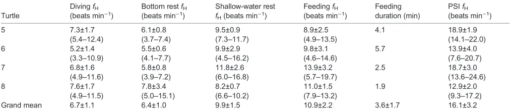
ECG data were successfully obtained in four turtles in 2013 and four turtles in 2015. To obtain fH, duration and water temperature of different conditions or events, instantaneousfHand TDR profiles (time, depth and water temperature) were plotted over a single 24-h period in Origin for each turtle. The number and duration of dives, maximum duration per turtle, PSI duration and mean dive water temperature were determined during the 24-h period for all eight turtles. Because R waves were not clearly discernible during movement in 2013 but were easily identified in 2015, only 2015 turtles were used for most fH analyses. However, minimum instantaneous diving fH was determined for the eight turtles as ECG signals were evident even in 2013 turtles when turtles rested on the bottom of the tank (Fig. S1A). For the 2015 turtles, feeding duration, total heart beats per dive and divingfH(total heart beats divided by duration) were calculated. In addition, in 2015 turtles, the meanfHand range offHwere determined for all (1) dives, (2) PSIs, (3) feeding events, (4) bottom rest periods and (5) shallow-water rest periods.
Statistics
We used linear mixed-effects models [ packages lme4, lmerTest and psycho (Bates et al., 2015; Kuznetsova et al., 2017; Makowski, 2018; https://cran.r-project.org/web/packages/nlme/index.html) implemented in R (version 3.2.3; https://www.r-project.org/)] to analyze the data. In all models, individual turtles were included as a random effect to account for repeated sampling. Water temperature was also included in all initial models as a fixed effect because it has a known effect on fH and duration (Bentivegna et al., 2003; Southwood et al., 2003). All models were fit by maximum likelihood using Akaike’s information criterion (AIC). Models were compared using Chi-squared distributed likelihood ratios.
P-values were calculated using Satterwaithe’s method to estimate denominator degrees of freedom fort-statistics (Kuznetsova et al., 2017). Marginal and conditionalR2 (R2
m and R2c, respectively) values were obtained to determine goodness of fit (Nakagawa and Schielzeth, 2013). Residuals were checked to confirm model requirements, including normality and homoscedasticity, for all models. Heart rates were compared under the five activity conditions (dive, bottom rest, shallow-water rest, feeding and PSI). Differences between conditions were compared with Tukey’s
post hocpairwise comparison. To assess changes infHin longer
dives and because the calculation of divingfH includes duration (total heart beats divided by dive duration), we evaluated the total number of heart beats as a function of dive duration and water temperature in 2015 turtles. We also analyzed the minimum instantaneous fH in relation to dive duration in all turtles. To investigate recovery after dives, we compared PSI mean fHand divingfHin 2015 turtles. Finally, we plotted PSI duration with dive duration in all turtles. Means are expressed ±s.d.
Journal
of
Experimental
RESULTS Behavior
Deployments ranged from 2 to 8 days. During the 24-h observation period, turtles typically submerged and rested on the bottom of the tank for up to 53 min (Table 1). Dives per turtle during the 24-h periods ranged from 29 to 58 for a total 328 dives (Table 1). Over 40% of dives were longer than 30 min and almost 60% were longer than 20 min (Fig. 1). cADLs ranged between 52 and 58 min for turtles at rest (Table 1). Mean and maximum dive durations for each turtle are reported in Table 1. Maximum dive durations were not followed by consistently high PSI durations; however, there was an evident increase in the minimum PSI duration in longer dives (Fig. 2). During shallow-water rest, turtles were left alone in a tank with water that reached the bottom of their carapace, but shallow enough so their head remained out of water. Although turtles were primarily motionless during both dive and shallow-water rest, there was some intermittent activity. Dives included movement to and from the surface and shallow-water rest included some turtles moving, including head movements potentially during breathing, although breathing was not quantified. The lack of movement during bottom rest periods was confirmed from the pitch and roll profiles of turtles lying on the bottom of tanks. When turtles were fed squid, they slowly swam underwater toward the squid and paused briefly as they consumed the squid. Feeding was the most active underwater condition as turtles moved almost continuously. During PSIs, turtles
swam continuously at the surface back and forth across the length of tanks, intermittently lifting their head to breathe. Turtles did not float at the surface or have periods of inactivity during PSIs.
DivingfHwas lower than surfacefH, but not different from shallow-water restfH
Heart rates during diving, bottom rest, shallow-water rest, feeding and PSIs are reported in Table 2. In the model comparingfHunder five conditions, water temperature did not have a significant effect and was removed in the final, best-fit model. Condition had a significant effect onfH(F4,12=18.3,P<0.001; Fig. 3) and explained 77.4% of variation under the model, whereas the effect of individual contributed 2.5% of explanatory power (R2
m=77.4,R2c=79.9). A
post hocpairwise comparison showed that PSIfHwas significantly
higher than diving fH(P<0.001, t=7.2, d.f.=12). However, there were no significant differences infHbetween shallow-water rest and dive (P=0.18,t=2.4, d.f.=12) or shallow-water rest and bottom rest (t=2.7, d.f.=14,P=0.12; Fig. 3).
[image:4.612.48.565.71.193.2]Heart rate increased as underwater activity levels increased When turtles moved underwater,fHincreased by 2- to 3-fold and occasionally reached PSI fH (Fig. 4, Table 2). Feeding fH was significantly higher than bottom restfH(P<0.05,t=3.5, d.f.=12) and
Table 1. Summary of data for each loggerhead sea turtle from 2013 (turtles 1–4) and 2015 (turtles 5–8)
Turtle Mass (kg)
Number of dives (PSIs)
Mean dive duration (min)
Maximum dive duration (min)
PSI duration (min)
cADL range (min)
Minimum dive instantaneous
fH(beats min−1)
Mean dive water temperature (°C)
1 88 36 (35) 25.7±10.2 46.1 3.3±1.9 54.4–115.1 4.5±1.3 22.8±0.8
2 61 41 (39) 24.8±13.7 42.4 8.9±5.0 51.7–109.4 2.5±0.5 22.6±0.3
3 79 41 (41) 31.0±8.7 44.7 4.2±2.1 51.5–109.1 3.6±0.8 23.4±0.8
4 66 46 (45) 29.0±11.3 45.1 4.9±3.3 52.7–111.5 3.4±1.0 22.5±0.8
5 41 32 (32) 35.4±15.3 52.5 5.7±5.1 56.5–119.6 3.6±1.2 19.9±0.2
6 57 45 (44) 15.3±8.0 32.4 5.1±4.0 57.7–122.2 3.1±1.0 20.5±0.5
7 38 29 (27) 26.7±13.5 45.9 8.5±7.2 55.8–118.1 3.6±1.3 19.9±0.7
8 38 58 (57) 13.0±9.8 34.1 8.7±6.5 56.1–118.8 5.6±1.6 19.8±0.9
Grand mean 58.5±18.9 41.0±9.1 24.1±13.4 42.9±6.7 6.2±5.1 54.5–115.4 3.8±1.5 21.4±1.5
Calculated aerobic dive limit (cADL) range for turtles is from 100% to 0% active (Kinoshita et al., 2018). Means are reported ±s.d. Mean dive water temperature is water temperature during dives. PSI, post-dive surface interval;fH, heart rate.N=8 turtles.
0 0
10 20 30 40 50
2 4 6 8 10 12 14 16 18 20
Percentage of dives (%)
[image:4.612.315.564.491.692.2]Dive duration (min)
Fig. 1. Frequency distribution of dive durations.n=8 turtles, 328 dives.
0 5 10 15 20 25 30 35 40 45 50 55 60 0
5 10 15 20 25
30 Turtle 1
Turtle 2 Turtle 3 Turtle 4 Turtle 5 Turtle 6 Turtle 7 Turtle 8
PSI duration (min)
Dive duration (min)
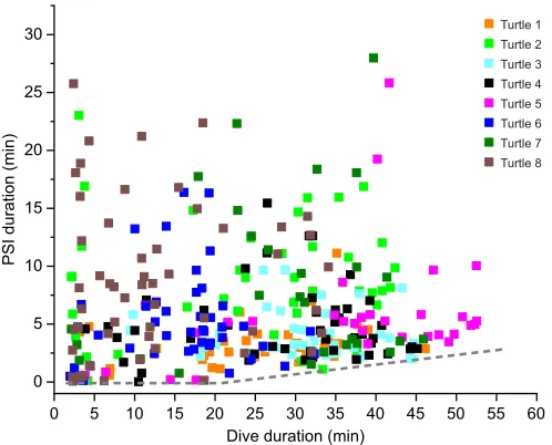
Fig. 2. Post-dive surface interval (PSI) duration versus dive duration.
Dives with PSIs in excess of 30 min were excluded. Dashed line represents minimum PSI duration. Individual turtles are represented by different colors
(see key).n=8 turtles, 317 dives.
Journal
of
Experimental
[image:4.612.52.296.522.720.2]diving fH(P<0.05, t=3.2, d.f.=12), but not shallow-water restfH (P=0.9,t=0.81, d.f.=12).
Relationship betweenfHand dive duration
The total number of heart beats was driven by dive duration and water temperature (P<0.001, χ2=476.3, d.f.=4, R2
m=0.87,
R2
c=0.97; Fig. 5A) as total beats increased by 5.4 and 15.0 beats for each additional minute submerged and 1°C increase in water temperature, respectively (duration: P<0.001, F1,2.8=373.2; water temperature: P<0.001, F1,158.4=62.5). Mean diving fH versus duration is plotted in Fig. 5B for illustration. Mean minimum instantaneousfHwas significantly related to dive duration and water temperature (P<0.001, χ2=187.3, d.f.=5, R2
m=0.30, R2c=0.77; Fig. 6). The best predictive model for mean minimum instantaneous diving fH included dive duration (P<0.05,
F1,20.5=7.1), water temperature (P<0.05, F1,95.4=38.9) and their interaction (P<0.01,F1,20.7=12.1).
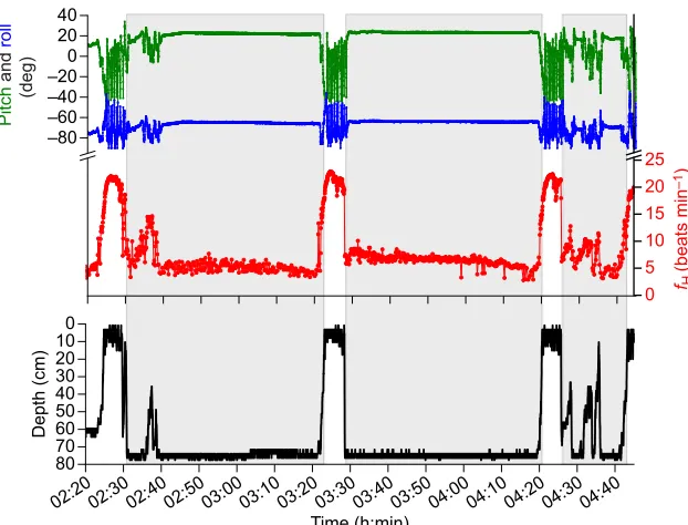
Heart rate patterns
When turtles began to descend during dives,fHdropped quickly from a range of 12 to 20 beats min−1 down to less than 10 beats min−1 (Fig. 4). While resting on the bottom, mean minimum instantaneous fH was less than 6 beats min−1 for all turtles (Table 1). There was no significant difference between bottom restfHand divingfH(P=1.0,t=0.29, d.f.=12; Fig. 3). After turtles returned to the surface,fHwas always the highestfHas turtles swam continuously during the PSI (Table 2, Fig. 3). In addition to being higher than divingfH, PSIfHwas significantly higher than
bottom rest fH (P<0.001, t=7.5, d.f.=12), shallow-water rest fH (P<0.01,t=4.8, d.f.=12) and feedingfH(P<0.05,t=4.0, d.f.=12). Dive duration and water temperature were significantly related to instantaneous mean PSIfH(χ2=91.3,P<0.001, d.f.=4, R2m=0.38,
R2
c=0.68, n=4; Fig. 7), such that for each additional minute submerged, post-dive fHincreased by 0.13 beats min−1 (P<0.05,
F1,3.6=13.3). An increase in water temperature also had a significant effect on PSI fH, raising fH by 1.7 beats min−1 °C−1 (P<0.001,
F1,156.6=37.9).
Effect of temperature
Mean water temperature was slightly higher in 2013 (23.0±0.4°C) than in 2015 (20.0±0.3°C) and varied among individual turtles (Table 1). Water temperature was not significant in all models, likely because it did not vary widely, especially among 2015 turtles (Table 1).
DISCUSSION
The most important findings in this study are that (a) divingfHand bottom restfHwere not significantly different fromfHwhen turtles were resting with their head out of water, (b) changes infHwere largely driven by activity, and (c)fH was not further reduced in longer dives.
DivingfHwas lower than PSIfH, but not different from shallow-water restfH
[image:5.612.47.562.70.183.2]Although it is often assumed that all air-breathing vertebrates have a diving bradycardia (e.g. Andersen, 1966), the question of whether
Table 2. Heart rate (fH) data for five conditions and feeding duration for the four 2015 turtles
Turtle
DivingfH
(beats min−1)
Bottom restfH
(beats min−1)
Shallow-water rest
fH(beats min−1)
FeedingfH
(beats min−1)
Feeding duration (min)
PSIfH
(beats min−1)
5 7.3±1.7 6.1±0.8 9.5±0.9 8.9±2.5 4.1 18.9±1.9
(5.4–12.4) (3.7–7.4) (7.3–11.7) (4.9–13.5) (14.1–22.0)
6 5.2±1.4 5.5±0.6 9.9±2.9 9.8±3.1 5.7 13.9±4.0
(3.3–10.9) (4.1–7.7) (4.5–16.2) (4.6–14.6) (7.6–20.7)
7 6.8±1.6 5.8±0.8 11.8±2.6 13.9±3.2 2.5 18.7±3.0
(4.9–11.6) (3.9–7.2) (6.0–16.8) (5.7–19.7) (13.6–24.6)
8 7.6±1.7 7.8±3.4 8.2±0.7 11.0±1.5 1.9 12.9±2.0
(4.9–11.5) (5.0–15.1) (6.6–10.2) (7.9–13.2) (9.3–17.2)
Grand mean 6.7±1.1 6.4±1.0 9.9±1.5 10.9±2.2 3.6±1.7 16.1±3.2
There was one bottom rest, shallow-water rest and feeding event per turtle. The numbers of dives and PSIs are different for each turtle and specified in Table 1. ForfHcolumns, the first line is the mean±s.d. and the second line is the range (in parentheses). PSI, post-dive surface interval;N=4 turtles.
Bottom rest Diving Shallow-water
rest
Underwater feeding
PSI surface swimming 2
0 4 6 8 10
fH
(beats min
–1
)
12 14 16 18 20
22 Fig. 3. Mean±s.d. heart rate (fH) for five routine activities of
loggerhead sea turtles from 2015.Bottom rest indicates
turtles resting motionless on the bottom of tanks, whereas shallow-water rest occurred when turtles were in shallow water with their head out of water. During underwater feeding, turtles swam (without breathing) after squid were placed in the tank. Dives began when the turtle’s head was underwater and ended when the turtle raised its head to breathe. Surface swimming includes breathing.n=4 turtles.
Journal
of
Experimental
[image:5.612.50.348.542.742.2]turtles experience a diving bradycardia is complicated by the control
fHwith which the divingfHis compared. If a diving bradycardia is defined as a lowerfHduring diving compared with at the surface, then loggerhead turtles experienced a diving bradycardia under the conditions of the present study. Diving fHwas less than half the PSI fH. In marine turtles freely diving at sea, leatherback turtles’ divingfH(17 beats min−1) was only moderately lower than post-dive surfacefH(25 beats min−1) (Southwood et al., 1999). However, it is difficult to compare these results because, in the present study, turtles were primarily resting on the bottom during dives, whereas leatherbacks were diving at sea and, thus, were potentially more active (Southwood et al., 1999). While freely diving at sea, loggerhead turtles may also have higherfH; however, to date there have been no studies examining divingfHin loggerhead turtles at sea. Conversely, if a diving bradycardia is defined as a significantly lowerfHduring dives compared withfHat the same activity level either at the surface or on land (Stephenson et al., 1986), then loggerhead turtles did not experience a diving bradycardia. There was no significant difference infHof these turtles either during the entire dive or during rest in the bottom phase of the dive compared withfHat rest in shallow water with the turtle’s head out of the water. Although this study will not resolve how to define a diving bradycardia in marine turtles, at a minimum, these results suggest thatfHis not further reduced by being submerged. These results are consistent with the lack of difference in restingfHfound in red-eared sliders, whether on land or underwater (Krosniunas and Hicks, 2003). The same study also found no difference in fH between walking on land, underwater swimming or diving in red-eared sliders (Krosniunas and Hicks, 2003).
The comparison between shallow-water rest and bottom rest or divingfHvalues is also complicated by the cost of ventilation during shallow-water rest. Although breathing was not monitored in the present study during shallow-water rest, the pitch and roll profiles indicated that, in some instances, breathing may have occurred. Ventilation in marine turtles requires the use of respiratory muscles to expand the lungs and neck muscles to raise the head, both of which should come at an energetic cost. However, O2consumption in resting loggerhead turtles did not increase during ventilation (Lutz et al., 1989). Similarly, estimates of the oxidative costs of
breathing in freshwater turtles suggest it is low (from 1% of resting metabolic rate) (Jackson et al., 1991). The lack of a significant difference between shallow-water restfHand divingfHor bottom rest fH supports the hypothesis that marine turtles exhibit low metabolic costs of ventilation or that the turtles in the present study were primarily apneic during the shallow-water rest periods.
Heart rate increased as underwater activity levels increased Changes in thefHof submerged loggerhead turtles were driven by activity, similar to terrestrial animals as part of the exercise response. When turtles were motionless on the bottom of tanks,fH was lowest at 6.3 beats min−1. However, with any discernible movement, fH increased (Fig. 4), at times nearing PSI fH levels. The highest underwater fH occurred as turtles swam underwater during feeding; it was significantly higher than bottom restfHand diving fH. While diving at sea, the most vigorous underwater swimming occurs as turtles begin to descend, when stroke frequency and amplitude are highest (Hays et al., 2007). Our findings suggest thatfHmay be highest during the descent phase of dives. This activity-related increase infHalso occurs in some diving endotherms. A number of marine mammals and birds have a graded dive response in which exercise appears to decrease the level of bradycardia (i.e. increasefH) in some, but not necessarily all, dives (Butler and Jones, 1997; Davis and Williams, 2012; Hindle et al., 2010; Noren et al., 2012; Signore and Jones, 1996; Williams et al., 2015). For example, in Steller sea lions,fHwas related to activity levels during shallow dives, but not deep dives (Hindle et al., 2010). Nonetheless, despite these varied responses,fHtypically remained below surface or resting levels in most dives of these other species. However, this effect of underwater activity on turtle fH is in stark contrast to effects in other marine mammals and birds, including emperor penguins diving at sea. During the bottom phases of the emperor penguin’s deepest dives at sea, stroke rate was the highest (Williams et al., 2012). Yet, Wright et al. (2014) found that during these deepest segments, emperor penguin fH was at its lowest. Similarly, during the initial post-release dives of narwhals,fHwas maintained near 10 beats min−1, independent of stroke activity (Williams et al., 2017). By uncoupling workload fromfH, these divers conserve O2for hypoxia-sensitive organs (the heart and brain). This
02:3002:4002:5003:0003:1003:2003:3003:4003:5004:0004:1004:2004:3004:40 02:20
80 70 60 50 40 30 20 10 0
Depth (cm)
Time (h:min) –80
–60 –40 –20 0 20 40
Pitch
and
roll
(deg)
0 5 10 15 20 25
fH
(beats min
–1
[image:6.612.50.361.56.293.2])
Fig. 4.fH(red), pitch (green), roll (blue) and depth
(black) profiles during three dives and three surface
intervals of turtle 5 on 8 September 2015.Shaded areas
indicate turtle is submerged. Heart rate during dives is low unless the turtle is moving, as indicated by the pitch and roll profiles.
Journal
of
Experimental
contrast between turtles and diving endotherms on the effect of activity was also observed in forced submersions of marine turtles and seals, where struggling elicited a significant increase infHin green turtles, but not in seals (Berkson, 1966; Scholander, 1940).
Like marine turtles, red-eared sliders also demonstrate differences infHrelated to their activity level (Krosniunas and Hicks, 2003).fH increased when red-eared sliders were active; however, in these turtles, the increases infHappeared to be constrained to a narrow range set by body temperature (Krosniunas and Hicks, 2003). In the present study, water temperature did not have a significant effect on
fHduring different activities, although there was only a minimal temperature difference among the turtles in 2015 (Table 1). Further research is needed to determine whether there is a similar effect of activity level onfHin marine turtles freely diving at sea and whether cardiovascular responses to activity at different water temperatures are similarly constrained by narrowfHranges.
Relationship betweenfHand dive duration
Marine mammals and emperor penguins frequently extend aerobic dive durations by reducingfH, which reduces O2 consumption in
perfusion dependent organs. In emperor penguins and grey seals diving at sea, the slopes of the relationship between total heart beats and dive duration begin with a linear relationship similar to that in loggerhead turtles (Fig. 5A), but then the number of heart beats flattens out in dives beyond 6 and 7 min, respectively (Thompson and Fedak, 1993; Wright et al., 2014). This plateau, which is consistent with greater reductions infHduring longer dives of seals and penguins, was not observed in the loggerhead turtles (Fig. 5). The lack of a plateau suggests these turtles did not need to further reduce fH and O2 consumption to maintain aerobic metabolism during the long dives in this study. Both this linear increase in total number of heart beats with dive duration and the fact that all dive durations were less than cADLs (Table 1) support the hypothesis that metabolism was primarily aerobic in these turtles.
It is possible that, in loggerhead turtles at sea,fHdecreases further in longer dives. The longest dives in the present study (30–50 min)
0 5 10 15 20 25 30 35 40 45 50 55 60 0
50 100 150 200 250 300 350 400
Turtle 5 Turtle 6 Turtle 7 Turtle 8
T
o
tal heart beats
0 5 10 15 20 25 30 35 40 45 50 55 60 0
1 2 3 4 5 6 7 8 9 10 11 12 13 14 15
Diving
fH
(beats min
–1
)
Dive duration (min)
A
[image:7.612.316.560.56.259.2]B
Fig. 5.fHduring dives of 2015 loggerhead turtles.(A) Total heart beats
versus dive duration. (B) DivingfHversus dive duration. Individual turtles are
indicated by different colors (see key).n=4 turtles, 164 dives.
0 5 10 15 20 25 30 35 40 45 50 55 60 0
1 2 3 4 5 6 7 8 9 10
Turtle 1 Turtle 2 Turtle 3 Turtle 4 Turtle 5 Turtle 6 Turtle 7 Turtle 8
Minimum instantaneous
fH
(beats min
–1
)
[image:7.612.49.297.56.464.2]Dive duration (min)
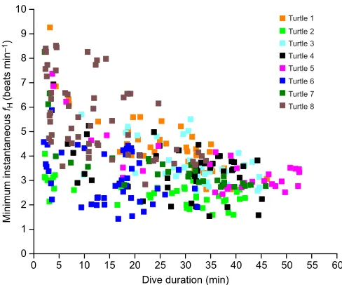
Fig. 6. Minimum instantaneousfHduring dives versus dive duration.
Individual turtles are represented by different colors (see key).n=8 turtles, 328 dives.
0 5 10 15 20 25 30 35 40 45 50 55 60 0
2 4 6 8 10 12 14 16 18 20 22 24 26
Turtle 5 Turtle 6 Turtle 7 Turtle 8
Mean PSI
fH
(beats min
–1
)
Dive duration (min)
Fig. 7. MeanfHduring surface intervals after dives compared with the
duration of the previous dive.Individual turtles are represented by different
colors (see key).n=4 turtles, 160 dives.
Journal
of
Experimental
[image:7.612.317.559.506.702.2]were considerably shorter than the longest dives of similarly sized loggerhead turtles diving at sea (80–150 min) in comparable temperatures (Hochscheid et al., 2007, 2005). Further, minimum instantaneousfHduring dives in the present study declined slightly, but significantly with dive duration (Fig. 6), raising the possibility that divingfHmay significantly decrease in dives over 50 min.
However, the shorter dive durations in the present study have several possible explanations with different implications for changes in divingfH. First, loggerhead turtles in the North Pacific may dive for shorter durations with higherfHbecause they have a higher metabolic rate than other populations. North Pacific juvenile loggerhead turtles maintain higher resting metabolic rates than Mediterranean loggerhead turtles, which results in lower cADLs of North Pacific turtles (Hochscheid et al., 2004; Kinoshita et al., 2018) (see Table S1 for differences inV̇O2and cADL calculated using equations derived from data of the two populations). Future heart rate studies comparing Mediterranean and North Pacific loggerhead turtles might reveal lower restingfHin Mediterranean turtles considering their lower metabolic rate. Second, anaerobic metabolism during dives at sea may contribute to longer dive durations, which would not necessitate lower fH in longer dives. Marine turtles have a high capacity for anaerobic metabolism; however, anaerobic metabolism is believed to be used during intense activities rather than to extend dive durations (Southwood et al., 2003, 2006). Third, dives in the present study could be shorter because turtles are diving with a reduced lung volume. To remain at their preferred depth, loggerhead turtles alter their depth to maintain neutral buoyancy or slightly negative buoyancy, which conserves O2 by reducing the work effort required to move underwater or rest on the ocean floor (Hays et al., 2004; Hochscheid et al., 2003; Minamikawa et al., 2000). Because turtles could not alter their depth in the shallow tanks, to become slightly negatively buoyant they would have to decrease their inhaled lung volume, reducing the lung O2store. As a result, loggerhead turtles diving at sea may have similar fH, but longer durations as a result of diving with a full lung volume, although extended dive durations were not shown in weighted loggerhead turtles diving in shallow tanks (Hochscheid et al., 2003). Thus, regardless of whether dive durations are shorter or longer at sea,fH may not be lower than in the present study because of differences in (1) metabolic rates between populations of loggerhead turtles, (2) preferred metabolic pathways at sea, or (3) diving lung volume. Additional research is needed to investigate fH in freely diving loggerhead turtles and determine if there is a relationship between at sea dive durations andfH.
PSIfHand minimum PSI duration increased after longer dive durations
Although loggerhead turtles did not extend dive durations by reducing fH, they did increase PSI fHafter longer dives (Fig. 7). A fasterfH will reload O2 stores and unload CO2 more quickly, minimize time at the surface and allow turtles to maintain high dive-duration-to-surface-interval ratios. Although the longest dives did not have the longest PSI durations, there was a clear increase in the minimum PSI duration as dive durations increased (Fig. 2), suggesting additional minimum recovery times were necessary for longer dives. However, the increase was minor as the longest dives only increased the minimum PSI by a few minutes.
Heart rate patterns
The profiles of loggerhead turtlefHare similar to those of other turtle studies in that underwater restingfHwas lower compared with swimmingfHor eupneicfH(Table 3). However, it is difficult to draw any other conclusions from comparisons with prior studies because of differences in measurement conditions, turtle size, species and water temperature (Table 3). Despite these differences, restingfH and swimming fH in the present study were much lower than in turtles from past studies, regardless of whether other turtles were bigger or smaller, in colder or warmer temperatures, or swimming in tanks or at sea. These exceptionally lowfHvalues were apparently adequate for maintenance of homeostasis out of water or underwater in these captive turtles.
The highest overallfHin the present study occurred when turtles were at the surface. The high PSIfHwas likely driven by ventilation, as well as activity. Activity at the surface included swimming and movements required for breathing. Marine turtles swim with their head underwater, but use their flippers to help lift their head to breathe (Prange, 1976). We could not separate the activity costs from ventilatory costs because turtles did not stop moving during PSIs, preventing a comparison between fH while floating at the surface and fH while surface swimming. However, a transient increase infHwas associated with breathing in green turtles (Butler et al., 1984; Davenport et al., 1982; West et al., 1992). Although ventilatory costs may be very low in marine turtles, increased ventilation likely contributes to a higher PSI fH because PSI fH increased with longer dives.
Conservation implications
[image:8.612.47.562.584.725.2]Mortality rates from turtle interactions with fisheries are high, with loggerhead turtles having interactions with fisheries more than other marine turtle species in the United States (Finkbeiner et al., 2011).
Table 3.fHof different marine turtle species during underwater rest and swimming or breathing
Marine turtle species
Mass (kg)
Water
temperature (°C) Study conditions
Sample size
Underwater rest
fH (beats min−1)
Swimming/eupneic
fH(beats min−1) Reference
Loggerhead 38–88 20–23 Data logger in tanks
8 6.4 10.9 Present study
Loggerhead 11.8 19.3–22.5 Flipper blood flow study
1 18 19 Hochscheid et al., 2002
Loggerhead 25 15.5 Attached to cord
in tank
1 7.5 15 Lanteri et al., 1980
Leatherback 172–377 25–27.5 Freely diving at sea
5 N/A 17.4 Southwood et al., 1999;
Southwood et al., 2005 Green (winter) 19–43 17 Data logger in
tanks
3–5 10.2 12.9 Southwood et al., 2003
Green (summer) 19–43 26 Data logger in tanks
3–5 19.6 24.0 Southwood et al., 2003
fHfrom present study is lower than marine turtles in other studies. Note differences in turtle size, water temperature and study conditions.
Journal
of
Experimental
Recent discovery of gas emboli in sea turtles suggest that turtles may suffer from decompression sickness after being entangled in nets at depth (García-Párraga et al., 2014). Although turtles typically lower heart rate during forced submergences (Berkson, 1966), the physiological response to net entanglement is likely different. Our results suggest that if turtles are struggling in nets,fHwill increase from vigorous activity, which will result in rapid oxygen depletion. A faster oxygen depletion can contribute to lactate accumulation, and turtles may suffer from lactic acidosis. Further, a higherfHwill increase pulmonary blood flow, which may contribute to increased nitrogen absorption, potentially resulting in the development of gas emboli, recently observed in marine turtles after being entangled in nets (García-Párraga et al., 2018). Future studies designed to increase our understanding of physiological responses to both baseline activities and net entanglement is vital for understanding how to protect and manage marine turtle populations.
Intravascular ECG electrodes resulted in clear ECG records even during activity
ECG signals in 2013 were clear when the turtles were resting on the bottom of the tanks, but became increasingly noisy as the turtle began to move, similar to what was observed in green turtles during movement (Southwood et al., 2003). The noise is likely due to interference from electromyography signals from muscle contractions during movements as it was highest when turtles were the most active–swimming at the surface. The use of a bipolar pacing catheter in 2015 resolved this problem, providing clear ECG signals during all activities, with only rare instances of unreadable signals (Fig. S1).
Conclusions
Our results thatfHwhile resting out of water was no different fromfH during dives or bottom rest indicate that, under the conditions of this study,fHis not further reduced by submergence. Further, our finding that fH also remained relatively constant in short and long dives suggests that loggerhead turtles do not rely on furtherfHreductions to reduce O2consumption and maintain aerobic metabolism during the longer dive durations of this study. Rather,fHappears driven by underwater activity levels as adjustments infHduring dives were in line with an exercise response. With their lack of elevated muscle O2 stores and the apparent absence of reduced muscle blood flow during forced submersions (Lutz and Bentley, 1985), loggerhead turtles must maintain high enough fH to support routine O2 consumption, as well as the increased O2requirements in muscle during activity. Thus, the increase infHduring feeding may be at least partially in response to the additional muscle O2demands from underwater swimming.
The fHvalues we report here highlight the value of recording ECG using self-contained data loggers. Our findings of lower overall fH compared with values in previously measured turtles suggest that, in some cases, fH in other studies may have been affected by the presence of investigators or the turtles being attached to external equipment. ThefHof undisturbed loggerhead turtles in our study provide a baseline for understanding cardiac responses to resting, surface swimming and underwater feeding. These are key activities for the loggerhead turtle, which is known to rest on the ocean bottom for over 7 h in cold temperatures (Hawkes et al., 2007; Hochscheid et al., 2007), spend significant time at the surface (traveling or foraging on surface prey) (Hochscheid et al., 2010; Polovina et al., 2004), and forage within the water column (Casale et al., 2008; Narazaki et al., 2013).fHat the surface was the highest likely because of increased O2demands from continuous swimming
and ventilation. PSIfHincreased as dive duration increased. This strategy allows loggerhead turtles to maximize underwater foraging time by minimizing recovery time at the surface.
These results are the firstfHrecords of undisturbed loggerhead turtles and provide valuable information to assess captive loggerhead turtles as well as to make comparisons with future studies on marine turtles at sea. Whether cardiac responses in loggerhead turtles freely diving at sea are different from the present study awaits further investigation.
Acknowledgements
We thank Takuya Fukuoka, Misaki Yamane and Chihiro Kinoshita for their assistance in the field. We also thank Tomoko Narazaki for valuable input on the initial design of the experiment, and James Hicks, Allyson Hindle, Graeme Hays and an anonymous reviewer for helpful comments on earlier versions of the manuscript. We are grateful to the volunteers from the Fisheries Cooperative Association of Funakoshi Bay, Hirota Bay, Kamaishi Bay, Kamaish-Tobu, Miyako, Ofunato, Okitai, Omoe, Shin-Otsuchi, Ryori, Sanriku-Yamada, Sasaki, Toni, Yamaichi and Yoshiama for providing the incidentally caught, wild loggerhead sea turtles used in this study.
Competing interests
The authors declare no competing or financial interests.
Author contributions
Conceptualization: C.L.W., P.J.P.; Methodology: C.L.W., K.S., P.J.P.; Software: C.L.W.; Validation: C.L.W., P.J.P.; Formal analysis: C.L.W.; Investigation: C.L.W., K.S., P.J.P.; Resources: K.S., P.J.P.; Writing - original draft: C.L.W.; Writing - review & editing: C.L.W., K.S., P.J.P.; Visualization: C.L.W.; Supervision: P.J.P.; Project administration: K.S., P.J.P.; Funding acquisition: C.L.W., P.J.P.
Funding
This study was funded by the National Science Foundation (IOS-1121324 to P.J.P.) and Tohoku Ecosystem-Associated Marine Science (TEAMS). C.L.W. was supported by a National Institutes of Health training grant (2T32AR047752 to V. J. Caiozzo). The present study was performed under the Cooperative Program of Atmosphere and Ocean Research Institute, the University of Tokyo. Deposited in PMC for release after 12 months.
Supplementary information
Supplementary information available online at
http://jeb.biologists.org/lookup/doi/10.1242/jeb.200824.supplemental
References
Andersen, H. T.(1966). Physiological adaptations in diving vertebrates.Physiol. Rev.46, 212-243. doi:10.1152/physrev.1966.46.2.212
Andrews, R. D., Jones, D. R., Williams, J. D., Thorson, P. H., Oliver, G. W., Costa, D. P. and Le Boeuf, B. J.(1997). Heart rates of northern elephant seals
diving at sea and resting on the beach.J. Exp. Biol.200, 2083-2095.
Bates, D., Mächler, M., Bolker, B. and Walker, S.(2015). Fitting linear
mixed-effects models using lme4.J. Stat. Softw.67, 1-48. doi:10.18637/jss.v067.i01
Bentivegna, F., Hochscheid, S. and Minucci, C.(2003). Seasonal variability in
voluntary dive duration of the Mediterranean loggerhead turtle,Caretta caretta.
Sci. Mar.67, 371-375. doi:10.3989/scimar.2003.67n3371
Berkson, H.(1966). Physiological adjustments to prolonged diving in the Pacific
green turtle (Chelonia mydas agassizii).Comp. Biochem. Physiol.18, 101-119.
doi:10.1016/0010-406X(66)90335-5
Butler, P. J. and Jones, D. R.(1997). Physiology of diving of birds and mammals.
Physiol. Rev.77, 837-899. doi:10.1152/physrev.1997.77.3.837
Butler, P. J., Milsom, W. K. and Woakes, A. J.(1984). Respiratory, cardiovascular and metabolic adjustments during steady state swimming in the green turtle,
Chelonia mydas.J. Comp. Physiol. B154, 167-174. doi:10.1007/BF00684141
Carr, A., Ogren, L. and McVea, C.(1980). Apparent hibernation by the Atlantic
loggerhead turtleCaretta carettaoff cape canaveral, Florida.Biol. Conserv.19,
7-14. doi:10.1016/0006-3207(80)90011-7
Casale, P., Abbate, G., Freggi, D., Conte, N., Oliverio, M. and Argano, R.(2008).
Foraging ecology of loggerhead sea turtles Caretta caretta in the central
Mediterranean Sea: evidence for a relaxed life history model.Mar. Ecol. Prog.
Ser.372, 265-276. doi:10.3354/meps07702
Davenport, J., Inagle, G. and Hughes, A. K.(1982). Oxygen uptake and heart rate
in young green turtles (Chelonia mydas).J. Zool.198, 399-412. doi:10.1111/j.
1469-7998.1982.tb02084.x
Davis, R. W. and Williams, T. M.(2012). The marine mammal dive response is
exercise modulated to maximize aerobic dive duration.J. Comp. Physiol. A
Journal
of
Experimental
Neuroethol. Sens. Neural Behav. Physiol.198, 583-591. doi:10.1007/s00359-012-0731-4
Di Bello, A., Valastro, C., Freggi, D., Saponaro, V. and Grimaldi, D.(2010). Ultrasound-guided vascular catheterization in loggerhead sea turtles (Caretta
caretta).J. Zoo Wildl. Med.41, 516-518. doi:10.1638/2008-0195.1
Felger, R. S., Cliffton, K. and Regal, P. J.(1976). Winter dormancy in sea turtles: independent discovery and exploitation in the Gulf of California by two local
cultures.Science191, 283-285. doi:10.1126/science.191.4224.283
Finkbeiner, E. M., Wallace, B. P., Moore, J. E., Lewison, R. L., Crowder, L. B. and Read, A. J.(2011). Cumulative estimates of sea turtle bycatch and mortality in
USA fisheries between 1990 and 2007.Biol. Conserv.144, 2719-2727. doi:10.
1016/j.biocon.2011.07.033
Gallivan, G. J., Kanwisher, J. W. and Best, R. C.(1986). Heart rates and gas exchange in the Amazonian manatee (Trichechus inunguis) in relation to diving.
J. Comp. Physiol. B156, 415-423. doi:10.1007/BF01101104
Garcıa-Pá ́rraga, D., Crespo-Picazo, J. L., de Quirós, Y. B., Cervera, V., Martı́ -Bonmati, L., Dı́az-Delgado, J., Arbelo, M., Moore, M. J., Jepson, P. D. and Fernández, A.(2014). Decompression sickness (‘the bends’) in sea turtles.Dis.
Aquat. Org.111, 191-205. doi:10.3354/dao02790
Garcıa-Pá ́rraga, D., Lorenzo, T., Wang, T., Ortiz, J.-L., Ortega, J., Crespo-Picazo, J.-L., Cortijo, J. and Fahlman, A.(2018). Deciphering function of the
pulmonary arterial sphincters in loggerhead sea turtles (Caretta caretta).J. Exp.
Biol.221, jeb179820. doi:10.1242/jeb.179820
Hawkes, L. A., Broderick, A. C., Coyne, M. S., Godfrey, M. H. and Godley, B. J.
(2007). Only some like it hot — quantifying the environmental niche of the
loggerhead sea turtle. Divers. Distrib.13, 447-457. doi:10.1111/j.1472-4642.
2007.00354.x
Hays, G. C., Metcalfe, J. D. and Walne, A. W.(2004). The implications of
lung-regulated buoyancy control for dive depth and duration.Ecology85, 1137-1145.
doi:10.1890/03-0251
Hays, G. C., Marshall, G. J. and Seminoff, J. A.(2007). Flipper beat frequency and
amplitude changes in diving green turtles,Chelonia mydas.Mar. Biol. 150,
1003-1009. doi:10.1007/s00227-006-0412-3
Hindle, A. G., Young, B. L., Rosen, D. A. S., Haulena, M. and Trites, A. W.(2010). Dive response differs between shallow- and deep-diving Steller sea lions
(Eumetopias jubatus). J. Exp. Mar. Biol. Ecol. 394, 141-148. doi:10.1016/j.
jembe.2010.08.006
Hochscheid, S., Bentivegna, F. and Speakman, J. R.(2002). Regional blood flow
in sea turtles: Implications for heat exchange in an aquatic ectotherm.Physiol.
Biochem. Zool.75, 66-76. doi:10.1086/339050
Hochscheid, S., Bentivegna, F. and Speakman, J. R.(2003). The dual function of
the lung in chelonian sea turtles: buoyancy control and oxygen storage.J. Exp.
Mar. Biol. Ecol.297, 123-140. doi:10.1016/j.jembe.2003.07.004
Hochscheid, S., Bentivegna, F. and Speakman, J. R.(2004). Long-term cold
acclimation leads to high Q10effects on oxygen consumption of loggerhead sea
turtlesCaretta caretta.Physiol. Biochem. Zool.77, 209-222. doi:10.1086/381472
Hochscheid, S., Bentivegna, F. and Hays, G. C.(2005). First records of dive
durations for a hibernating sea turtle.Biol. Lett.1, 82-86. doi:10.1098/rsbl.2004.
0250
Hochscheid, S., Bentivegna, F., Bradai, M. N. and Hays, G. C. (2007).
Overwintering behaviour in sea turtles: dormancy is optional.Mar. Ecol. Prog.
Ser.340, 287-298. doi:10.3354/meps340287
Hochscheid, S., Bentivegna, F., Hamza, A. and Hays, G. C. (2010). When surfacers do not dive: multiple significance of extended surface times in marine
turtles.J. Exp. Biol.213, 1328-1337. doi:10.1242/jeb.037184
Jackson, D. C., Singer, J. H. and Downey, P. T.(1991). Oxidative cost of breathing
in the turtleChrysemys picta bellii.Am. J. Physiol. Regul. Integr. Comp. Physiol.
261, R1325-R1328. doi:10.1152/ajpregu.1991.261.5.R1325
Kinoshita, C., Fukuoka, T., Niizuma, Y., Narazaki, T. and Sato, K.(2018). High resting metabolic rates with low thermal dependence induce active dives in
overwintering Pacific juvenile loggerhead turtles.J. Exp. Biol.221, jeb.175836.
doi:10.1242/jeb.175836
Kooyman, G. L., Wahrenbrock, E. A., Castellini, M. A., Davis, R. W. and Sinnett, E. E. (1980). Aerobic and anaerobic metabolism during voluntary diving in Weddell seals: evidence of preferred pathways from blood chemistry and
behavior.J. Comp. Physiol. B Biochem. Syst. Environ. Physiol.138, 335-346.
doi:10.1007/BF00691568
Kooyman, G. L., Castellini, M. A., Davis, R. W. and Maue, R. A.(1983). Aerobic
diving limits of immature Weddell seals.J. Comp. Physiol. B Biochem. Syst.
Environ. Physiol.151, 171-174. doi:10.1007/BF00689915
Krosniunas, E. H. and Hicks, J. W.(2003). Cardiac output and shunt during
voluntary activity at different temperatures in the turtle, Trachemys scripta.
Physiol. Biochem. Zool.76, 679-694. doi:10.1086/377745
Kuznetsova, A., Brockhoff, P. B. and Christensen, R. H. B.(2017). lmerTest
package: tests in linear mixed effects models.J. Stat. Softw.82, 1-26. doi:10.
18637/jss.v082.i13
Lanteri, A., Lloze, R. and Roussel, H.(1980). Diving and heart beat compounds in
the marine turtleCaretta caretta(LINNÉ) (Reptilia, Testudines).Amphibia-Reptilia
1, 337-341. doi:10.1163/156853881x00429
Lutcavage, M. E., Bushnell, P. G. and Jones, D. R.(1990). Oxygen transport in the
leatherback sea turtleDermochelys coriacea. Physiol. Zool.63, 1012-1024.
doi:10.1086/physzool.63.5.30152626
Lutcavage, M. E., Bushnell, P. G. and Jones, D. R.(1992). Oxygen stores and
aerobic metabolism in the leatherback sea turtle.Can. J. Zool.70, 348-351.
doi:10.1139/z92-051
Lutz, P. L. and Bentley, T. B.(1985). Respiratory physiology of diving in the sea
turtle.Copeia1985, 671-679. doi:10.2307/1444761
Lutz, P. L., LaManna, J. C., Adams, M. R. and Rosenthal, M.(1980). Cerebral
resistance to anoxia in the marine turtle.Respir. Physiol.41, 241-251. doi:10.
1016/0034-5687(80)90074-2
Lutz, P. L., Bergey, A. and Bergey, M.(1989). Effects of temperature on gas
exchange and acid-base balance in the sea turtleCaretta carettaat rest and
during routine activity.J. Exp. Biol.144, 155-169.
Makowski, D.(2018). The psycho package: an efficient and publishing-oriented
workflow for psychological science.J. Open Source Software3, 470. doi:10.
21105/joss.00470
McDonald, B. I. and Ponganis, P. J.(2014). Deep-diving sea lions exhibit extreme
bradycardia in long-duration dives.J. Exp. Biol.217, 1525-1534. doi:10.1242/jeb.
098558
Meir, J. U., Stockard, T. K., Williams, C. L., Ponganis, K. V. and Ponganis, P. J. (2008). Heart rate regulation and extreme bradycardia in diving emperor
penguins.J. Exp. Biol.211, 1169-1179. doi:10.1242/jeb.013235
Meir, J. U., Champagne, C. D., Costa, D. P., Williams, C. L. and Ponganis, P. J. (2009). Extreme hypoxemic tolerance and blood oxygen depletion in diving
elephant seals.Am. J. Physiol. Regul. Integr. Comp. Physiol.297, R927-R939.
doi:10.1152/ajpregu.00247.2009
Minamikawa, S., Naito, Y., Sato, K., Matsuzawa, Y., Bando, T. and Sakamoto, W. (2000). Maintenance of neutral buoyancy by depth selection in the loggerhead
turtleCaretta caretta.J. Exp. Biol.203, 2967-2975.
Nakagawa, S. and Schielzeth, H. (2013). A general and simple method for
obtainingR2
from generalized linear mixed-effects models.Methods Ecol. Evol.4,
133-142. doi:10.1111/j.2041-210x.2012.00261.x
Narazaki, T., Sato, K., Abernathy, K. J., Marshall, G. J. and Miyazaki, N.(2013). Loggerhead turtles (Caretta caretta) use vision to forage on gelatinous prey in
mid-water.PLoS ONE8, e66043. doi:10.1371/journal.pone.0066043
Narazaki, T., Sato, K. and Miyazaki, N.(2015). Summer migration to temperate
foraging habitats and active winter diving of juvenile loggerhead turtlesCaretta
carettain the western North Pacific.Mar. Biol.162, 1251-1263. doi:10.1007/
s00227-015-2666-0
Noren, S. R., Kendall, T., Cuccurullo, V. and Williams, T. M.(2012). The dive response redefined: underwater behavior influences cardiac variability in freely
diving dolphins.J. Exp. Biol.215, 2735-2741. doi:10.1242/jeb.069583
Polovina, J. J., Balazs, G. H., Howell, E. A., Parker, D. M., Seki, M. P. and Dutton, P. H.(2004). Forage and migration habitat of loggerhead (Caretta caretta) and olive ridley (Lepidochelys olivacea) sea turtles in the central North Pacific Ocean.
Fish. Oceanogr.13, 36-51. doi:10.1046/j.1365-2419.2003.00270.x
Ponganis, P. J.(2015).Diving Physiology of Marine Mammals and Seabirds. Cambridge University Press.
Ponganis, P. J., Stockard, T. K., Meir, J. U., Williams, C. L., Ponganis, K. V., van Dam, R. P. and Howard, R.(2007). Returning on empty: extreme blood O2
depletion underlies dive capacity of emperor penguins. J. Exp. Biol. 210,
4279-4285. doi:10.1242/jeb.011221
Prange, H. D.(1976). Energetics of swimming of a sea turtle.J. Exp. Biol.64, 1-12. Prange, H. D. and Jackson, D. C.(1976). Ventilation, gas exchange and metabolic
scaling of a sea turtle. Respir. Physiol. 27, 369-377.
doi:10.1016/0034-5687(76)90065-7
Scholander, P. F.(1940). Experimental investigations on the respiratory function in
diving mammals and birds.Hvalradets Skrifter22, 1-131.
Signore, P. E. and Jones, D. R.(1996). Autonomic nervous control of heart rate
in muskrats during exercise in air and under water. J. Exp. Biol. 199,
1563-1568.
Southwood, A. L., Andrews, R. D., Lutcavage, M. E., Paladino, F. V., West, N. H., George, R. H. and Jones, D. R.(1999). Heart rates and diving behavior
of leatherback sea turtles in the eastern Pacific Ocean.J. Exp. Biol. 202,
1115-1125.
Southwood, A. L., Darveau, C. A. and Jones, D. R.(2003). Metabolic and cardiovascular adjustments of juvenile green turtles to seasonal changes in
temperature and photoperiod.J. Exp. Biol. 206, 4521-4531. doi:10.1242/jeb.
00689
Southwood, A. L., Andrews, R. D., Paladino, F. V. and Jones, D. R.(2005). Effects of diving and swimming behavior on body temperatures of Pacific
leatherback turtles in tropical seas.Physiol. Biochem. Zool.78, 285-297. doi:10.
1086/427048
Southwood, A. L., Reina, R. D., Jones, V. S., Speakman, J. R. and Jones, D. R. (2006). Seasonal metabolism of juvenile green turtles (Chelonia mydas) at Heron
Island, Australia.Can. J. Zool.84, 125-135. doi:10.1139/z05-185
Stephenson, R., Butler, P. and Woakes, A.(1986). Diving behaviour and heart rate
in tufted ducks (Aythya fuligula).J. Exp. Biol.126, 341-359.
Journal
of
Experimental
Thompson, D. and Fedak, M. A.(1993). Cardiac responses of grey seals during
diving at sea.J. Exp. Biol.174, 139-164.
West, N. H., Butler, P. J. and Bevan, R. M.(1992). Pulmonary blood flow at rest and
during swimming in the green turtle,Chelonia mydas.Physiol. Zool.65, 287-310.
doi:10.1086/physzool.65.2.30158254
Williams, C. L. and Hicks, J. W. (2016). Continuous arterial PO2 profiles in
unrestrained, undisturbed aquatic turtles during routine behaviors.J. Exp. Biol.
219, 3616-3625. doi:10.1242/jeb.141010
Williams, C. L., Meir, J. U. and Ponganis, P. J.(2011). What triggers the aerobic dive limit? Patterns of muscle oxygen depletion during dives of emperor penguins.
J. Exp. Biol.214, 1802-1812. doi:10.1242/jeb.052233
Williams, C. L., Sato, K., Shiomi, K. and Ponganis, P. J. (2012). Muscle energy stores and stroke rates of emperor penguins: implications for muscle
metabolism and dive performance.Physiol. Biochem. Zool.85, 120-133. doi:10.
1086/664698
Williams, T. M., Fuiman, L. A., Kendall, T., Berry, P., Richter, B., Noren, S. R., Thometz, N., Shattock, M. J., Farrell, E., Stamper, A. M. et al.(2015). Exercise at depth alters bradycardia and incidence of cardiac anomalies in deep-diving
marine mammals.Nat. Commun.6, 6055. doi:10.1038/ncomms7055
Williams, T. M., Blackwell, S. B., Richter, B., Sinding, M.-H. S. Heide-Jørgensen, M. P.(2017). Paradoxical escape responses by narwhals (Monodon monoceros).
Science358, 1328-1331. doi:10.1126/science.aao2740
Wright, A. K., Ponganis, K. V., McDonald, B. I. and Ponganis, P. J.(2014). Heart rates of emperor penguins diving at sea: implications for oxygen store
management.Mar. Ecol. Prog. Ser.496, 85-98. doi:10.3354/meps10592