www.elsevier.com/locate/brainres
Available online at www.sciencedirect.com
Research Report
Decoding signal processing in thalamo-hippocampal
circuitry: implications for theories of memory and
spatial processing
Marian Tsanov
a,b, Shane M. O’Mara
a,b,n aTrinity College Institute of Neuroscience, Trinity College Dublin, Ireland b
School of Psychology, Trinity College Dublin, Ireland
a r t i c l e i n f o
Article history:
Accepted 1 December 2014
Keywords: Hippocampus Anterior thalamus Head direction cells Theta rhythm Place cells
a b s t r a c t
A major tool in understanding how information is processed in the brain is the analysis of neuronal output at each hierarchical level through which neurophysiological signals are propagated. Since the experimental brain operation performed on Henry Gustav Molaison (known as patient H.M.) in 1953, the hippocampal formation has gained special attention, resulting in a very large number of studies investigating signals processed by the hippo-campal formation. One of the main information streams to the hippohippo-campal formation, vital for episodic memory formation, arises from thalamo-hippocampal projections, as there is extensive connectivity between these structures. This connectivity is sometimes overlooked by theories of memory formation by the brain, in favour of theories with a strong cortico-hippocampalflavour. In this review, we attempt to address some of the complexity of the signals processed within the thalamo-hippocampal circuitry. To understand the signals encoded by the anterior thalamic nuclei in particular, we review key findings from electrophysiological, anatomical, behavioural and computational studies. We include recent findings elucidating the integration of different signal modalities by single thalamic neurons; we focus in particular on the propagation of two prominent signals: head directionality and theta rhythm. We conclude that thalamo-hippocampal processing provides a centrally important, substantive, and dynamic input modulating and moderating hippocampal spatial and mnemonic processing.
This article is part of a Special Issue entitled SI: Brain and Memory.
&2014 Published by Elsevier B.V.
1.
Introduction
The purpose of the present review is to dissect some of the complexity of the signals propagated from anterior thalamus to the hippocampal formation in order to try and understand
the importance of this information processing for the coding properties of individual neurons in the hippocampus.“ Ante-rior thalamus”includes the anterodorsal, anteroventral and dorsolateral thalamic nuclei, which form the thalamic com-ponent of the limbic system (the ‘thalamic level’of Papez's
http://dx.doi.org/10.1016/j.brainres.2014.12.003 0006-8993/&2014 Published by Elsevier B.V.
nCorresponding author.
E-mail address:[email protected](S.M. O'Mara).
circuit). Functionally, this circuit is involved in episodic memory formation; dysfunction in this circuit results in the anterograde amnesic syndrome, known as Korsakoff's syn-drome (Aggleton and Sahgal, 1993; Gold and Squire, 2006; Harding et al., 2000). Lesion experiments have also confirmed the profound importance of these nuclei for hippocampal-dependent learning (Parker and Gaffan, 1997; Warburton et al., 2001). A key point in this review is to emphasize the importance of hippocampally-directed thalamic processing. The anterior thalamic nuclei are abundantly connected with the hippocampal formation, and have the capacity to pro-foundly shape hippocampal spatial and mnemonic informa-tion processing, a key point sometimes be overlooked in analyses favouring of hippocampally-directed cortical processing.
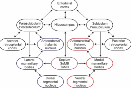
Information processing within the diencephalic portion of the Papez's circuit involves the propagation of two main signals: theta rhythm and head directionality (for reviews: (Vann and Aggleton, 2004;Vertes et al., 2004). The pathways mediating these signals share similar connectivity, arising from Gudden's tegmental nuclei-mammillary bodies-anterior tha-lamic nuclei-retrosplenial/entorhinal cortices-hippocampal formation (the latter including the hippocampus and the subicular cortices (Fig. 1) (Gonzalo-Ruiz et al., 1997; Shibata, 1993;Swanson and Cowan, 1977;Van Groen and Wyss, 1995; van Groen et al., 1999; Witter et al., 1990). The anatomical substrates of these parallel pathways at a thalamic level involves the anterodorsal and anteroventral nuclei, respec-tively. The two pathways interact at multiple levels within Papez's circuit, and there is an indication that the integration of theta and head-direction processing occurs also in anterior thalamus (Tsanov et al., 2011a). Lesions of anterior thalamus produce impairments on spatial memory tasks, including
radial-arm maze, T-maze and water maze (Aggleton et al., 1996;Byatt and Dalrymple-Alford, 1996;van Groen et al., 2002; Wilton et al., 2001). These memory deficits are less severe after selective lesions of individual anterior thalamic nuclei (Aggleton et al., 1996; Byatt and Dalrymple-Alford, 1996; van Groen et al., 2002). This additive effect indicates that the head direction and theta systems individually only have a partial effect on hippocampal spatial representation. Thus, it is likely that these parallel signals require functional integration to support hippocampal spatial learning. The integration of idio-thetic signals might explain the additive effects of separate and individual lesions of the anterior thalamic nuclei. Head-direction and theta pathways converge at the cortical levels of the Papez's circuitry (entorhinal and subicular cortices) where neurons possess both theta and head-directional properties (Boccara et al., 2010;Cacucci et al., 2004). The additional effects of the thalamic lesions demonstrate the functional significance of this signal integration.
The firing patterns of the neurons encoding the head-direction signal as well as those that encode theta rhythmicity derive from two main factors: afferent inputs and biophysical cellular properties. The first factor depends on the synaptic inputs, and defines what information the cell is conveying, while the second factor is based on the properties of membrane channels, and determines how the cell will propagate this information. Here, we will examine the nature of the head-direction signal as well as the biophysical properties of ante-rodorsal thalamic neurons. Next, we will discuss the tuning curve features that are shared between head-direction and hippocampal place cells. We will discuss the effects of theta rhythm and self-motion on the directional signal. Finally, we will review the integration of theta and directionality on thalamo-hippocampal information processing.
2.
Integration of internal idiothetic and
external sensory signals
Head-direction neurons encode the animal's directional heading in the horizontal and vertical planes (Stackman et al., 2000; Taube et al., 1990; Taube, 2011). The head-direction signal carries highly-processed sensory informa-tion. The signals maintaining the head directional represen-tation integrate external sensory (landmark) inputs as well as internal idiothetic signals (Knierim et al., 1998; Kudrimoti et al., 1996;Stackman and Taube, 1997). The activity of head-direction cells recorded in behaving animals is updated constantly, even in total darkness, due to idiothetic signals (Goodridge et al., 1998; McNaughton et al., 1991; Mizumori and Williams, 1993). These idiothetic signals are driven by a combination of vestibular, proprioceptive and motor effer-ence inputs (Stackman and Taube, 1997; Stackman et al., 2003;van der Meer et al., 2007). The generation of the head direction signal has been attributed particularly to vestibular signals. Lesions of the vestibular labyrinth abolish the head-direction signal in the anterodorsal nucleus of the thalamus (Stackman and Taube, 1997). Similarly, vestibular inactivation impairs the directionality properties of postsubicular head-direction neurons (Stackman et al., 2002) and thisfinding is not surprising considering the direction of head direction
Fig. 1–Schematic representation of the main connections in
the Papez's circuit. The hippocampal formation includes hippocampus and subicular cortices. The structures marked in blue mediate head-direction signals, while the structures in red mediate theta rhythm. The structures marked in the purple exert a neuromodulatory control on hippocampo-diencephalic circuitry: medial septum, supramammillary (SuMB) and tuberomammillary (TuMB) nuclei. Minor connections among the presented regions are not shown. For a detailed review of the afferents and efferents of the anterior thalamic nuclei, see (Jankowski et al., 2013).
[image:2.595.53.282.467.623.2]signal from anterior thalamus to the parahippocampal region; anterodorsal thalamic lesions completely abolish directional signal propagation within the parahippocampal region (for review: (Taube, 2007). Recent data also suggest the presence of a population of head-direction cells in the nucleus reuniens of the thalamus (Jankowski et al., 2014). The nucleus reuniens head-direction cells are similar to the head-direction cells of the anterodorsal and anteroventral thalamic nuclei, lateral mammillary nucleus (Taube, 2007), and some parahippocampal regions. Nucleus reuniens receives widespread cortical and subcortical afferent inputs (McKenna and Vertes, 2004). Nucleus reuniens is not part of the more usual head direction circuit, which is regarded as deriving principally from the dorsal tegmental nucleus of Gudden and the lateral mammillary nucleus (Cassel et al., 2013; Taube, 2007)(see Fig. 1). Nucleus reuniens sends a strong excitatory projection to hippocampal formation (Vertes et al., 2007), thus offering yet another route by which thalamic activity may also modulate hippocampal signal processing (Jankowski et al., 2014). Further research is needed to investigate whether the vestibular signal in nucleus reuniens is a parallel or alternative to anterior thalamus directional information. The presence of this rostral thalamic spatial input to the hippocampus offers alternative hypoth-eses for the generation of spatial signals in the hippocampus. It also highlights the widespread dispersion of head direc-tional processing in the brain, as compared with other spatial signals.
The significance of vestibular signals for thalamo-hippocampal information processing is underlined by the finding that vestibular inactivation disrupts the location-specificfiring of hippocampal place cells (Calton et al., 2003; Russell et al., 2003;Stackman et al., 2002). Hippocampal place cellsfire in response to a rodent's spatial location (O'Keefe, 1976), using sensory stimuli as a directional reference to provide a rodent's orientation in space. Hippocampal place cells are also closely coupled to head-direction cells, and hippocampal spatial representation is functionally related to the directional system (Knierim et al., 1995; Knierim et al., 1998). From external sensory signals, visual (landmark) infor-mation is the most potent source maintaining and updating activity of head-direction cells (Yoder et al., 2011a). The current hypothesis of how visual landmark cues control directional tuning suggests that the postsubiculum (Goodridge and Taube, 1997) and the retrosplenial cortex (Clark et al., 2010) might transfer visual landmark informa-tion to spatial signals within the limbic system (Yoder et al., 2011b).
3.
Integration of intrinsic biophysical
properties and extrinsic directional signals
Sensory and motor neurons in primary sensory and motor cortices express their responsiveness to stimuli with a Gaus-sian tuning curve. The means of the tuning curves vary linearly with the stimulus intensity, where the neuronfires with highest frequency at the mean or the peak of the curve. As the sensory signal is processed across secondary, tertiary
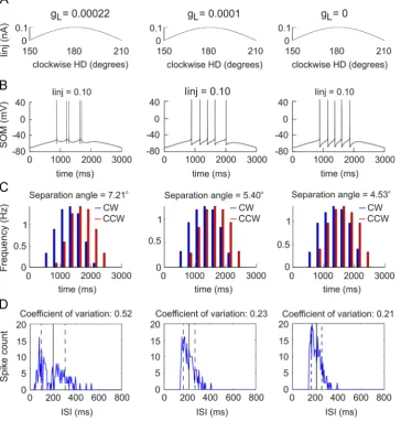
and further association zones the responsiveness of the neurons there becomes less tuned to single sensory stimuli (and more difficult to study). One of the reasons that the neurons in Papez's and hippocampal formation circuit have been frequently studied is the presence of neurons whose responsiveness is also expressed by a Gaussian tuning curve. The tuning curve of these neurons allows us to measure how their activity is related to incoming, highly-processed signals. The most-explored neuron types are the head-direction cells with a one-dimensional tuning curve, and place cells and grid cells with two-dimensional tuning curves (although the three types of neurons are known now to encompass one more dimension, namely the vertical plane (Taube and Shinder, 2013)). The neuronal activity of head-direction cells is repre-sented by a Gaussian curve of the firing rate versus the animal's heading direction over 360 degrees. The peak of the curve is defined as the preferredfiring direction (Taube and Muller, 1998). Thefiring patterns from the tuning curve show variability in the means of the firing frequencies and inter-spike intervals (ISI), both between regions and between neurons in the anterodorsal thalamic nucleus (Taube, 2010). To understand how such variability affects the encoding of the extrinsic directional signal, we must first address the intrinsic biophysical properties of neurons. The ionic nature of different types of conductance has been well-established for cortical and thalamic neurons (Crill, 1996; Llinas, 1988; Schwindt et al., 1988a; Schwindt et al., 1988b). Generally speaking, the spiking properties of thalamic neurons range from tonic or regular firing to bursting or phasic (Steriade, 2001). However, such systematization does not implicate distinctly different categories: thalamic neurons are able to switch between tonic and burst modes. The firing mode is determined by the conductance of voltage-gated calcium channels of the somatic and the axonal membranes (Jahnsen and Llinas, 1984a; Jahnsen and Llinas, 1984b). Furthermore,firing mode strongly affects the propagation of the sensory signal (Ramcharan et al., 2000;Ramcharan et al., 2005;Sherman, 2001). Head-direction neurons in anterodorsal thalamic nucleus normally do not express bursting proper-ties; however, suchfiring modes appear after disconnection of the afferent input that processes the vestibular signal (Muir et al., 2009; Stackman and Taube, 1997; Yoder and Taube, 2009) but see also (Stackman et al., 2002). The variability of thefiring patterns between the head-direction neurons dur-ing tonic spikdur-ing is expressed by their inter-spike intervals (Taube, 2010) and recent data suggest that ISI variability affects the encoding of the directional signal (Tsanov et al., 2014a). ISI variability is linked particularly to one of the unresolved features of the tuning curve, namely, its depen-dence on the direction of angular displacement, also known as the ‘separation angle’. The separation angle reflects the difference in the preferredfiring direction between clockwise and counter-clockwise head turns of the animal in the horizontal plane (Blair and Sharp, 1995). Analysis of firing activity across the tuning curve of thalamic head-direction neurons shows a positive correlation between the separation angle and the spiking variability of thalamic head-direction cells, as expressed by the coefficient of variation (Tsanov et al., 2014a). ISI variability is evaluated by the coefficient of variation (the ratio between the standard deviation to the
mean of ISI) where low values indicate regularfiring, while high values reflect irregular pattern of the spikes (Softky and Koch, 1993). This relationship is particularly expressed when the ISIs from the tuning curve's ascending and descending slopes are included in analyses. The role of the intrinsic biophysical properties that link ISI diversity and separation angle was demonstrated by a single-cell Hodgkin–Huxley type model (Tsanov et al., 2014a) that investigated ISI pat-terns ranging from irregularly- to regularly-firing type cells (Pospischil et al., 2008). These findings show that the depolarization-triggered calcium currents, together with the
adaptation currents, evoke irregularfiring, leading to a higher firing rate on the ascending slope of a sinusoidal depolariza-tion (Fig. 2). Modelled sinusoidal current injections mimic the Gaussian tuning curve of the head-direction signal in thalamic neurons (Fig. 2A). Importantly, spike trains mediated by calcium and slow potassium channels occur preferentially on the ascending slope of the input signal (Gabbiani et al., 1996;Guido et al., 1992;Kepecs et al., 2002) and therefore the irregularly-spiking neurons may detect the rising slope of input signals with greater precision, compared to regularly-spiking neurons (Metzner et al., 1998;Sherman,
Fig. 2–A model of an irregular spiking anterior thalamic head-direction.A, Sinusoidal current injection (Iinj) to a neuron with a high-threshold calcium channel conductance ofgL¼0.00022 S/cm2. (left),gL¼0.0001 S/cm2(middle) andgL¼0 S/cm2(right). The trace represents the current application, where the start is considered at 1501for clockwise simulations and 2101for counter-clockwise simulations.B, Evoked spiking with the sinusoidal current injection to the neuron withgL¼0.00022 S/cm2. (left),gL¼0.0001 S/cm2 (middle) andgL¼0 S/cm2(right).C,Histograms of averagedfiring frequency for 30 simulations with small I
injvariations (0.001 nA step) for clockwise (blue) and counter-clockwise (red) simulations forgL¼0.00022 S/cm2(left),gL¼0.0001 S/cm2(middle) andgL¼0 S/cm2 (right). The values of the separation angle, calculated as the difference between clockwise and counter-clockwise mean directions, are represented on the top of the histograms.E, Inter-spike interval (ISI) histograms of averagedfiring frequency for 30 simulations with small Iinjvariations (0.001 nA step) forgL¼0.00022 S/cm2(left),gL¼0.0001 S/cm2(middle) andgL¼0 S/cm2(right). The vertical black line denotes the mean ISI, while the dashed black lines represent the standard variation. The values of the coefficient of variation, calculated as the ratio between the standard deviation to the mean of inter-spike intervals, are represented on the top of the histograms. The plots are from (Tsanov et al., 2014a).
[image:4.595.116.481.230.613.2]2001). These data show that the conductance of high-threshold calcium channels together with the conductance of slow potassium channels evokes irregular spiking, which results in a phase lead to modelled sinusoidal current injec-tion (Fig. 2B). The phase lead is expressed by higherfiring rate on the ascending phase compared to the descending phase of a sinusoidal input. The phase lead creates dependence on the direction of angular displacement, which results (function-ally) in a separation angle between clockwise and counter-clockwise simulations (Fig. 2C). Thisfinding suggests that one of the main factors determining separation angle in the head-direction signal is the degree offiring irregularity of head-direction cells, which in turn is a function of their intrinsic biophysical properties (Fig. 2D). The role of inter-spike dynamics (and particularly of spike-frequency adaptation) might not be unique to the neurons supporting thalamic directional signal processing in the formation of the separa-tion angle. A computasepara-tional study described a similar effect at the level of the vestibular nucleus using spike rate adaptation and the post-inhibitory rebound of vestibular nucleus neurons (van der Meer et al. 2007). This study explored the formation of anticipatory time interval (ATI) and the authors challenged the idea that ATI actively predicts direction by anticipating a future directional signal. ATI is a parameter closely related to the separation angle and is defined as the time displacement for which a cell'sfiring rate is best correlated with the directional position of the rat's head (Blair and Sharp, 1995; Blair et al., 1997). ATI values differ substantially across the Papez's circuit: however, the presence of ATI does not mean that there is a particular information processing pattern across monosynaptically-connected regions. ATI is almost absent for the dorsal tegmental nucleus of Gudden; it is highly-expressed at the next hierarchical level in the mammillary bodies (40–75 ms) and less-expressed at the next hierarchical level of thalamic cells (25 ms). Further, at the neocortical level, the ATI in the retrosplenial cortex (25 ms) differs from the ATI of post-subicular cortex with values close to 0 (Blair and Sharp, 1995; Blair et al., 1998; Cho and Sharp, 2001; Sharp et al., 2001;
Stackman et al., 2003;Taube and Muller, 1998). There is no evidence showing a link between ATI and the vestibular signal, which is proposed to follow a bottom-up direction (Taube, 2007), or between ATI and the motor efference copy (Bassett et al., 2005). One of the possible explanations is that ATI does not reflect a process of anticipation, but rather represents the intrinsic neuronal properties of the neurons across the regions that encode directional signals in the context of the direction of angular displacement.
4.
Parallels between the encoding of thalamic
head-direction cells and hippocampal place cells
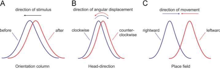
The head-direction signal is not unique in terms of tuning curve dependence on the direction of signal displacement. This is a common feature of sensory and motor systems that encode their signal with a Gaussian distribution. The orientation selectivity of neurons in the primary visual cortex shift in their orientation tuning as a result of experience (Fu et al., 2002;Yao and Dan, 2001). Repetitive pairing of visual stimuli at two orientations induced a shift in the orientation tuning of cat cortical neurons, and the direction of the curve shift is in the opposite direction to the test stimulus (Fig. 3A). This backward shift results from asymmetric visual stimulation of cat visual cortex, and is believed to involve spike-timing dependent plasticity (Yao and Dan, 2001), allowing sensory circuits to adjust to the visual stimuli (Brenner and Bialek, 2000;Muller et al., 1999). Importantly, the backward shift of the receptivefield tuning curve, which is in the opposite direction to the test stimulus, shares the same rule observed in the head-direction tuning curve, which is in the opposite head-direction to the angular displacement (Fig. 3B). Hippocampal placefield encoding confirms the same backwards pattern during experience-dependent activity. When an animal is engaged in stereotyped trajectories on uniform one-dimensional tracks, the hippocampal tuning curves of forward and backward journeys can be uncorre-lated (the cell is active only in one direction) or correuncorre-lated (bidirectional cells) (Gothard et al., 1996; Markus et al., 1995; McNaughton et al., 1983; Muller et al., 1994). The bidirectional
Fig. 3–Experience-dependent backward shift signal-encoding of tuning curves. A, Backward shift of the perceived orientation
for a visual receptivefield from primary visual cortex. The orientation tuning response evoked by a visual test stimulus is
shown before (blue) and after (red) conditioning. The tuning curve shift is opposite to the direction of the test stimulus (black
arrow).B,Shift of the preferredfiring direction opposite to the direction of angular displacement (shown with curved arrows)
for anterodorsal thalamic head-direction cells. The difference between clockwise turns (blue) and counter-clockwise turns
(red) is known as the separation angle.C, Bidirectional placefields from hippocampal CA1 region during a left-bound journey
(red) and a right-bound journey (blue). The centres of masses for both directions are displaced backward with respect to the
rat's direction (shown with arrow). Thefigure includes schematic representations from thefindings of (Battaglia et al., 2004;
Blair and Sharp, 1995;Yao and Dan, 2001).
[image:5.595.120.479.533.646.2]place field centres are displaced rearward or backward with respect to the rat's direction of motion (Battaglia et al., 2004) (Fig. 3C). In linear, cue-rich, tracks, hippocampal placefield location depends on the animal's direction of motion (Battaglia et al., 2004), showing the same backward shift of the tuning curve as observed with head-direction tuning curves. Similar to the shift in orienta-tion tuning of the visual receptive fields, the backward shift of hippocampal placefield is shown to be experience-dependent, where hippocampal placefields shift backwards in the course of several journey repeats from the animal in the same linear track (Mehta et al., 1997;Mehta et al., 2000). The subsequent shift of the centres of the placefields is known as‘centre of mass difference’ (ΔCOM) (Knierim, 2002;Lee et al., 2004;Lee and Knierim, 2007). There is sub-regional-specific functional differentiation between CA1 and CA3 hippocampal placefield centre of mass shifts (Lee et al., 2004;Roth et al., 2012); however, both shifts are backwards relative to the animals’linear movement. There is little informa-tion available about the effect of experience on the backward shift of head-direction curve and the formation of separation angle or anticipatory time intervals. The head-direction tuning curve has been found to shift backwards, similar to the COM shifting observed in the simultaneously recorded place cells, in animals repeatedly running in a circular linear track (Yu et al., 2006). However, the authors did not examine the dynamics of the separation angle or the anticipatory time interval of the recorded head-direction cells. Further investigations should investigate if the head-direction ISI properties, which are correlated with the separation angle (Tsanov et al., 2014a), change with experience. The head direction tuning curves shift backwards in ways that are similar, but not identical, to the hippocampal place fields. The differences between these cells included shift patterns relative to the cue-altered conditions, the proportion of backward versus forward shifts, and the time course of shift resetting (Yu et al., 2006). Also, unlike the hippocampal spatial relation to proximal and distal cues, the thalamic head direction representation main-tains an internally-coherent representation that is strongly bound to the distal landmarks (Yoganarasimha et al., 2006). Despite the differences between head-direction and placefield properties, it is widely accepted that both systems functionally interact at the level of episodic memory networks. The head-direction system is proposed to provide highly-processed directional signal that integrates idiothetic information together with visual (landmark) information to hippocampal circuitry where the direction of motion is integrated into a spatial representation of the environ-ment (McNaughton et al., 1996;McNaughton et al., 2006;Muller et al., 1996; O'Keefe and Burgess, 2005; Sargolini et al., 2006; Yoganarasimha and Knierim, 2005).
5.
Integration of angular velocity and theta
signal from thalamic head-direction cells
The head-direction tuning curve is modulated by incoming idiothetic signals, suggesting that these signals are integrated during head-direction information processing. Angular velo-city is one of these signals, and changes of angular head velocity are related to the neuronal activity of head-direction cells (Blair and Sharp, 1995; Taube, 1995). The tuning curve peak of anterodorsal thalamic head-direction neurons is
higher during faster passive rotations (Zugaro et al., 2002). This finding suggests the role of angular velocity in the maintenance of the directional signal, and recent data pro-pose a link between the effect of angular velocity on thalamic head direction neurons and their biophysical properties (Tsanov et al., 2014a). The angular velocity signal is believed to be processed by the lateral mammillary bodies, which are one of the main inputs to anterodorsal thalamic nucleus. The lateral mammillary nuclei contains neurons sensitive to an animal's angular head velocity (Blair et al., 1998; Stackman and Taube, 1998). The lateral mammillary nucleus also receives projections from the dorsal tegmental nucleus (which contain neurons which respond to angular velocity) (Bassett and Taube, 2001; Sharp et al., 2001); in turn, the lateral mammillary nucleus sends ascending inputs to the anterodorsal thalamic nucleus.
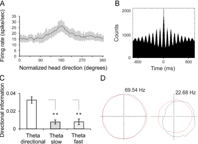
Another idiothetic signal that may regulate head-direction activity is self-motion or linear velocity. The head-direction tuning curve is affected by self-motion signals (Shinder and Taube, 2014;Zugaro et al., 2001), but see (Shinder and Taube, 2011), disrupting the directional path integration by the head direction cells (Stackman et al., 2003). Head-direction cells discharge at a slightly higher rate during exploration periods when the animal is moving faster than when it is motionless (Taube and Muller, 1998;Zugaro et al., 2001). Running speed (linear velocity) has a strong effect, with afiring rate increase of about 10% of the cell's peakfiring rate, compared to the angular velocity, which accounts for only about 1% of the firing rate variance (Taube, 1995; Taube and Muller, 1998; Zugaro et al., 2001). The effect of self-motion on the head-direction neurons might be explained by the link between self-motion and theta rhythm (the limbic local field oscilla-tion in the range of 5–12 Hz). Spatial paths represented by theta sequences in rats depend solely on the running speed (Geisler et al., 2007) and the length of the path is directly related to the length of the theta cycle (Gupta et al., 2012). The efficiency of signal processing is enhanced during theta oscillatory periods in the hippocampal formation (Fox et al., 1986) and in the neocortex (Delacour et al., 1990). In the Papez's circuit, linear velocity is integrated by the oscillatory and spiking patterns of the medial mammillary bodies (Sharp and Turner-Williams, 2005). Neuronal populations that oscil-late at theta frequency are also found in the anteroventral thalamic nucleus, a functional-anatomical interface between thalamic oscillatory activity, and limbic theta activity during vigilant behaviour (Tsanov et al., 2011b;Vertes et al., 2001). Our recent preliminary work found that theta periods express a higher peakfiring rate for the preferredfiring direction of heading, compared to the peakfiring rate during non-theta periods (Tsanov et al., 2011c). We examined the effect of theta rhythm in the anteroventral thalamic nucleus on the head-direction firing rate of the neurons in the adjacent antero-dorsal nucleus during theta and non-theta periods. We analysed theta power from anteroventral localfield potential (LFP) during pellet-chasing recordings, and defined the theta periods as epochs with theta power 450% of the maximal theta power. Head-direction spiking frequency was normal-ized as a function of the peakfiring rate, and compared theta versus non-theta periods. The averaged tuning curve for theta periods (Fig. 4A, black line) was significantly higher
for the preferred direction of heading, compared to the non-theta periods (Fig. 4A,red line). The average peakfiring rate during non-theta periods (0.8770.02) was about 13% lower than the average peakfiring rate for theta periods (Fig. 4A).
To test if the lower peakfiring rate during the non-theta periods was not simply a result of spiking adaptation to prolonged exposure in a single directional heading (which is more common during the non-theta periods (Taube and Muller, 1998)), we analysed only short-term spiking episodes (Fig. 4B), i.e. with a duration r1 s, preceded by Z1 s-long pauses, in order to compare the HD tuning curve between theta and non-theta episodes (Fig. 4C,D) for the preferred heading direction. The averaged tuning curve for theta epochs (Fig. 4B, black line) was significantly higher for the preferred direction of heading, compared to non-theta epochs (Fig. 4B, red line). Similar to the previous full time-scale analysis, the average peak firing rate of non-theta epochs (0.8770.03) was about 13% lower than the average peakfiring rate for theta epochs (Fig. 4B).
To demonstrate that the firing rate of the head directional signal is linked to the changes in thalamic theta power, we
correlated theta power with the normalized head-direction rate for the entire duration of the pellet-chasing recordings (Fig. 4E). We found a significant positive correlation between theta power and head-directionfiring rate (Fig. 4E). We next tested if thalamic theta rhythm relates to running speed (a property of the hippocampal theta frequency). The speed was low-pass filtered to eliminate episodes reflecting only the rat's head movement, and to include all episodes of running (Geisler et al., 2007). The analysis shows a highly-significant positive correlation between theta power and running speed (Fig. 4F). These correlates demonstrate that the head-direction signal integrates information about theta power and motion. One possible explanation for the effect of self-motion signal suggests that projections from the lateral habenula and interpeduncular nucleus to the dorsal tegmental nuclei might comprise a motor efference pathway (Sharp et al., 2006). A small proportion of neurons modulated by linear velocity are found in the habenula and the interpeduncular nucleus (Sharp et al., 2006). This hypothesis is supported by thefinding that damage to the interpeduncular nucleus disrupts anterodorsal head-direction sta-bility (Clark et al., 2009). A parallel explanation for the effect of theta on the tuning curve of the head-direction cells is that
Fig. 4–HDfiring rate is higher for theta compared to non-theta periods for the preferred direction of heading.A, Firing rate versus head-direction tuning plot of all HD cells for theta periods (-30 to 0 dB, black line) and non-theta periods (60 to30 dB, red line). ANOVA, F(1, 36)¼8.12,n¼23, **Po0.01. The difference in the peakfiring rate is significant in the range of thirty degrees - from 170 to 190 normalized degrees, Newman–Keuls test,po0.01.B, Firing rate versus head-direction tuning plot includingr1 s long spike trains, preceded byZ1 s long pauses for theta periods (black line) and non-theta periods (red line). ANOVA, F(1, 13)¼7.34,po0.01, n¼23, **Po0.01. The difference in the peakfiring rate is significant in the range of thirty degrees - from 170 to 190 normalized
degrees; Newman–Keuls test,po0.01.C, Sample colour-coded power spectrograms representing recording of the LFP in the
anteroventral thalamic nucleus for theta (left panels) and non-theta (right panels) signals.D, Upper panels: the LFP trace for theta (left,
in black) and non-theta (right, in red) epochs, corresponding to the power spectrograms inC. The spikes of the simultaneously
recorded HD unit are plotted below the LFP trace. Middle panels: parallel representation of the head directionality (degrees) for theta (left, in black) and non-theta (right, in red) epochs. Lower panels: the speed of the animal for the same time period (cm/s) for theta (left, in black) and non-theta (right, in red) epochs.E. The normalizedfiring rate (percent of the maximalfiring rate in Hz) correlates positively with the normalized theta power (amplitude of 8–12 Hz spectral power per recording session in dB). Pearson,n¼22, r¼0.2534, F(1,1089),po0.001.F. The running speed of the animal (cm/s) correlates positively with the normalized theta power (dB). Pearson,n¼22,r¼0.5059, F(1,1089),po0.001.
thalamic responsiveness is controlled by the activation of voltage-dependent intrinsic mechanisms. Such mechanisms may involve the neuromodulatory effect of the acetylcholine on thalamic responsiveness (Pare et al., 1987; Pare et al., 1990). The medial septum/diagonal band of Broca complex is the key structure involved in the integration of limbic theta oscillations and somatosensory signals (Kay, 2005; Tsanov et al., 2014b). Septal efferents diverge across several limbic structures, where they regulate the amplitude and frequency of localfield theta oscilla-tions as well as neuronal responsiveness. The septal cholinergic projections might affect the anterodorsal thalamus indirectly through descending inputs from postsubiculum or possibly through ascending inputs through the mammillary complex (Vann and Aggleton, 2004;Vertes et al., 2004).
6.
Integration of directional signal from
thalamic theta bursting cells
Together with the head-direction signal, theta rhythm is the other main signal that propagates within the thalamo-hippocampal circuitry. Theta rhythm appears to serve a critical role for spatial and non-spatial mnemonic functions of the limbic system (Burgess et al., 2002;Buzsáki, 2005). The Papez's pathways mediating theta rhythm share remarkably similar connectivity to those mediating head-direction sig-nals. Both pathways involve the tegmental nuclei, mammil-lary bodies, anterior thalamic nuclei and subicular cortices
(Gonzalo-Ruiz et al., 1997;Shibata, 1993;Swanson and Cowan, 1977; Van Groen and Wyss, 1995; van Groen et al., 1999; Witter et al., 1990). Thalamo-hippocampal circuitry involved in theta rhythm is represented by the anterovetral thalamic nucleus and subiculum together with medial entorhinal cortex. Unlike the head-direction signal, which is crucially dependent on afferent synaptic inputs, theta rhythm might be solely generated by intrinsic networks, as a result of the biophysical properties of the neurons. The intrinsic genera-tion of theta oscillagenera-tions is evoked by rhythmicfluctuations of the membrane potential, and the membrane properties of the involved neurons continuously maintain this rhythm through the excitatory and inhibitory synaptic interactions within the network (Buzsáki, 2002;Colgin and Moser, 2009). Local theta generators are observed in hippocampus (Goutagny et al., 2009), and similarly, neurons in the entorh-inal cortex have intrinsically oscillating membranes with natural frequencies in the theta range (Alonso and Llinas, 1989). However, there is no evidence that the neurons of the anteroventral thalamic nucleus are capable of supporting independent local generation of the theta rhythm. The lack of local GABAergic interneurons within the anterior thalamic nuclei (Wang et al., 1999) suggests that there is no developed local inhibitory network maintaining an independent thala-mic theta oscillation. This is supported by thefinding that inactivation of medial septum eliminates theta activity in the mammillary bodies (Kirk et al., 1996), which are the main afferent input to the anteroventral nucleus. The medial
Fig. 5–Directional modulation of anteroventral theta cells.A, Firing rate versus head-direction tuning plot for directional theta
cell. The x-axis represents the normalized head direction degrees that set the peak of the tuning curve for all units to 1800.
The y-axis represents absolutefiring frequency in spikes per sec (meann7sem).B, Sample 1000 ms autocorrelogram for
directional theta cell.C, Comparison of the directional information for the directional theta, slow-spiking theta and
fast-spiking theta cells (t-test,Po0.01).D, Polar plot examples reveal the head directionality of a directional theta unit (right) compared to fast-spiking theta cell (left). The polar plot for each cell represents the distribution of time heading in different directions across all time bins of the trial (red) and the distribution of head directions for time bins when a spike was recorded
from the cell (black). The coordinate system denotes maximalfiring frequency of the recorded unit with 0 Hz in the centre and
69.54/22.68 Hz in the periphery. The plots are from (Tsanov et al., 2011a).
[image:8.595.134.459.411.647.2]septum has pacemaker properties, and its activity is linked to the power and frequency generation of theta rhythm throughout the limbic system (Vinogradova, 1995). A plausi-ble hypothesis is that the diencephalic theta signal might synchronize the ascending mammillary and descending hip-pocampal theta signals (Tsanov et al., 2011d;Tsanov et al., 2011e). Concurrently, the thalamic theta signal may also integrate septal theta oscillations and idiothetic signals. Anteroventral thalamic neurons, which express theta-bursting and theta-modulated properties (Tsanov et al., 2011b; Vertes et al., 2001), have recently been shown to encode directional signals (Tsanov et al., 2011a). This study identified a population of anteroventral theta-bursting cells that undergo directional modulation of their spiking in the horizontal plane (Fig. 5A,B). The difference between maximal and averagefiring frequency for preferred head direction of these cells reached 40% and their directional information was significantly higher than the directional information (Fig. 5C, D). The crossover between theta and head directional signals is also found in the theta modulation of the anteroventral population of head-direction cells (Tsanov et al., 2011a). These cells exhibit rhythmic spiking in the theta range, and this mode of spiking was closely related to thefiring activity of local theta-bursting cells. While the anteroventral thala-mus does not receive direct inputs from the lateral mammil-lary nucleus (Shibata, 1992), the populations of directionally-tuned neurons in the anteroventral thalamus (Tsanov et al., 2011a;Yoganarasimha et al., 2006) might process descending directional information from the retrosplenial cortex (Wyss and Van Groen, 1992), postsubiculum (van Groen and Wyss, 1990) or from potential direct intra-thalamic projections. This line of research supports the hypothesized interaction of theta and head directional processing within the structures of the Papez's circuit (Kocsis and Vertes, 1994;Vertes et al., 2001). Oscillating networks are considered to provide tem-poral windows for single cells to suppress or facilitate their synaptic inputs in a coordinated manner (Buzsaki, 2010; Csicsvari et al., 1999). Thus, experience-dependent modifi ca-tion of this circuitry are possible through the integraca-tion of activity within critical time windows during appropriate phases of network oscillations (Ekstrom et al., 2001; Mehta et al., 2000).
7.
Conclusion
The anterior thalamus provides highly-processed informa-tion to the hippocampal formainforma-tion. The head-direcinforma-tion signal integrates processed idiothetic and external sensory informa-tion. The idiothetic signals integrate vestibular and self-motion signals, while sensory external stimuli include visual (landmark) information. Concurrently, theta rhythm affects the linear and angular velocity modulation of head-direction cell responsiveness. Reciprocally, the directional signal is integrated by a population of theta-bursting thalamic neu-rons, which propagate the theta signal across the Papez's circuit. The anatomical location of the anterior thalamus in the limbic system suggests that these nuclei act as a func-tional crossroads between ascending hypothalamic, descend-ing parahippocampal and septal neuromodulatory inputs.
Damage to the anterior thalamus results in episodic memory impairment and this may be function of lost thalamo-hippocampal information transfer. The complexity of thala-mic signals and their contribution to the encoding of experience-dependent memory traces in hippocampal for-mation needs further investigation, as signal processing in the hippocampal formation does not always follow a corti-cofugal route, but is also affected profoundly by thalamofugal signals.
Acknowledgements
This work was supported by Science Foundation Ireland, the Health Research Board and the Wellcome Trust Biomedical Research Partnership grants to Marian Tsanov and Shane O'Mara.
r e f e r e n c e s
Aggleton, J.P., Sahgal, A., 1993. The contribution of the anterior thalamic nuclei to anterograde amnesia. Neuropsychologia 31, 1001–1019.
Aggleton, J.P., Hunt, P.R., Nagle, S., Neave, N., 1996. The effects of selective lesions within the anterior thalamic nuclei on spatial memory in the rat. Behav. Brain Res. 81, 189–198.
Alonso, A., Llinas, R.R., 1989. Subthreshold Naþ-dependent theta-like rhythmicity in stellate cells of entorhinal cortex layer II. Nature 342, 175–177.
Bassett, J.P., Taube, J.S., 2001. Neural correlates for angular head velocity in the rat dorsal tegmental nucleus. J. Neurosci. 21, 5740–5751.
Bassett, J.P., Zugaro, M.B., Muir, G.M., Golob, E.J., Muller, R.U., Taube, J.S., 2005. Passive movements of the head do not abolish anticipatory firing properties of head direction cells. J. Neurophysiol. 93, 1304–1316.
Battaglia, F.P., Sutherland, G.R., McNaughton, B.L., 2004. Local sensory cues and place cell directionality: additional evidence of prospective coding in the hippocampus. J. Neurosci. 24, 4541–4550.
Blair, H.T., Sharp, P.E., 1995. Anticipatory head direction signals in anterior thalamus: evidence for a thalamocortical circuit that integrates angular head motion to compute head direction. J. Neurosci. 15, 6260–6270.
Blair, H.T., Lipscomb, B.W., Sharp, P.E., 1997. Anticipatory time intervals of head-direction cells in the anterior thalamus of the rat: implications for path integration in the head-direction circuit. J. Neurophysiol. 78, 145–159.
Blair, H.T., Cho, J., Sharp, P.E., 1998. Role of the lateral mammillary nucleus in the rat head direction circuit: a combined single unit recording and lesion study. Neuron 21, 1387–1397. Boccara, C.N., Sargolini, F., Thoresen, V.H., Solstad, T., Witter, M.P.,
Moser, E.I., Moser, M.B., 2010. Grid cells in pre- and parasubiculum. Nat. Neurosci. 13, 987–994.
Brenner, N., Bialek, W., de Ruyter van Steveninck, R., 2000. Adaptive rescaling maximizes information transmission. Neuron 26, 695–702.
Burgess, N., Maguire, E.A., O’Keefe, J., 2002. The human hippocampus and spatial and episodic memory. Neuron 35, 625–641.
Buzsaki, G., 2010. Neural syntax: cell assemblies, synapsembles, and readers. Neuron 68, 362–385.
Buzsa´ki, G., 2002. Theta oscillations in the hippocampus. Neuron 33, 325–340.
Buzsa´ki, G., 2005. Theta rhythm of navigation: link between path integration and landmark navigation, episodic and semantic memory. Hippocampus 15, 827–840.
Byatt, G., Dalrymple-Alford, J.C., 1996. Both anteromedial and anteroventral thalamic lesions impair radial-maze learning in rats. Behav. Neurosci. 110, 1335–1348.
Cacucci, F., Lever, C., Wills, T.J., Burgess, N., O’Keefe, J., 2004. Theta-modulated place-by-direction cells in the hippocampal formation in the rat. J. Neurosci. 24, 8265–8277.
Calton, J.L., Stackman, R.W., Goodridge, J.P., Archey, W.B., Dudchenko, P.A., Taube, J.S., 2003. Hippocampal place cell instability after lesions of the head direction cell network. J. Neurosci. 23, 9719–9731.
Cassel, J.C., Pereira de Vasconcelos, A., Loureiro, M., Cholvin, T., Dalrymple-Alford, J.C., Vertes, R.P., 2013. The reuniens and rhomboid nuclei: neuroanatomy, electrophysiological characteristics and behavioral implications. Prog. Neurobiol. 111, 34–52.
Cho, J., Sharp, P.E., 2001. Head direction, place, and movement correlates for cells in the rat retrosplenial cortex. Behav. Neurosci. 115, 3–25.
Clark, B.J., Sarma, A., Taube, J.S., 2009. Head direction cell instability in the anterior dorsal thalamus after lesions of the interpeduncular nucleus. J. Neurosci. 29, 493–507.
Clark, B.J., Bassett, J.P., Wang, S.S., Taube, J.S., 2010. Impaired head direction cell representation in the anterodorsal thalamus after lesions of the retrosplenial cortex. J. Neurosci. 30, 5289–5302. Colgin, L.L., Moser, E.I., 2009. Hippocampal theta rhythms follow
the beat of their own drum. Nat. Neurosci. 12, 1483–1484. Crill, W.E., 1996. Persistent sodium current in mammalian central
neurons. Annu. Rev. Physiol. 58, 349–362.
Csicsvari, J., Hirase, H., Czurko, A., Mamiya, A., Buzsaki, G., 1999. Oscillatory coupling of hippocampal pyramidal cells and interneurons in the behaving rat. J. Neurosci. 19, 274–287. Delacour, J., Houcine, O., Costa, J.C., 1990. Modifications of the
responses of barrel field neurons to vibrissal stimulation during theta in the awake and undrugged rat. Neuroscience 37, 237–243.
Ekstrom, A.D., Meltzer, J., McNaughton, B.L., Barnes, C.A., 2001. NMDA receptor antagonism blocks experience-dependent expansion of hippocampal “place fields”. Neuron 31, 631–638. Fox, S.E., Wolfson, S., Ranck Jr., J.B., 1986. Hippocampal theta
rhythm and the firing of neurons in walking and urethane anesthetized rats. Exp. Brain Res. 62, 495–508.
Fu, Y.X., Djupsund, K., Gao, H., Hayden, B., Shen, K., Dan, Y., 2002. Temporal specificity in the cortical plasticity of visual space representation. Science 296, 1999–2003.
Gabbiani, F., Metzner, W., Wessel, R., Koch, C., 1996. From stimulus encoding to feature extraction in weakly electric fish. Nature 384, 564–567.
Geisler, C., Robbe, D., Zugaro, M., Sirota, A., Buzsaki, G., 2007. Hippocampal place cell assemblies are speed-controlled oscillators. P. Natl. Acad. Sci. USA 104, 8149–8154.
Gold, J.J., Squire, L.R., 2006. The anatomy of amnesia: neurohistological analysis of three new cases. Learn Mem 13, 699–710.
Gonzalo-Ruiz, A., Morte, L., Lieberman, A.R., 1997. Evidence for collateral projections to the retrosplenial granular cortex and thalamic reticular nucleus from glutamate and/or aspartate-containing neurons of the anterior thalamic nuclei in the rat. Exp. Brain Res. 116, 63–72.
Goodridge, J.P., Taube, J.S., 1997. Interaction between the postsubiculum and anterior thalamus in the generation of head direction cell activity. J. Neurosci. 17, 9315–9330. Goodridge, J.P., Dudchenko, P.A., Worboys, K.A., Golob, E.J., Taube,
J.S., 1998. Cue control and head direction cells. Behav. Neurosci. 112, 749–761.
Gothard, K.M., Skaggs, W.E., McNaughton, B.L., 1996. Dynamics of mismatch correction in the hippocampal ensemble code for
space: interaction between path integration and environmental cues. J. Neurosci. 16, 8027–8040.
Goutagny, R., Jackson, J., Williams, S., 2009. Self-generated theta oscillations in the hippocampus. Nat. Neurosci. 12, 1491–1493. Guido, W., Lu, S.M., Sherman, S.M., 1992. Relative contributions of burst and tonic responses to the receptive field properties of lateral geniculate neurons in the cat. J. Neurophysiol. 68, 2199–2211.
Gupta, A.S., van der Meer, M.A., Touretzky, D.S., Redish, A.D., 2012. Segmentation of spatial experience by hippocampal theta sequences. Nat. Neurosci. 15, 1032–1039.
Harding, A., Halliday, G., Caine, D., Kril, J., 2000. Degeneration of anterior thalamic nuclei differentiates alcoholics with amnesia. Brain 123, 141–154 (Pt 1).
Jahnsen, H., Llinas, R., 1984a. Electrophysiological properties of guinea-pig thalamic neurones: an in vitro study. J. Physiol. 349, 205–226.
Jahnsen, H., Llinas, R., 1984b. Ionic basis for the electro-responsiveness and oscillatory properties of guinea-pig thalamic neurones in vitro. J. Physiol. 349, 227–247.
Jankowski, M.M., Ronnqvist, K.C., Tsanov, M., Vann, S.D., Wright, N.F., Erichsen, J.T., Aggleton, J.P., O’Mara, S.M., 2013. The anterior thalamus provides a subcortical circuit supporting memory and spatial navigation. Front Syst. Neurosci 7, 45. Jankowski, M.M., Islam, M.N., Wright, N.F., Vann, S.D., Erichsen,
J.T., Aggleton, J.P., O’Mara, S.M., 2014. Nucleus reuniens of the thalamus contains head direction cells. Elife, e03075. Kay, L.M., 2005. Theta oscillations and sensorimotor
performance. P. Natl. Acad. Sci. USA 102, 3863–3868. Kepecs, A., Wang, X.J., Lisman, J., 2002. Bursting neurons signal
input slope. J. Neurosci. 22, 9053–9062.
Kirk, I.J., Oddie, S.D., Konopacki, J., Bland, B.H., 1996. Evidence for differential control of posterior hypothalamic,
supramammillary, and medial mammillary theta-related cellular discharge by ascending and descending pathways. J. Neurosci. 16, 5547–5554.
Knierim, J.J., Kudrimoti, H.S., McNaughton, B.L., 1995. Place cells, head direction cells, and the learning of landmark stability. J. Neurosci. 15, 1648–1659.
Knierim, J.J., Kudrimoti, H.S., McNaughton, B.L., 1998. Interactions between idiothetic cues and external landmarks in the control of place cells and head direction cells. J. Neurophysiol. 80, 425–446.
Knierim, J.J., 2002. Dynamic interactions between local surface cues, distal landmarks, and intrinsic circuitry in hippocampal place cells. J. Neurosci. 22, 6254–6264.
Kocsis, B., Vertes, R.P., 1994. Characterization of neurons of the supramammillary nucleus and mammillary body that discharge rhythmically with the hippocampal theta rhythm in the rat. J. Neurosci. 14, 7040–7052.
Kudrimoti, H.S., Knierim, J.J., McNaughton, B.L., 1996. Dynamics of visual cue control over head direction cells. Ann. N.Y. Acad. Sci 781, 642–644.
Lee, I., Rao, G., Knierim, J.J., 2004. A double dissociation between hippocampal subfields: differential time course of CA3 and CA1 place cells for processing changed environments. Neuron 42, 803–815.
Lee, I., Knierim, J.J., 2007. The relationship between the field-shifting phenomenon and representational coherence of place cells in CA1 and CA3 in a cue-altered environment. Learn Mem. 14, 807–815.
Llinas, R.R., 1988. The intrinsic electrophysiological properties of mammalian neurons: insights into central nervous system function. Science 242, 1654–1664.
Markus, E.J., Qin, Y.L., Leonard, B., Skaggs, W.E., McNaughton, B.L., Barnes, C.A., 1995. Interactions between location and task affect the spatial and directional firing of hippocampal neurons. J. Neurosci. 15, 7079–7094.
McKenna, J.T., Vertes, R.P., 2004. Afferent projections to nucleus reuniens of the thalamus. J. Comp. Neurol. 480, 115–142. McNaughton, B.L., Barnes, C.A., O’Keefe, J., 1983. The contributions of
position, direction, and velocity to single unit activity in the hippocampus of freely-moving rats. Exp. Brain Res. 52, 41–49. McNaughton, B.L., Chen, L.L., Markus, E.J., 1991. “Dead
reckoning,” landmark learning, and the sense of direction: a neurophysiological and computational hypothesis. J. Cogn. Neurosci. 3, 190–201.
McNaughton, B.L., Barnes, C.A., Gerrard, J.L., Gothard, K., Jung, M.W., Knierim, J.J., Kudrimoti, H., Qin, Y.L., Skaggs, W.E., Suster, M.S., Weaver, K., 1996. Deciphering the hippocampal polyglot: the hippocampus as a path integration system. J. Exp. Biol. 199, 173–185.
McNaughton, B.L., Battaglia, F.P., Jensen, O., Moser, E.I., Moser, M. B., 2006. Path integration and the neural basis of the ’cognitive map. Nat. Rev. Neurosci. 7, 663–678.
Mehta, M.R., Barnes, C.A., McNaughton, B.L., 1997. Experience-dependent, asymmetric expansion of hippocampal place fields. P. Natl. Acad. Sci. USA 94, 8918–8921.
Mehta, M.R., Quirk, M.C., Wilson, M.A., 2000. Experience-dependent asymmetric shape of hippocampal receptive fields. Neuron 25, 707–715.
Metzner, W., Koch, C., Wessel, R., Gabbiani, F., 1998. Feature extraction by burst-like spike patterns in multiple sensory maps. J. Neurosci. 18, 2283–2300.
Mizumori, S.J., Williams, J.D., 1993. Directionally selective mnemonic properties of neurons in the lateral dorsal nucleus of the thalamus of rats. J. Neurosci. 13, 4015–4028.
Muir, G.M., Brown, J.E., Carey, J.P., Hirvonen, T.P., Della Santina, C. C., Minor, L.B., Taube, J.S., 2009. Disruption of the head direction cell signal after occlusion of the semicircular canals in the freely moving chinchilla. J. Neurosci. 29, 14521–14533. Muller, J.R., Metha, A.B., Krauskopf, J., Lennie, P., 1999. Rapid
adaptation in visual cortex to the structure of images. Science 285, 1405–1408.
Muller, R.U., Bostock, E., Taube, J.S., Kubie, J.L., 1994. On the directional firing properties of hippocampal place cells. J. Neurosci. 14, 7235–7251.
Muller, R.U., Stead, M., Pach, J., 1996. The hippocampus as a cognitive graph. J. Gen. Physiol. 107, 663–694.
O’Keefe, J., 1976. Place units in the hippocampus of the freely moving rat. Exp. Neurol. 51, 78–109.
O’Keefe, J., Burgess, N., 2005. Dual phase and rate coding in hippocampal place cells: theoretical significance and relationship to entorhinal grid cells. Hippocampus 15, 853–866.
Pare, D., Steriade, M., Deschenes, M., Oakson, G., 1987. Physiological characteristics of anterior thalamic nuclei, a group devoid of inputs from reticular thalamic nucleus. J. Neurophysiol. 57, 1669–1685.
Pare, D., Steriade, M., Deschenes, M., Bouhassira, D., 1990. Prolonged enhancement of anterior thalamic synaptic responsiveness by stimulation of a brain-stem cholinergic group. J. Neurosci. 10, 20–33.
Parker, A., Gaffan, D., 1997. The effect of anterior thalamic and cingulate cortex lesions on object-in-place memory in monkeys. Neuropsychologia 35, 1093–1102.
Pospischil, M., Toledo-Rodriguez, M., Monier, C., Piwkowska, Z., Bal, T., Fregnac, Y., Markram, H., Destexhe, A., 2008. Minimal Hodgkin-Huxley type models for different classes of cortical and thalamic neurons. Biol. Cybern. 99, 427–441.
Ramcharan, E.J., Cox, C.L., Zhan, X.J., Sherman, S.M., Gnadt, J.W., 2000. Cellular mechanisms underlying activity patterns in the monkey thalamus during visual behavior. J. Neurophysiol. 84, 1982–1987. Ramcharan, E.J., Gnadt, J.W., Sherman, S.M., 2005. Higher-order
thalamic relays burst more than first-order relays. P. Natl. Acad. Sci. USA 102, 12236–12241.
Roth, E.D., Yu, X., Rao, G., Knierim, J.J., 2012. Functional
differences in the backward shifts of CA1 and CA3 place fields in novel and familiar environments. PLoS One 7, e36035. Russell, N.A., Horii, A., Smith, P.F., Darlington, C.L., Bilkey, D.K.,
2003. Long-term effects of permanent vestibular lesions on hippocampal spatial firing. J. Neurosci. 23, 6490–6498. Sargolini, F., Fyhn, M., Hafting, T., McNaughton, B.L., Witter, M.P.,
Moser, M.B., Moser, E.I., 2006. Conjunctive representation of position, direction, and velocity in entorhinal cortex. Science 312, 758–762.
Schwindt, P.C., Spain, W.J., Foehring, R.C., Chubb, M.C., Crill, W.E., 1988a. Slow conductances in neurons from cat sensorimotor cortex in vitro and their role in slow excitability changes. J. Neurophysiol. 59, 450–467.
Schwindt, P.C., Spain, W.J., Foehring, R.C., Stafstrom, C.E., Chubb, M.C., Crill, W.E., 1988b. Multiple potassium conductances and their functions in neurons from cat sensorimotor cortex in vitro. J. Neurophysiol. 59, 424–449.
Sharp, P.E., Tinkelman, A., Cho, J., 2001. Angular velocity and head direction signals recorded from the dorsal tegmental nucleus of gudden in the rat: implications for path integration in the head direction cell circuit. Behav. Neurosci. 115, 571–588. Sharp, P.E., Turner-Williams, S., 2005. Movement-related
correlates of single-cell activity in the medial mammillary nucleus of the rat during a pellet-chasing task.
J. Neurophysiol. 94, 1920–1927.
Sharp, P.E., Turner-Williams, S., Tuttle, S., 2006. Movement-related correlates of single cell activity in the interpeduncular nucleus and habenula of the rat during a pellet-chasing task. Behav. Brain Res. 166, 55–70.
Sherman, S.M., 2001. Tonic and burst firing: dual modes of thalamocortical relay. Trends Neurosci. 24, 122–126. Shibata, H., 1992. Topographic organization of subcortical
projections to the anterior thalamic nuclei in the rat. J. Comp. Neurol. 323, 117–127.
Shibata, H., 1993. Efferent projections from the anterior thalamic nuclei to the cingulate cortex in the rat. J. Comp. Neurol. 330, 533–542.
Shinder, M.E., Taube, J.S., 2011. 2011. Active and passive movement are encoded equally by head direction cells in the anterodorsal thalamus. J. Neurophysiol. 106, 788–800. Shinder, M.E., Taube, J.S., 2014. Self-motion improves head
direction cell tuning. J. Neurophysiol. 111, 2479–2492. Softky, W.R., Koch, C., 1993. The highly irregular firing of cortical
cells is inconsistent with temporal integration of random EPSPs. J. Neurosci. 13, 334–350.
Stackman, R.W., Taube, J.S., 1997. Firing properties of head direction cells in the rat anterior thalamic nucleus: dependence on vestibular input. J. Neurosci. 17, 4349–4358. Stackman, R.W., Taube, J.S., 1998. Firing properties of rat lateral
mammillary single units: head direction, head pitch, and angular head velocity. J. Neurosci. 18, 9020–9037.
Stackman, R.W., Tullman, M.L., Taube, J.S., 2000. Maintenance of rat head direction cell firing during locomotion in the vertical plane. J. Neurophysiol. 83, 393–405.
Stackman, R.W., Clark, A.S., Taube, J.S., 2002. Hippocampal spatial representations require vestibular input. Hippocampus 12, 291–303.
Stackman, R.W., Golob, E.J., Bassett, J.P., Taube, J.S., 2003. Passive transport disrupts directional path integration by rat head direction cells. J. Neurophysiol. 90, 2862–2874.
Steriade, M., 2001. Impact of network activities on neuronal properties in corticothalamic systems. J. Neurophysiol. 86, 1–39.
Swanson, L.W., Cowan, W.M., 1977. An autoradiographic study of the organization of the efferent connections of the
hippocampal formation in the rat. J. Comp. Neurol. 172, 49–84.
Taube, J.S., Muller, R.U., Ranck Jr., J.B., 1990. Head-direction cells recorded from the postsubiculum in freely moving rats. I. Description and quantitative analysis. J Neurosci. 10, 420–435.
Taube, J.S., 1995. Head direction cells recorded in the anterior thalamic nuclei of freely moving rats. J. Neurosci. 15, 70–86. Taube, J.S., Muller, R.U., 1998. Comparisons of head direction cell
activity in the postsubiculum and anterior thalamus of freely moving rats. Hippocampus 8, 87–108.
Taube, J.S., 2007. The head direction signal: origins and sensory-motor integration. Annu. Rev. Neurosci. 30, 181–207.
Taube, J.S., 2010. Interspike interval analyses reveal irregular firing patterns at short, but not long, intervals in rat head direction cells. J. Neurophysiol. 104, 1635–1648.
Taube, J.S., 2011. Head direction cell firing properties and behavioural performance in 3-D space. J. Physiol. 589, 835–841. Taube, J.S., Shinder, M., 2013. On the nature of three-dimensional
encoding in the cognitive map: commentary on Hayman, Verriotis, Jovalekic, Fenton, and Jeffery. Hippocampus 23, 14–21. Tsanov, M., Chah, E., Vann, S.D., Reilly, R.B., Erichsen, J.T.,
Aggleton, J.P., O’Mara, S.M., 2011a. Theta-modulated head direction cells in the rat anterior thalamus. J. Neurosci. 31, 9489–9502.
Tsanov, M., Chah, E., Wright, N., Vann, S.D., Reilly, R., Erichsen, J.T., Aggleton, J.P., O’Mara, S.M., 2011b. Oscillatory entrainment of thalamic neurons by theta rhythm in freely moving rats. J. Neurophysiol. 105, 4–17.
Tsanov, M., Chah, E., Wright, N., Vann, S.D., Reilly, R., Erichsen, J.T., Aggleton, J.P., O’Mara, S.M., 2011c. Theta modulation of the head-direction cells in the rat anterior thalamus. Society for Neuroscience Conference, Washington DC. Abstract 938.22.
Tsanov, M., Vann, S.D., Erichsen, J.T., Wright, N., Aggleton, J.P., O’Mara, S.M., 2011d. Differential regulation of synaptic plasticity of the hippocampal and the hypothalamic inputs to the anterior thalamus. Hippocampus 21, 1–8.
Tsanov, M., Wright, N., Vann, S.D., Erichsen, J.T., Aggleton, J.P., O’Mara, S.M., 2011e. Hippocampal inputs mediate theta-related plasticity in anterior thalamus. Neuroscience 187, 52–62.
Tsanov, M., Chah, E., Noor, M.S., Egan, C., Reilly, R.B., Aggleton, J.P., Erichsen, J.T., Vann, S.D., O’Mara, S.M., 2014a. The irregular firing properties of thalamic head direction cells mediate turn-specific modulation of the directional tuning curve. J. Neurophysiol. 112, 2316–2331.
Tsanov, M., Chah, E., Reilly, R., O’Mara, S.M., 2014b. Respiratory cycle entrainment of septal neurons mediates the fast coupling of sniffing rate and hippocampal theta rhythm. Eur. J. Neurosci. 39, 957–974.
van der Meer, M.A., Knierim, J.J., Yoganarasimha, D., Wood, E.R., van Rossum, M.C., 2007. Anticipation in the rodent head direction system can be explained by an interaction of head movements and vestibular firing properties. J. Neurophysiol. 98, 1883–1897.
van Groen, T., Wyss, J.M., 1990. The connections of presubiculum and parasubiculum in the rat. Brain Res. 518, 227–243. Van Groen, T., Wyss, J.M., 1995. Projections from the anterodorsal
and anteroventral nucleus of the thalamus to the limbic cortex in the rat. J. Comp. Neurol. 358, 584–604.
van Groen, T., Kadish, I., Wyss, J.M., 1999. Efferent connections of the anteromedial nucleus of the thalamus of the rat. Brain Res. Brain Res. Rev. 30, 1–26.
van Groen, T., Kadish, I., Michael Wyss, J., 2002. Role of the anterodorsal and anteroventral nuclei of the thalamus in spatial memory in the rat. Behav. Brain Res. 132, 19–28.
Vann, S.D., Aggleton, J.P., 2004. The mammillary bodies: two memory systems in one?. Nat. Rev. Neurosci 5, 35–44. Vertes, R.P., Albo, Z., Viana Di Prisco, G., 2001. Theta-rhythmically
firing neurons in the anterior thalamus: implications for mnemonic functions of Papez’s circuit. Neuroscience 104, 619–625.
Vertes, R.P., Hoover, W.B., Viana Di Prisco, G., 2004. Theta rhythm of the hippocampus: subcortical control and functional significance. Behav. Cogn. Neurosci. Rev. 3, 173–200. Vertes, R.P., Hoover, W.B., Szigeti-Buck, K., Leranth, C., 2007.
Nucleus reuniens of the midline thalamus: link between the medial prefrontal cortex and the hippocampus. Brain Res. Bull. 71, 601–609.
Vinogradova, O.S., 1995. Expression, control, and probable functional significance of the neuronal theta-rhythm. Prog. Neurobiol. 45, 523–583.
Wang, B., Gonzalo-Ruiz, A., Sanz, J.M., Campbell, G., Lieberman, A.R., 1999. Immunoelectron microscopic study of gamma-aminobutyric acid inputs to identified thalamocortical projection neurons in the anterior thalamus of the rat. Exp. Brain Res. 126, 369–382.
Warburton, E.C., Baird, A., Morgan, A., Muir, J.L., Aggleton, J.P., 2001. The conjoint importance of the hippocampus and anterior thalamic nuclei for allocentric spatial learning: evidence from a disconnection study in the rat. J. Neurosci. 21, 7323–7330.
Wilton, L.A., Baird, A.L., Muir, J.L., Honey, R.C., Aggleton, J.P., 2001. Loss of the thalamic nuclei for “head direction“ impairs performance on spatial memory tasks in rats. Behav. Neurosci. 115, 861–869.
Witter, M.P., Ostendorf, R.H., Groenewegen, H.J., 1990. Heterogeneity in the dorsal subiculum of the rat. Distinct neuronal zones project to different cortical and subcortical targets. Eur. J. Neurosci. 2, 718–725.
Wyss, J.M., Van Groen, T., 1992. Connections between the retrosplenial cortex and the hippocampal formation in the rat: a review. Hippocampus 2, 1–11.
Yao, H., Dan, Y., 2001. Stimulus timing-dependent plasticity in cortical processing of orientation. Neuron 32, 315–323. Yoder, R.M., Taube, J.S., 2009. Head direction cell activity in mice:
robust directional signal depends on intact otolith organs. J. Neurosci. 29, 1061–1076.
Yoder, R.M., Clark, B.J., Brown, J.E., Lamia, M.V., Valerio, S., Shinder, M.E., Taube, J.S., 2011a. Both visual and idiothetic cues contribute to head direction cell stability during navigation along complex routes. J. Neurophysiol. 105, 2989–3001.
Yoder, R.M., Clark, B.J., Taube, J.S., 2011b. Origins of landmark encoding in the brain. Trends Neurosci. 34, 561–571. Yoganarasimha, D., Knierim, J.J., 2005. Coupling between place
cells and head direction cells during relative translations and rotations of distal landmarks. Exp. Brain Res. 160, 344–359. Yoganarasimha, D., Yu, X., Knierim, J.J., 2006. Head direction cell
representations maintain internal coherence during conflicting proximal and distal cue rotations: comparison with hippocampal place cells. J. Neurosci. 26, 622–631. Yu, X., Yoganarasimha, D., Knierim, J.J., 2006. Backward shift of
head direction tuning curves of the anterior thalamus: comparison with CA1 place fields. Neuron 52, 717–729. Zugaro, M.B., Tabuchi, E., Fouquier, C., Berthoz, A., Wiener, S.I.,
2001. Active locomotion increases peak firing rates of anterodorsal thalamic head direction cells. J. Neurophysiol. 86, 692–702.
Zugaro, M.B., Berthoz, A., Wiener, S.I., 2002. Peak firing rates of rat anterodorsal thalamic head direction cells are higher during faster passive rotations. Hippocampus 12, 481–486.