warwick.ac.uk/lib-publications
Original citation:
Baier, Waltraud, Norman, Daniel, Warnett, Jason, Payne, Mark, Harrison, Nigel P., Hunt, Nicholas, Burnett, Brian and Williams, M. A. (Mark A.). (2017) Novel application of three-dimensional technologies in a case of dismemberment. Forensic Science International, 270 . pp. 139-145.
Permanent WRAP URL:
http://wrap.warwick.ac.uk/84416
Copyright and reuse:
The Warwick Research Archive Portal (WRAP) makes this work by researchers of the University of Warwick available open access under the following conditions. Copyright © and all moral rights to the version of the paper presented here belong to the individual author(s) and/or other copyright owners. To the extent reasonable and practicable the material made available in WRAP has been checked for eligibility before being made available.
Copies of full items can be used for personal research or study, educational, or not-for-profit purposes without prior permission or charge. Provided that the authors, title and full bibliographic details are credited, a hyperlink and/or URL is given for the original metadata page and the content is not changed in any way.
Publisher’s statement:
© 2017, Elsevier. Licensed under the Creative Commons Attribution-NonCommercial-NoDerivatives 4.0 International http://creativecommons.org/licenses/by-nc-nd/4.0/
A note on versions:
The version presented here may differ from the published version or, version of record, if you wish to cite this item you are advised to consult the publisher’s version. Please see the ‘permanent WRAP url’ above for details on accessing the published version and note that access may require a subscription.
1
Novel application of three-dimensional technologies in a case of
dismemberment
Abstract
This case study reports the novel application of three-dimensional technologies such as micro-CT and 3D printing to the forensic investigation of a complex case of dismemberment. Micro-CT was successfully employed to virtually align severed skeletal elements found in different locations, analyse tool marks created during the dismemberment process, and virtually dissect a charred piece of evidence. High resolution 3D prints of the burnt human bone contained within were created for physical visualisation to assist the investigation team. Micro-CT as a forensic radiological method provided vital information and the basis for visualisation both during the investigation and in the subsequent trial making it one of the first examples of such technology in a UK court.
1. Introduction
Although rare in the UK, cases of complete dismemberment pose a significant challenge to the police [1]. In most cases, the individual body parts are deposited separately making them difficult to locate and thereby obscuring the victim’s identity. Fortunately, from an evidentiary perspective, such cases offer a great potential, as the likelihood of finding traces at the dismemberment or deposition site is high due to the inherently visceral nature of dismemberment [1]. On the body itself, further evidence such as characteristic tool marks can be identified during an anthropological examination. With this potential wealth of evidence, the overall success of the investigation can often depend on the police’s ability to exploit all available resources during their inquiry.
Traditionally, dismembered bodies are analysed by forensic anthropologists who typically employ destructive methods in order to examine the evidence [2]. However, with modern advances in forensic radiology, evidence destruction can now often be avoided by employing innovative imaging techniques such as high resolution X-ray computed tomography (CT) [3,4] which allows non-invasive examination [5]. More commonly applied, ‘medical’ grade CT (resolution >300µm) is useful for detecting gross skeletal injury but can obscure important anatomical detail below the scanning resolution [6]. Micro-CT however, provides much higher resolutions (0.5 - 120µm) which allows the depiction of minute features such as micro-fractures which is beneficial for forensic examination [7].
2
2. Case background
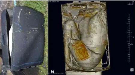
When canal workers removed a suitcase from a canal in the West Midlands, its weight raised suspicions about its contents and the police were called to the site. The suitcase was taken to the University Hospital Coventry and Warwickshire (UHCW) where prior to opening, it was CT scanned using ‘medical’ grade CT (GE Medical Systems). The scan revealed tightly packed human body parts whose initial undisturbed state of discovery was digitally preserved by the CT data. Upon opening, the initial CT scan was verified as the decomposing remains of an adult male (Figure 1b). These remains were physically examined confirming that the head, arms, and the left lower leg of the individual were absent. The cause of death could not be determined due to the advanced state of decomposition. Given the complex findings from the suitcase multiple parallel lines of investigation were taken with four main objectives: to find the remaining body parts; identify the victim; identify the original deposition site; and identify the perpetrator(s).
The first line of enquiry saw a systematic search of the canal upstream of the initial discovery point, working under the hypothesis that the remaining elements were disposed of in the same location. This point of origin provided a starting point from where to search for the perpetrator(s). Working with a flowing body of water made the interpretation of the suitcase’s movements in the canal difficult but the search strategy proved successful nevertheless and a second suitcase containing further body elements was recovered (Figure 2) along with several tools including a saw, a kitchen knife, a hammer, and a chisel (Figure 3). A CT scan of the second suitcase (Figure 2b) revealed the missing leg, arms, and head of the victim but the left shoulder joint remained unaccounted for. Positive identification was achieved by analysing the victim’s DNA, fingerprints, and characteristic tattoos. DNA samples were taken from both suitcases to confirm that they pertain to the same individual.
Figure 1: a) Crime scene photograph of first suitcase. b) CT scan of the same suitcase containing the torso, right leg, and partial left leg.
[image:3.595.75.526.284.535.2]3
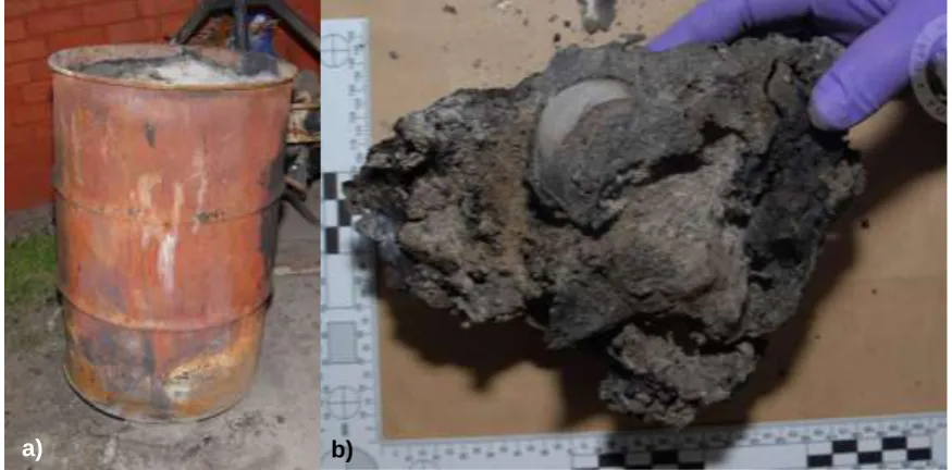
[image:4.595.80.546.95.361.2]With the victim’s identity known, police inquiries focussed on the victims’ social relationships, witness statements and other evidence which then pointed towards the victim’s former housemate and their partner as the prime suspects. A warranted search of their property was conducted during which miniscule blood stains of the victim were detected linking him to that property. However, as he had lived at the address, the evidentiary value thereof was limited. In the back yard an old oil drum (Figure 4) was recovered which contained the remnants of a Figure 3: Scene photographs of the tools recovered from the canal by divers. a) A knife, hammer, and chisel. b) A saw. An electric carving knife was disposed of by the offender shortly after the murder.
a)
b)
Figure 2: a) Crime scene photograph of suitcase 2, once dragged on land. b) CT scan of same suitcase, containing the head, arms, and left lower leg. It had been weighed down by concrete paving slabs, visible in the centre of the
suitcase. a)
[image:4.595.65.550.405.633.2]4
bonfire. Fire brigade records indicated that the bonfire took place around the time of the suspected murder which was determined from CCTV and telecommunications evidence. The entire oil drum was taken to the lab where careful archaeological excavation of the contents produced large amounts of charred debris, documents, and clothes, as well as a fused lump appearing to contain bone (Figure 4). The bone was suspected to be the missing left shoulder joint and micro-CT examination was undertaken to investigate the sample non-invasively prior to the physical excavation. This step proved crucial as the bone partially disintegrated upon dissection.
3. Materials and methods
A few centimetres of bone either side of the dismemberment locations were freed from soft tissue and retained by the pathologist during post-mortem. The individual samples were submitted for high-resolution micro-CT imaging at WMG, University of Warwick. The post-processing of the scans included the production of animations, a court presentation, and 3D prints of some elements.
Eleven bone samples relating to the dismemberment areas, including the charred lump, underwent micro-CT imaging. The samples found in suitcase one were the left proximal femur, left acromion, left lateral scapula, sixth cervical vertebra, right proximal humerus and right medial clavicle. Samples from suitcase two were the; left distal femur, left humerus and right humerus. The charred sample from the oil drum was also scanned.
All samples were scanned separately using a Nikon XT H 225/320LC micro-CT scanner (Nikon Metrology, UK) with parameters being adjusted appropriately for each sample to ensure optimal image contrast. This resulted in scan parameters of power between 24-36W, voltage at 200kV and exposure between 1.4-2s yielding voxel resolutions between 36-67µm. The visualisation software VGStudio MAX 2.2 was used with four objectives: examine the internal structure of the charred lump through virtually dissection; match fragments of dismemberment areas where possible; examine any tool marks found on the bone; and generate a 3D printable model of the virtually dissected bone.
Virtual dissection: The contents of the charred lump recovered from the oil drum were virtually segmented by material density and visualised. This information was then used to identify a
Figure 4: a) Oil drum recovered from the suspects’ backyard, containing remains of a substantial bonfire. b) The charred lump which appeared to contain human tissue and burnt plastic sheets.
[image:5.595.80.518.191.407.2]5
suitable site for DNA extraction as well as assist the forensic anthropologist in developing a suitable strategy for physical excavation of the bone from the charred lump.
Virtual part-fitting: Given re-approximation of dismembered remains is strongly advised for forensic anthropological analysis [13], 3D scans of the paired fragments of each side of the dismembered location were virtually aligned in VGStudio. Following visual alignment, a proprietary automated best fit algorithm allowed further alignment at the voxel level.
Tool mark analysis - The voxel resolution of approximately 50µm enabled an accurate visualisation of the tool marks created during dismemberment. The measurement tools in VGStudio were used to obtain precise dimensions of features, such as their length, depth, and width. The validity of such dimensional measurements on micro-CT scans has been confirmed in a study by Kumar et al [14].
3D printing: Also known as Additive Layer Manufacturing, 3D printing is the process of creating three-dimensional objects by depositing and fusing layers of liquid polymers [13]. It is a commonly employed in industrial rapid prototyping but it is also gaining increasing use in biomedical research [14] and forensic research [17, 18, 19] as the technology advances [20]. The system used for the present case was an Objet 260 Connex printer printing at a resolution of approximately 20µm.
4. Results
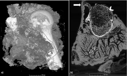
Virtual dissection - The virtual dissection of the charred lump revealed the missing proximal end of the left humerus and fragments of the scapula surrounded by burnt soft tissue, along with layers of burnt plastic (Figure 5). As the sample was extremely fragile, non-destructive imaging was crucial to plan further steps such as DNA extraction required for positive identification. The sample was recovered directly from the location likely to have suffered the least heat damage which was an area adjacent to the bone where intact blood vessels were seen in the CT image. Knowing the internal structure of the lump also assisted the forensic archaeologist in their physical segmentation of the different materials within which caused
[image:6.595.99.515.474.724.2]6
some damage to the delicate sample, thus emphasising the benefit of the initial non-destructive examination.
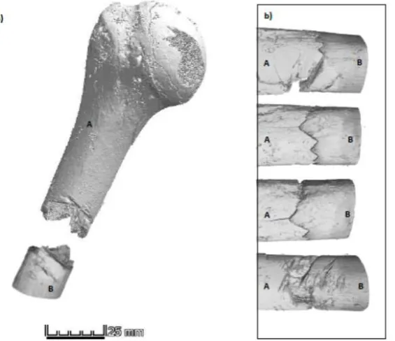
Virtual part-fitting - The charred proximal humerus could be virtually extracted from the mass and aligned with the other fragment of the left humerus found in suitcase 2, which provided a near perfect geometrical match (Figure 6). This match confirmed that all fragments pertained to the same individual which was a vital insurance in case the DNA test had not produced any results due to heat damage. The fact that parts of the body were discovered in the suspects’ property was another vital link between them and the crime. Further positive matches were achieved between the fragments of the left distal femur and those of the right proximal humerus.
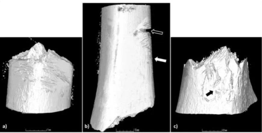
Tool mark analysis - Micro-CT offers several advantages over standard light microscopy in the examination of tool marks in bone since it is non-invasive and allows advanced visualisation of small features [7]. The current findings support this position as the analysis of the cut marks revealed a variety of characteristics with fine detail which could be visualised accurately on the three-dimensional CT image. The cut morphology and the presence of numerous false starts shown in Figure 7a, in particular on the left femur and humerus, was initially thought to indicate the use of a power tool [21]. This corresponded to the later information that the suspect had owned an electric carving knife which he disposed of after the murder. Other marks could be explained by the tools discovered by the divers in the vicinity of suitcase 2. There were superficial V-shaped, narrow grooves possibly created by the knife [22] as well as approximately U-shaped wider ones, possibly indicating a saw [23] (Figure 7b). The extensive shattering around the dismemberment locations suggests that the hammer found amongst the tools was also employed in an attempt to separate the limbs [24]. Many of the dismemberment sites fractured instead of cut (Figure 7c). The examination of the marks on the bone paint a vivid picture of the assailant trying to dismember the body but running into unexpected difficulties, resulting in the trial of numerous different tools, which is not unusual in such cases [13].
[image:7.595.162.445.229.473.2]7
[image:8.595.89.525.113.335.2]3D printing - The main piece of evidence was the charred bone found in the oil drum which was emphasised by creating actual-scale 3D prints of the burnt bone and the matching fragment from the suitcase (Figure 9). These two parts were used during case reviews with the police and the Crown Prosecution Service to demonstrate how well the bones fit and to provide an impression of the actual bone dimensions and the force that would be required to separate it, evident in the numerous cut marks also visible on the print. The benefit of 3D prints in the interpretation of trauma as experienced during these case review meetings has also been demonstrated by Kettner et al [25] but there is hardly any record of the use of 3D prints in UK courtrooms which this case study hopes to change for the future.
Figure 7 a-c: A variety of different tool marks was observed on samples. a) depicts numerous false starts on the humerus segment found in suitcase 2; b) displays the left distal femur with a deep U-shaped kerf probably caused by a saw (open arrow), and shallower V-shaped grooves (solid arrow) probably caused by the knife retrieved from the canal; c) the arrow indicates the area of extensive shattering which suggests the involvement of the hammer in the dismemberment process.
[image:8.595.95.510.392.537.2]8
5. Discussion/Impact
The use of CT imagery in forensic contexts has two main areas of application, which are the ongoing police investigation and the evidence presentation in court. Both applications were successfully employed in the current case. The scan results, especially that of the charred element, significantly influenced subsequent steps of the police inquiry, providing supporting information to enable linking the perpetrators to the crime. While the burnt bone specimen was ultimately linked by DNA analysis to the skeletal remains found in the suitcases, this case provided an opportunity to validate the use of CT in such a scenario for cases where DNA is not available. Furthermore, the extraction of the DNA sample was based on the CT data, thus increasing the probability of its success. While CT is not the only method to study tool marks, it is a useful instrument for non-invasive analysis, minimising evidence handling and potential contamination and allowing detailed examination even in cases where a physical examination is prohibited by practical [12] or ethical [26] constraints.
The models produced using micro-CT and 3D printing assisted the examination of the forensic samples themselves, but they also served a more psychological purpose. Being confronted with such detailed visual depictions of their crime, the suspect, who had kept silent during previous interviews, succumbed to the overwhelming evidence and confessed to killing and dismembering the victim.
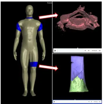
[image:9.595.110.499.130.324.2]Once the investigation period was closed, there were still further advantages of CT data over traditional methods as the three-dimensional images proved a powerful tool for presenting evidence to the jury during trial. Presenting medical or pathological information can be, even if accompanied by photographs, difficult to grasp for non-medically trained laymen. Being able to support this kind of evidence with interactive 3D images offers jurors assistance and allows them to make a more informed decision [27]. However, presentation of this realistic 3D evidence can be potentially upsetting for a jury or family present during trial and therefore appropriate measure should be taken to sanitise the images [28]. In the current case, a standardised human dummy model was used to show the dismemberment locations as
9
simple coloured regions (Figure 10). These regions could then be presented in more detail by opening a hyperlink to an animation demonstrating how the skeletal elements fitted
together following dismemberment. To reduce the emotive impact, both fragments were coloured different (blue and green) thereby adding clarity and further abstraction. Given that the evidence was presented by the pathologist, it was important to make the presentation interactive to allow them to incorporate it in their statement in a logical manner.
6. Conclusion
[image:10.595.103.460.107.469.2]This case study demonstrates the novel application of micro-CT as an invaluable tool for police investigations of major crimes. The case outlined in this report has shown that an integrated forensic strategy and application of new technology in evidence collection and presentation lends itself to a successful investigation. To date, there are only few cases which employ such an approach but their success in establishing the truth will hopefully ease the way for future cases. The main perpetrator in the case was found guilty and sentenced to 19 years imprisonment for murder, their accomplice to 2 ½ years for assisting an offender. The positive impact of WMG’s work was commended by the judge in their closing comments.
10
References
[1] G.N. Rutty and S.V. Hainsworth, The dismembered body, in: G.N. Rutty (Ed.), Essentials of Autopsy Practice. Advances, Updates, and Emerging Technologies. Springer, London, 2014, pp. 59-87.
[2] S. Black and E. Ferguson (eds.) Forensic anthropology: 2000 to 2010. (2011) London: CRC Press.
[3] J.P. Kruth, M. Bartscher, S. Carmignato, R. Schmitt, L. De Chiffre and A. Weckenmann, Computed tomography for dimensional metrology, CIRP Annals- Manufacturing Technology. 60:2 (2011) 821-842.
[4] L.W. Goldman, Principles of CT and CT technology, Journal of Nuclear Medicine Technology. 35:3 (2007) 115-128.
[5] M.J. Thali, U. Taubenreuther, M. Karolczak, M. Braun, W. Brueschweiler, W.A. Kalender, R. Dirnhofer, Forensic microradiology: micro-computed tomography (Micro-CT) and analysis of patterned injuries inside of bone. Journal of Forensic Sciences. 48:6 (2003) 1336-42. [6] D. Norman, A. Getgood, J.A. Thornby, J. Bird, G.A. Turley, T. Spalding, M.A. Williams, Quantitative topographic anatomy of the femoral ACL footprint : a micro-CT analysis. Medical & Biological Engineering & Computing, Volume 52:11 (2014), 985-995.
[7] G.N. Rutty, A. Brough, M.J.P. Biggs, C. Robinson, S.D.A. Lawes and S.V. Hainsworth, The role of micro-computed tomography in forensic investigations, Forensic Science International. 225 (2013) 60-66.
[8] C. Giraudo, P. Fais, G. Pelletti, A. Viero, D. Miotto, R. Boscolo-Berto, G. Viel, M. Montisci, G. Cecchetto, S.D. Ferrara, Micro-CT features of intermediate gunshot wounds covered by textiles. International Journal of Legal Medicine. 130:5 (2016) 1257-64.
[9] D.J. Pounder, and L.J. Sim, Virtual casting of stab wounds in cartilage using micro-computed tomography. The American journal of forensic medicine and pathology. 32:2 (2011) 97-99.
[10] S.M. Bello, S.A. Parfitt, and C. Stringer, Quantitative micromorphological analyses of cut marks produced by ancient and modern handaxes. Journal of Archaeological Science. 36:9 (2009) 1869-80.
[11] P. Fais, C. Giraudo, A. Viero, D. Miotto, F. Bortolotti, F. Tagliaro, M. Montisci, G. Cecchetto, Micro computed tomography features of laryngeal fractures in a case of fatal manual strangulation. Legal Medicine. 18 (2016) 85-89.
[12] M. Kettner, S. Potente, B. Schulz, P. Knauff, P.H. Schmidt, F. Ramsthaler, Analysis of laryngeal fractures in decomposed bodies using microfocus computed tomography (mfCT). Forensic Science, Medicine, and Pathology. 10:4 (2014) 607-12
11
[14] J. Kumar, A. Attridge, P.K.C. Wood and M.A. Williams, Analysis of the effect of cone-beam geometry and test object configuration on the measurement accuracy of a computed tomography scanner used for dimensional measurement, Measurement Science and Technology. 22 (2011) 1-15.
[15] S.H. Huang, P. Liu, A. Mokasdar and L. Hou, Additive manufacturing and its societal impact: a literature review, The International Journal of Advanced Manufacturing
Technology. 67:5 (2013) 1191-1203.
[16] S. Bose, S. Vahabzadeh and A. Bandyopadhyay, Bone tissue engineering using 3D printing, Materials Today. 16:12 (2013) 496-504.
[17] M. Kettner, P. Schmidt, S. Potente, F. Ramsthaler, and M. Schrodt, Reverse engineering—rapid prototyping of the skull in forensic trauma analysis. Journal of forensic sciences. 56:4 (2011) 1015-1017.
[18] K. Woźniak, E. Rzepecka-Woźniak, A. Moskała, J. Pohl, K. Latacz, and B. Dybała, Weapon identification using antemortem computed tomography with virtual 3D and rapid prototype modeling—A report in a case of blunt force head injury. Forensic Science International. 222:1-3 (2012) e29-e32.
[19] L.C. Ebert, M.J. Thali, and S. Ross, Getting in touch—3D printing in Forensic Imaging. Forensic Science International. 211:1 (2011) e1-e6.
[20] M. Vaezi, S. Chianrabutra, B. Mellor and S. Yang, Multiple material additive manufacturing. Part 1: a review, Virtual and Physical Prototyping. 8:1 (2013) 19-50. [21] S.A. Symes, E.N. Chapman, C.W. Rainwater, L.L. Cabo and S.M.T. Myster, Knife and toolmark analysis in bone: A manual designed for the examination of criminal mutilation and dismemberment. US Department of Justice. December 2010. Document number 232864. pp. 1-47. Available online at
https://www.ncjrs.gov/App/Publications/abstract.aspx?ID=254311 [Accessed 07/09/2015].
[22] R.J. Kooi and S.I. Fairgrieve, SEM and Stereomicroscopic analysis of cut marks in fresh and burned bone, Journal of Forensic Sciences. 58:2 (2013) 452-458.
[23] S.A. Symes, Morphology of saw marks in human bone: Identification of class characteristics. PhD thesis, University of Tennessee. 1992. Available online at http://trace.tennessee.edu/cgi/viewcontent.cgi?article=2578&context=utk_graddiss [Accessed 10/04/2016].
[24] N.C. Lovell, Analysis and interpretation of skeletal trauma, in: M.A. Katzenberg and S.R. Saunders (Eds.) Biological Anthropology of the Human Skeleton, 2nd Edition, Hoboken,
John Wiley & Sons, 2008, pp 341-386.
[25] M. Kettner, P. Schmidt, S. Potente, F. Ramsthaler and M. Schrodt, Reverse
engineering- rapid prototyping of the skull in forensic trauma analysis, Journal of Forensic Sciences. 56:4 (2011) 1015-1017.
[26] S. Grabherr, B.A. Stephan, U. Buck, S. Näther, A. Christe, L. Oesterhelweg, S. Ross, R. Dirnhofer, and M.J. Thali, Virtopsy–radiology in forensic medicine. Imaging Decisions. 11:1 (2007) 2-9.
12