STRUCTURAL AND FUNCTIONAL STUDY OF THE NITRATE TRANSPORTER, NRTA
Ingrid Rodrigues Nunes da Silva
A Thesis Submitted for the Degree of MPhil at the
University of St Andrews
2013
Full metadata for this item is available in Research@StAndrews:FullText
at:
http://research-repository.st-andrews.ac.uk/
Please use this identifier to cite or link to this item:
http://hdl.handle.net/10023/3740
Structural and Functional Study of the
Nitrate Transporter, NrtA
Ingrid Rodrigues Nunes da Silva
This thesis is submitted in partial fulfilment for the degree of MPhil
at the
University of St Andrews
Declarations
1. Candidate’s declarations:
I, Ingrid Rodrigues Nunes da Silva, hereby certify that this thesis, which is approximately 42, 000 words in length, has been written by me, that it is the record of work carried out by me and that it has not been submitted in any previous application for a higher degree. I was admitted as a research student in September, 2004 and as a candidate for the degree of PhD in the School of Biology in May 2010; the higher study for which this is a record was carried out in the University of St Andrews between 2004 and 2010.
Date: 29th May 2013 signature of candidate:
2. Supervisor’s declaration:
I hereby certify that the candidate has fulfilled the conditions of the Resolution and Regulations appropriate for the degree of MPhil in the University of St Andrews and that the candidate is qualified to submit this thesis in application for that degree.
Date: signature of supervisor:
3. Permission for electronic publication:
In submitting this thesis to the University of St Andrews I understand that I am giving permission for it to be made available for use in accordance with the regulations of the University Library for the time being in force, subject to any copyright vested in the work not being affected thereby. I also understand that the title and the abstract will be published, and that a copy of the work may be made and supplied to any bona fide library or research worker, that my thesis will be electronically accessible for personal or research use unless exempt by award of an embargo as requested below, and that the library has the right to migrate my thesis into new electronic forms as required to ensure continued access to the thesis. I have obtained any third-party copyright permissions that may be required in order to allow such access and migration, or have requested the appropriate embargo below. The following is an agreed request by candidate and supervisor regarding the electronic publication of this thesis:
Access to printed copy and electronic publication of thesis through the University of St Andrews.
Date: 29th May 2013
Signature of candidate:
i
Acknowledgements
I owe my loving thanks to my mother and relatives in Brasil. They and I
have lost a lot for this study. Without their encouragement and understanding it
ii
Abstract
Membrane proteins play a crucial role in most cellular processes. Transporter
integral proteins carry a whole variety of solute molecules across biological
membranes. While the structure of thousands of soluble proteins has been
determined, very few integral membrane proteins have been solved. The structures
of several major facilitator superfamily (MFS) proteins that have been determined
revealed structural similarities but these proteins exhibit strict substrate specificity
and the structural and functional basis of this specificity is poorly understood. In
this study, a membrane protein from the saprotrophic ascomycete Aspergillus
nidulans NrtA, which is involved in the nitrate transport was investigated. This
high-affinity nitrate transporter is a member of the MFS and shares similar
structural features to other MFS proteins that have been fully characterised. In
order to reach a better understanding of the function and the structure of NrtA
certain amino acids - that could play a critical role in nitrate transport - were
changed using oligonucleotide mediated site-directed mutagenesis. NrtA
homologue sequences facilitated the identification of certain conserved and highly
conserved amino acids for mutational studies. Alanine scanning mutagenesis was
used to analyse highly conserved glycine residues in the conserved NRT2 motif,
which were found to play important roles but not essential as mutants were able to
uptake nitrate. Three other residues located adjacent to this highly conserved motif
were also analysed, revealing that a phenylalanine at position 457 probably play an
important, albeit not crucial, structural role, an asparagine at position 459 is critical
for NrtA function, whereas a leucine at position 460 is not essential. A comparative
study of NrtA residues that have been replaced by their residue-equivalent in NrtB
showed that a slight change in the hydrophobicity around TM2 highly conserved
arginine at position 87 and also around TM8 highly conserved arginine at position
368 can potentially be the cause of differences between NrtA and NrtB 6-fold rate
of transport and 10-fold affinity to nitrate. A variety of conserved and highly
conserved aromatic residues were analysed revealing a variety of roles. A residue
located at the proposed nitrate binding site was also analysed and substitutions to
other residues were not tolerated and no alteration to enzyme kinetics in mutant
N364Q was found when using the radioactive tracer ¹³NO₃⁻. Understanding the
role of certain amino acids on nitrate uptake should in turn enable development of
iii
Table of Contents
1. INTRODUCTION 1-30
1.1 General Introduction Background:--- 1
1.2 Nitrate (NO3-):--- 1-2 1.3 Nitrate Contamination:--- 2-5 1.4 Nitrate Assimilation:--- 5-7 1.5 Nitrate Transport Systems:--- 7-9 1.6 A. nidulans as a Model Organism:--- 9-13 1.7 Membrane Proteins:--- 13-17 1.7.1. Why Study Membrane Proteins?:--- 14-15 1.7.2. Membrane Protein Classification Systems:--- 16-17 1.8 Major Facilitator Superfamily:--- 17-20 1.8.1. NRT1also known as Proton-dependent Oligopeptide Transporter (POT/OPT):--- 18-19 1.8.2. The Importance of 3D Structure Determination:--- 18-19 1.8.3. High affinity Nitrate Transporter Protein, NrtA:--- 19-20 1.9 Amino Acids Characteristics:--- 21-26 1.9.1. Acidic Amino Acids:--- 22
1.9.2. Basic Amino Acids:--- 22-23 1.9.3. Aromatic Amino Acids:--- 23
1.9.4. Sulphur-containing Amino Acids:--- 23-24 1.9.5. Uncharged Hydrophilic Amino Acids:--- 24
1.9.6. Inactive Hydrophobic Amino Acids:--- 24-25 1.9.7. Proline:--- 25-26 1.10 Aims of this study:--- 26-28 1.10.1. Strategy 1: Site-directed mutagenesis:--- 27-28 1.10.2.Strategy 2: Kinetic Analysis:--- 28
1.11 Conclusion:--- 28-29 2. MATERIALS AND METHODS 31-52 2.1 Equipment:--- 31
2.2 Molecular biology and cloning:--- 31-41 2.2.1.Polymerase Chain reaction (PCR):--- 31-33 2.2.2. Rapid PCR Purification System:--- 33-34 2.2.3. Electrophoresis of DNA:--- 34
2.2.4. Recovery of DNA fragments:--- 34-35 2.2.5. DNA Concentration by UV Spectrophotometry:--- 35
2.2.6. Restriction Enzyme Digestion of DNA:--- 35
2.2.7. Dephosphorylation of plasmid DNA:--- 36
2.2.8. Ligation and sub-cloning of DNA fragments:--- 36
2.2.9. Plasmid Vectors:--- 36-38 2.2.10. Preparation of plasmid DNA:--- 38
2.2.11. Fungal DNA Isolation using Nucleon Kit:--- 38-39 2.2.12. Fungal genomic DNA isolation:--- 39-40 2.2.13. Fungal DNA purification:--- 40-41 2.3 Organisms:--- 41-
2.3.1. Fungal Strains Characteristics:--- 41-42 2.3.2. Bacterial Strains:--- 42
iv
2.5 Southern Blotting:--- 45-47 2.6 Net Nitrate Uptake Assay:--- 47-48 2.7 Net Nitrate Uptake Assays Kinetic Determination:--- 48-49 2.8 Uptake Assay using the tracer 13NO3-:--- 49-50 2.9 Sequencing, alignments and computational methods:--- 50-52
2.9.1. DNA Sequencing:--- 50
2.9.2. Computational methods:--- 50-52
3. The use of alanine canning mutagenesis on NrtA 2nd Nitrate Signature
53-75
3.1 General Introduction:--- 53
3.1.1. Importance of Alanine Scanning Strategy:--- 53 3.1.2. Glycine and Alanine Characteristics:--- 54
3.1.3. Gene Duplication:--- 55-56 3.1.4. Nitrate Signature (NS):--- 56-59
3.2 Results:--- 60-65
3.2.1. Residue Replacements:--- 60-61 3.2.2. Phenotypical Analysis:--- 62-63 3.2.3. Characterization of Net NitrateUptake in Residue Replacements:--- 63-64 3.2.4. Helix Wheel Analysis of TM5 and TM11:--- 64-65 3.3 Discussion:--- 66-75
4. NrtA/NrtB Comparative Study 76-94
4.1 Introduction:--- 76-82
4.1.1. NrtA and NrtB possible gene duplication event:--- 79-80 4.1.2. NrtA/NrtB sequence comparison:--- 80-82 4.2 Results:--- 82-
4.2.1. Residue Replacements:--- 82 4.2.2. Phenotypical Analysis:--- 82-84 4.2.3. Helical Wheel analysis of residues analysed in NrtA and its NrtB
equivalent:---
84-87
4.2.4. Characterization of net nitrateuptake in residue replacements:--- 86-87
4.3 Discussion:--- 87-93
5. The study of highly conserved NrtA aromatic residues 95-113 5.1 Introduction:--- 95-97 5.2 Results:--- 97-109
5.2.1. Residue Replacements:--- 97 5.2.2. Phenotypical Analysis:--- 98-100 5.2.3. Helical Wheel analysis of aromatic residues studied:--- 101
5.2.4. Characterization of net nitrateuptake in residue replacements:--- 101-103 5.2.5. Kinetic Analysis:--- 103-109
5.2.5.1 Analysis of variance (ANOVA t-test) for Kcat values:--- 107
5.2.6. Uptake Assay using the tracer 13NO3-:--- 107-109
5.3 Discussion:--- 109-113
6. Nitrate binding site study involving the residue N364 located at TM8
114-128
6.1 Introduction:--- 114-115 6.2 Results:--- 116-124
6.2.1. Residue Replacements:--- 116
v
Figures
Chapter 1 Introduction
Figure 1.1 Comparison between global and European manure production and fertilizer consumption levels
3
Figure 1.2 Worldwide freshwater usages 4
Figure 1.3 A. nidulans nitrate assimilation pathway 6
Figure 1.4A Sexual cycle of A. nidulans 11
Figure 1.4B Asexual cycle of A. nidulans 12
Figure 1.4C Parasexual cycle of A. nidulans 12
Figure 1.5 Example of a typical MFS protein 17
Figure 1.6 Provisional secondary structure model of NrtA 20
Chapter 2 Materials and Methods
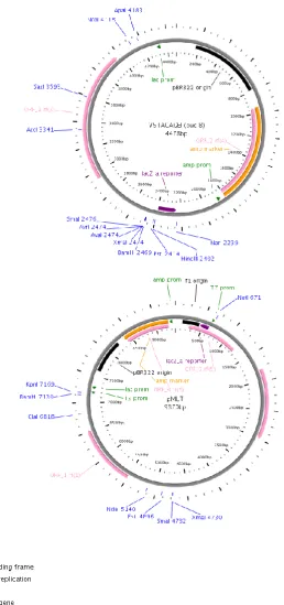
Figure 2.1 V5TAGAGE1 and pMUTv5wt vector 37
Figure 2.2 Direct selection of transformants example 44 Figure 2.3 Indirect selection of transformants example 45
Chapter 3 The use of alanine scanning mutagenesis on the NrtA 2nd
Nitrate Signature
Figure 3.1 Section of the proposed 2D structure of NrtA (Kinghorn et al. 2005) illustrating two conserved regions similar to the ones found ubiquitously in all MFS proteins
55
Figure 3.2 Proposed 3D model of NrtA illustrating the opposing nitrate signatures
58
Figure 3.3 Proposed 3D model of NrtA illustrating the residues analysed 59
6.2.4. Characterization of net nitrateuptake in residue replacements:--- 118-119 6.2.5. Kinetic analysis:--- 119-124
6.2.5.1 Analysis of variance (ANOVA t-test) for Kcat values:--- 122
6.2.6. Uptake Assay using the tracer 13NO3-:--- 123-124
6.3 Discussion:--- 124-128
7. CONCLUSION 129-134
Files on CD
Appendix 1: Homology table
Appendix 2: Buffers, Solution, Media Composition and Suppliers
Appendix 3: Transformation
Appendix 4: Raw data
vi
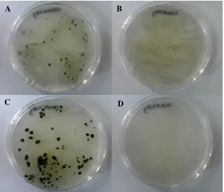
Figure 3.4 Pictures of single colonies tested phenotypically on NaNO3 under two different temperatures and ClO-3
62
Figure 3.5 Helical wheel displays on TM5 and TM11 65
Figure 3.6 Hypothetical 3D image of TM11 65
Figure 3.7 Hypothetical 3D model illustrating G461A substitution 68 Figure 3.8 Hypothetical 3D model illustrating F457 aromatic ring facing the
hydrophobic core
70
Chapter 4 NrtA and NrtB comparative study
Figure 4.1 A simplified 2D structure of NrtA and NrtB 77 Figure 4.2 A hypothetical 3D model for NrtA and NrtB superimposed with 4
other MFS structurally determined proteins
78
Figure 4.3 NrtA and NrtB UniprotKB alignment 80
Figure 4.4 Pictures of single colonies tested phenotypically on NaNO3 under two different temperatures and on ClO-3
84
Figure 4.5 Helical transmembrane segments rotational angle prediction comparison between NrtA/NrtB TM2, TM8 and TM11
85
Figure 4.6 Illustration of L84 and L88 side-chains in comparison with NrtB 89
Chapter 5 The study of highly conserved NrtA aromatic residues
Figure 5.1 Pictures of single colonies tested phenotypically on NaNO3 under two different temperatures and on ClO-3
98
Figure 5.2 Helical wheel transmembrane segments rotational angle display 101 Figure 5.3 Influx values for the wild-type strain T454 104 Figure 5.4 Influx values for the mutant strain F47Y 105 Figure 5.5 Influx values for the mutant strain F457M 106 Figure 5.6 13NO3- Influx values for the wild-type T454 108 Figure 5.7 13NO3- Influx values for the mutant strain F47Y 109 Figure 5.8 Hydrogen bond interaction between Y51 and E330 111
Chapter 6 Nitrate binding site study involving the residue N364 located at
TM8
Figure 6.1 Superimposition of NrtA, GlpT and LacY 115
Figure 6.2 Pictures of single colonies tested phenotypically on NaNO3 under two different temperatures and on ClO-3
116
Figure 6.3 Helical transmembrane segments rotational angle prediction for N364
118
Figure 6.4 Influx values of the mutant strain N364A 120 Figure 6.5 Influx values of the mutant strain N364Q 121 Figure 6.6 13NO3-Influx values for the mutant strain N364Q 123 Figure 6.7 Hypothetical polar interactions between N364, R368 and T421 126 Figure 6.8 Possible polar interactions between N364 substitutions, R368 and
T421
vii
Tables
Chapter 1 Introduction
Table 1.1 NR and NiR biochemical reactions 7
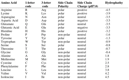
Table 1.2 Amino acids main nomenclature, code and properties 21
Chapter 2 Materials and Methods
Table 2.1 Primers 32-33
Chapter 3 The use of alanine scanning mutagenesis on the NrtA 2nd Nitrate Signature
Table 3.1 Residue conservation percentage in the second nitrate signature 59 Table 3.2 Description of mutant strain with codon changes, net nitrate
uptake, phenotypical test and protein expression
61
Table 3.3 Net nitrate uptake rates for mutant and wild-type strains 63 Table 3.4 Comparison of the amino acid composition on both nitrate
signatures
74
Chapter 4 NrtA and NrtB comparative study
Table 4.1 Amino acid composition of NrtA and NrtB 81
Table 4.2 Description of mutant strain with codon changes, net nitrate uptake, phenotypical tests and protein expression
83
Table 4.3 Net nitrate uptake rates for mutant and wild-type strains 86
Chapter 5 The study of highly conserved NrtA aromatic residues
Table 5.1 Aromatic residues conservation based on 694 homologue sequences
97
Table 5.2 Description of mutant strain with codon changes, net nitrate uptake, phenotypical tests and protein expression
99-100
Table 5.3 Net nitrate uptake rates for mutant and wild-type strains 102 Table 5.4 Kinetic constants and statistical analysis for NaNO3 influx by the
wild-type strain T454
103-104
Table 5.5 Kinetic constants and statistical analysis for NaNO3 influx by the mutant strain F47Y
105
viii mutant strain F457M
Table 5.7 Estimated mean valuesof the kinetic parameters 107
Table 5.8 Descriptive Statistics 107
Table 5.9 Estimated mean valuesof the kinetic parameters 108
Chapter 6 Nitrate binding site study involving the residue N364 located at TM8
Table 6.1 Description of mutant strain with codon changes, net nitrate uptake, phenotypical tests and protein expression
164
Table 6.2 Net nitrate uptake rates for mutant and wild-type strains 166 Table 6.3 Kinetic constants and statistical analysis for NaNO3 influx by the
mutant strain N364A
167
Table 6.4 Kinetic constants and statistical analysis for NaNO3 influx by the mutant strain N364Q
168
Table 6.5 Estimated mean valuesof the kinetic parameters 169
Table 6.6 Estimates of Marginal Means 169
Table 6.7 Descriptive Statistics 170
Table 6.8 Parameter estimates with 95% confidence interval of Kcat values 170
Table 6.9 Pairwise Comparisons (PC) 170
Table 6.10 Estimated mean valuesof the kinetic parameters 171
Chapter 7 The role of positively charged residues on nitrate transport
Table 7.1 Description of mutant strain with codon changes, net nitrate uptake, phenotypical tests and protein expression
180
Table 7.2 Net nitrate uptake rates for mutant and wild-type strains 182
Chapter 8 The structural importance of A20, P113 and G328 on NrtA’s nitrate transport
Table 8.1 Description of mutant strain with codon changes, net nitrate uptake, phenotypical tests and protein expression
190
ix
Abbreviation List
α alpha
A adenine
A alanine
APS ammonium persulphate
ATP adenosine triphosphate
bp base pair
β beta
BSA bovine serum albumin
C-terminus carboxy terminus
°C degrees centigrade
cDNA complementary DNA
C cysteine
C cytosine
D aspartic acid
Da Dalton
DNA deoxyribonucleic acid
dNTP deoxynucleoside triphosphate dH2O distilled water
DTT dithiothreitol
DW dry weight
E glutamic acid
EDTA ethylenediaminetetraacetic acid et al et alia (and others)
et cetera and other things
EtBr ethidium bromide
F phenylalanine
G glycine
G guanine
g gram
H histidine
h hour
HIV-1 human immunodeficiency virus-1 HRP horseradish peroxidase
I isoleucine
K lysine
kb kilobase
kDa kiloDalton
Kg kilogram
Km Michaelis-Menten constant
λ lambda
L litre
L leucine
LB Luria broth
Ltd limited
μg microgram
μl microlitre
μm micrometre
μM microMolar
M methionine
x
MM minimal media
mg milligram
min minutes
ml millilitre
mM milliMolar
mm millimetre
mRNA messenger RNA
N asparagine
N-terminus amino terminus
ng nanogram
nM nanoMolar
NR nirate reductase
OD optical density
ORF open reading frame
P proline
PCR polymerase chain reaction PEG polyethylene glycol 3350
pH Potential of hydrogen
PMSF phenylmethylsulphonyl fluoride
Q glutamine
R arginine
RNA ribonucleic acid
rpm revolutions per minute
S serine
s seconds
SDS sodium dodecyl sulphate
SDS-PAGE sodium dodecyl sulphate-polyacrylamide gel electrophoresis
SE standard error
SEM standard error of the mean
SOB super optimal broth
SOC super optimal catabolite repression broth SSC saline-(tri) sodium citrate
SSPE sodium.chloride-sodium.phosphate-EDTA
sp species
T threonine
T thymine
TBE tris-borate-EDTA
TE tris-EDTA
TEMED tetramethylethylenediamine
TM transmembrane domain
Tris tris(hydroxymethyl) methylamine
Tris-HCl tris(hydroxymethyl) aminomethane hydrochloride Triton X-100 t-Octylphenoxypolyethoxyethanol
Tween Polyoxyethylenesorbitan, monolaurate Polyoxyethylenesorbitan, monolaurate
W tryptophan
u unit
UB universal bottle
UV ultraviolet
UV-Vis UltraViolet-Visible Spectroscopy
xi
Vmax maximal velocity (in Boltzmann equation)
v/v volume per volume
W tryptophan
w/v weight per volume
2D second dimension
1 CHAPTER 1:
Introduction
1.1 General Introduction Background
Nitrate is the main nitrogen source for most bacteria, algae, fungi, plants and
yeast. This key nutrient absorbed from soil is crucial for the growth, development and
metabolism of these organisms (Unkles et al. 1991; Crawford and Glass 1998). Ever
since the first nitrate gene was isolated 20 years ago from the fungus, Aspergillus
nidulans (Unkles et al. 1991) nitrate transporters have been studied extensively, and
recent advances in molecular biology alongside the advent of computer technology,
have had a massive impact, resulting in the identification of genes encoding
high-affinity and low-high-affinity transport systems in Arabidopsis thaliana and in other
species. Nevertheless, major issues are still to be elucidated including the roles and
interactions between genes encoding proteins that regulate the transport of nitrate.
NrtA is the protein responsible for high-affinity nitrate transport in A. nidulans
and the functional role of its amino acids is not thoroughly understood. Moreover, the
analysis of structure and function of NrtA is particularly important as it has the
potential to provide information on other anionic transporters. Additionally, analysis
of this protein responsible for the transport of nitrate has the potential to provide
mitigating strategies for future improvement of nitrogen use by agriculture. Nitrate is
the main component of fertilizers and excess nitrate has the potential to leach into
groundwater causing detrimental effects on the environment and human health, thus
hindering agriculture and ecological sustainability. Hence the importance of extending
our understanding of this high-affinity nitrate transporter protein.
In order to gain further insight into the structure and function of this important
class of nitrate permease a whole range of targeted site-directed mutagenesis were
carried out. This mutational strategy combined with biochemical analysis has the
potential to elucidate the role and importance of individual residues in the movement
of nitrate across the membrane.
1.2 Nitrate (NO3- )
Nitrate is a naturally occurring polyatomic ion with a molecular mass of 62 g/mol.
Nitrate is a negatively charged ion containing one molecule of nitrogen and three of
oxygen. It has a conjugated base of strong acid with a pKa of -1.3 and is very soluble in
2
particularly important for the development of plants (Crawford and Glass 1998).
Nitrogen compounds are produced naturally in our environment by the activity of
lightning strikes (Rakov and Uman 2007) and via anthropogenic effects such as waste
from industrial processes, motor vehicles and synthetic fertilizers used in agriculture.
Nitrate is used mainly in inorganic fertilizers for agriculture and also in the production
of gunpowder and explosives (Addiscott 2005). Additionally, nitrate is used in glass
manufacturing and as food preservatives for cured meats (WHO 2004).
1.3 Nitrate Contamination
According to the last World Bank population indicator, the world’s population is
fast approaching 6.8 billion (World Bank 2011) and despite falling birth rates the
world population is growing at a rate of 74 million people per year. By 2050 the
world’s population is projected by UN demographers to reach 9.1 billion (World
population prospect, UN 2009). This worldwide population boom especially in the last
two centuries is due to medical and scientific advancements as well as significant
developments in agriculture.
In order to increase food supplies we rely on the use of artificial fertilizers to
enhance natural nitrogen found in soil. In fact, 48% of the world’s population is fed
today thanks to the introduction of synthetic fertilizers (Erisman et al. 2008).
According to the International Fertilizer Industry Association (IFA 2009) global
agricultural demand for fertilizers is bound to increase between 50-80% above today’s
level by 2050. Developing countries particularly in Asia are today the main consumers
of fertilizers accounting for almost half of the world’s nitrogen fertilizer consumption.
Although, European consumption of fertilizers is declining, production of manure
through N excretion from animals is increasing annually (Figure 1.1) (Erisman et al.
2011; Velthof et al. 2011). Europe’s dependence on agricultural imports of goods is
also increasing, for instance 71% of Europe’s agricultural imports come from
developing countries fuelling their emerging agriculture industry (CAP 2010).
The agriculture industry is the largest source of nitrogen pollution in
groundwater. Excess application of inorganic nitrogenous fertilizers is washed away
due to precipitation or irrigation then leaching from soil into groundwater (Velthof et
al. 2011). Nitrates from livestock and septic waste systems, automobile exhaust
emissions and other fossil-fuel combustion add to this excess pollution. Excess
amounts of nitrate can pollute groundwater through the soil naturally by rain or
anthropogenically via irrigation, and also via wells that are wrongly constructed or
3
Figure 1.1 Comparison between global and Europe’s manure production and
fertilizer consumption levels
Left-hand graph: World and European’s manure production and fertilizer
consumption (Erisman et al. 2011); right-hand pie chart: Nitrogen fertilizer
consumption by region based on Potash Corp (the world’s largest fertilizer
corporation) 2009 estimations (Potash 2009).
The fate of nitrate in soil varies according to soil type, aerobic or anaerobic
conditions, presence of high or low water tables, amount of precipitation, presence of
organic materials and other physico-chemical characteristics. In soil, organic nitrogen
is decomposed to give ammonia, and then oxidized to NO3- and NO2-. Plants are
responsible for most of the nitrate reduction in surface water via the uptake of NO3-
(Velthof et al. 2011). Nitrate is a highly soluble molecule and excess nitrate can
easily pass through soil and contaminate ground water. The main sources of nitrate
contamination are from agriculture run-offs and sewage treatment systems. Pollutants
such as nitrate are usually filtered by the soil and porous spaces of rocks; however,
due to the physical characteristics of soils and rocks some pollutants such as nitrates
might not be filtered well enough, and thus leaching into groundwater (Williams et al.
1998; Velthof et al. 2011). Soils have different drainage features due to permeability
rates; soils composed mainly of gravel and sand filter fluids down to the aquifer at a
much faster rate than soils comprised of compact particles such as clay (Velthof et al.
2011). Furthermore, nitrate is more susceptible to leaching because soils have a
tendency to adsorb more cations than anions, moreover nitrate has a weak affinity to
form complexes with soil minerals (Strahm and Harrisson 2006).
The concentration of nitrate found in drinking groundwater is increasing
4
lack of proper septic waste treatment, wastewater from industries, natural atmospheric
fixation, precipitation and more significantly by the rising amount of fertilizers used
by the agriculture industry (WHO 2004). Nitrate concentration levels in drinking
water have increased linearly in many EU countries. In the past 20 years nitrate
concentration levels have doubled, for instance, in the UK an increase of 0.7 mg/l per
year has been observed in some rivers. The World Health Organisation (WHO)
guideline limit value for nitrate concentration level in groundwater for the European
Union (EU) is 50 mg/l (WHO 2004).
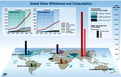
Groundwater provides 30% of the world’s drinking water supply. Water
withdrawals and consumption are projected to increase 18% in developed countries
and 50% in countries in development by 2025 (UNEP 2007) (Figure 1.2).According
to the Global Environment Outlook (GEO-4) report, agriculture accounts for 70% of
all water usage and if mitigating steps are not put into place a projection of 10-20%
global increase in fluvial nitrate contamination flow towards coastal ecosystems is
[image:18.595.121.516.386.637.2]predicted by 2050 (UNEP 2007).
Figure 1.2 Worldwide freshwater usages
Illustration of the worldwide freshwater withdrawal and consumption from the
1900s including projections up to 2025 and the world’s top 20 water consumers
(UNEP 2002).
Nitrogen sources such as nitrate found in high concentrations have a substantial
ecological, agricultural and medical impact. Nitrate contamination causes detrimental
5
turning the water darker and hypoxic due to algal blooms, affecting the tourism
industry, and killing fish and other aquatic life (Schindler and Vallentyne 2008).
Eutrophication of waters is believed to cost the US government approximately $2.2
billion dollars annually (Dodds et al. 2008). Nitrate contamination is also intrinsically
linked to salinization of surface water affecting aquatic ecosystems, and to an increase
in altered soil erosion and sedimentation patterns (Palaniappan et al. 2010).
Nitrate contamination has the potential to cause health problems such as cancers
of the digestive tract (Morales et al. 1995), congenital anomalies, goitre, hypertension,
genotoxicity, diabetes, also methaemoglobinaemia, an oxygen deficiency triggered by
the blood's inability to transport oxygen to cells and tissues, also known as the “blue
baby syndrome”, as infants are more vulnerable and develop a blue coloration,
particularly around their mucous membranes (Palaniappan et al. 2010). Both acquired
and environmentally-caused form of methaemoglobinaemia can be fatal or cause
serious damage to digestive and respiratory systems (Ash-Bernal et al. 2004).
However, according to Powlson et al. (2008) methaemoglobinaemiacan be caused by
gastroenteritis and in a review of about 50 epidemiological studies from the early
1970s onwards they suggested no relation between nitrate in drinking water and
stomach cancers. There is also evidence of possible benefits from nitrate in
cardiovascular health (Wink and Paolocci 2008). Several studies generated
inconclusive outcomes resulting in much debate over opposing views on the current
data. In order for this debate to be resolved careful consideration is vital as the issue of
nitrate contamination has a substantial economic, health and environmental impact.
1.4 Nitrate Assimilation
Nitrate assimilation is an important biological system carried out by most
bacteria, yeast, filamentous fungi and plants involved in uptake and delivery of nitrate
from the environment into cellular processes (Unkles et al. 2004a). Furthermore,
nitrate assimilation markedly affects plant growth, productivity and biomass, and a
deficit in nitrate results in a gradual decrease in structural components such as stunted
growth (Dechorgnat et al. 2011). The transport of nitrate into cells has been largely
studied using different microorganism models such as Neurospora crassa (Schloemer
and Garrett 1974), Chlamydomonas reinhardtii (Rexach et al. 2002) and Hansenula
polymorpha (Navarro et al. 2003), A. thaliana (Filleur and Daniel-Vedele 1999), A.
nidulans (Unkles et al. 1991) and the moss, Physcomitrella patens (Tsujimoto et al.
6
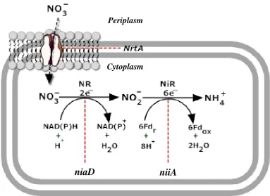
reduction of nitrate to ammonium, catalysed by the enzyme nitrate reductase (Figure
[image:20.595.137.521.127.406.2]1.3).
Figure 1.3A. nidulans nitrate assimilation pathway
Schematic diagram of A. nidulans nitrate assimilation pathway. The figure
shows the genes responsible for nitrate assimilation regulation marked in italics:
nrtA-niaD-niiA. Nitrate reduction to ammonium is mediated by two enzymes
(NR and NiR). NR converts nitrate to nitrite via a reduction of 2e− and Nir
converts nitrite to ammonium via a reduction of 6e−. Redox reactions of
ferredoxins (Fd) is indicated by r (reduced) and ox (oxidised). This image was
based on the original Kinghorn (1989) nitrate assimilation pathway modified
using SmartDraw trial version.
Nitrate assimilation by bacteria, fungi, algae, some yeast and higher plants
occurs via a sequential reduction of nitrate to nitrite by the enzyme nitrate reductase
(NR) (EC 1.7.1.1) and nitrite to ammonium by the nitrite reductase (NiR) (EC
1.7.2.1). In A. nidulans nitrate is transported through the permease, NrtA by a
sequential reaction involving NR (regulated by niaD) which catalyses the 2e−
reduction converting nitrate to nitrite, followed by NiR (regulated by niiA) which
catalyses the 6e− reduction converting nitrite into ammonium (Table 1.1) (Kinghorn
1989).
Cytoplasm Periplasm
7
Table 1.1 NR and NiR biochemical reactions
Enzyme Reaction
NR NO3-+NAD(P)H+H++2e-=NO2-+NAD(P)+H2O
NiR NO2-+Fdr+8H++6e-=NH4++Fdox+2H2O
Enzymes and biochemical reactions for A. nidulans nitrate assimilation
Abbreviations: Fdr: reduced ferredoxin and Fox: oxidised ferredoxin (Kinghorn
1989).
NR is able to convert nitrate to nitrite because of the presence of reduced
coenzymes NAD and NADP (Campbell 1999). NR-nicotinamide adenine dinucleotide
phosphate (NADPH; EC 1.7.1.3) is an iron-sulphur molybdenum flavoprotein
whereas, Nir-reduced nicotinamide adenine dinucleotide (phosphate) ([NAD(P)H];
EC 1.7.1.4) is an iron-sulphur heme flavoprotein which contains a siroheme group
(Kinghorn and Campbell 1989). The regulation of fungal nitrate uptake is regulated
by the induction of nitrate and feedback repression due to glutamine (Unkles et al.
2004b and references therein). NR is fundamental for nitrate uptake activity in the
cells of fungi but according to Unkles et al. (2004a) NR activity is not essential for
nitrate uptake by plant cells. Using mutant organisms lacking NR activity Unkles et
al. (2004a) demonstrated that Pichia pastoris and A. nidulans failed to accumulate
nitrate, as opposed to A. thaliana mutants that despite lacking NR activity were able to
uptake nitrate.
1.5 Nitrate Transport Systems
Nitrate is an essential nitrogen source to many organisms, it is also involved in
the metabolism and development of plants (Crawford and Glass 1998; Zhou and
Miller 2000), albeit not exclusively, as plants in rice paddies and in the arctic tundra
prefer to uptake ammonia and amino acids (Glass 2009). Additionally, nitrate stored
in the leaves of plants serves in osmoregulation by playing an important role in the
maintenance of cation-anion balance (Carrow et al. 2001). Nitrate uptake is regulated
via a network of active transport systems (King et al. 1992). Nevertheless, the main
dynamics of function and structural features of nitrate transport in NrtA are not clearly
understood.
Two families of nitrate transporters have been identified namely NRT1 and
8
oligopeptide transporter), NRT1 is a low-affinity nitrate transporter, whereas NRT2 is
a high affinity nitrate transporter, previously known as NNP (Nitrate-Nitrite Porter)
(Crawford 1995; Trueman et al. 1996). The NRT1 family is composed of members
that transport nitrate or histidine (Rexach et al. 1999), whereas NRT2 is composed of
large proteins that catalyses nitrate uptake or nitrite efflux (Pao et al. 1998).
There are two kinds of nitrate transport systems in plants, a high-affinity
transport system (HATS), activated when the external nitrate concentration is low, and
a low-affinity transport system (LATS), activated when the external nitrate
concentration is high. Both systems are characterised by whether they are substrate
induced (iHATS, iLATS) or constitutively active (cHATS, cLATS). cHATS and
cLATS are expressed in the absence of nitrate whose activity is stimulated by nitrate
treatment and have higher affinity to nitrate, whereas iHATS and iLATS are regulated
by products of nitrate uptake such as amino acids and ammonia via negative feedback
and have greater capacity for nitrate uptake (Crawford and Glass 1998; Forde 2000;
Orsel et al. 2002a).
HATS operates at external nitrate concentration < than 1mmol L-1 (Williams and
Miller 2001) and is regarded as the H+/anion co-transport carrier mechanism whose
activity is regulated by cellular energy supply. Fungal proteins of HATS encode genes
of functional similarities to plants (Filleur and Daniel-Vedele 1999). LATS seemingly
has a signalling role as it recognises nitrate entry into the cell in order to induce
expression but only above a particular threshold as LATS functions linearly at nitrate
concentration > than 1mmol L-1 (Crawford and Glass 1998; Lea 2001).
AtNRT1.1 (formerly named CHL1) was the first A. thaliana nitrate transporter
gene belonging to the NRT1 family cloned from the screening of mutants resistant to
chlorate (Doddema and Telkamp 1979). In 1993 the dual affinity AtNRT1.1 gene from
Xenopus oocytes was isolated by T-DNA activation tagging and functional expression
analysis revealed that the protein encodes a nitrate inducible form of LATS (Tsay et
al. 1993). In the late 1990s a second A. thaliana gene, AtNRT1.2 was cloned by Huang
et al. (1999). A. thaliana’s genome contains 53 NRT1 genes but only 8 members have
been functionally characterised, AtNRT1.1-AtNRT1.8, encoding low-affinity
transporters except AtNRT1.1 which has dual affinity to nitrate (Tsay et al. 2007;
Glass 2009). Recently, Wang and Tsay (2011) reported AtNRT1.9 as a low-affinity
nitrate transporter responsible for distributing nitrate from the root to the phloem.
The first eukaryote member of the NRT2 family cloned was the nrtA (previously
known as crnA) gene from A. nidulans (Unkles et al. 1991) followed by the nrtB gene
(Unkles et al. 2001). In higher plants 7 high-affinity transporters AtNRT2.1- AtNRT
9
et al. 1996), Nicotiana plumbaginifolia (Quesada et al. 1997) and Glycine max
(Amarasinghe et al. 1998). Other NRT2 members were isolated from C. reinhardtii
(Quesada et al. 1994) and Cyanobacterium synechococcus (Sakamoto et al. 1999).
Higher plants and algae nrt2 genes are 30% identical to their fungal homologues
(Tsay et al. 2007). NRT2 have conserved genes amongst plants, fungi and algae
(Yokoyama et al. 2001), however NRT1 is not present in fungi (Crawford 1995). The
vast majority of the nrt2 genes have a nitrate inducible expression (Rexach et al.
1999) and share greater sequence similarity with each other in comparison with the
NRT1 members. In higher plants the nrt3 genes (AtNRT3.1 and AtNRT3.2) are not
nitrate transporters per se but are required for the nitrate transport of other NRT2
members (Okamoto et al. 2006).
1.6 A. nidulans as a Model Organism
Fungi are heterotrophic organisms that play an essential role in the Earth
ecosystems; they play a crucial role in degradation of plant material and recycling
nitrogen as well as the remineralisation of organic matter. The fungal kingdom is
composed of many taxa, currently comprising 7 phyla: chytridiomycota,
glomeromycota, microsporidia, neocallimastigomycota, ascomycota, basidiomycota
and blastocladiomycota (Hibbett et al. 2007).
Ascomycota is the largest phylum with over 64,000 species identified to date
(Kirk et al. 2008) but it is estimated that more than 1.5 million fungal species could
potentially be identified in the future (Neubert et al. 2006). Within ascomycota,
Aspergillus is a ubiquitous filamentous group that underwent over 200 million years
of evolution (Galagan et al. 2005). This group is of particular relevance as several
species have been used as model organisms on a wide range of subjects such as
genetics, molecular cell biology, pathogenesis and metabolism. Most notably A.
nidulans, a model organism studied in eukaryotic cell biologyfor over 60 years, which
has helped to elucidate the understanding of many fundamental cellular processes
(Martinelli and Kinghorn 1994; Galagan et al. 2005).
A. nidulans is also an excellent biological model used for comparative studies of
other more economically/commercially important species such as Aspergillus niger
and Aspergillus oryzae for example, used in the production of industrial enzymes e.g.
amylases (David et al. 2008). A. nidulans is one of the few species of its genus able to
form sexual spores via meiosis, which enables the manipulation of sexual-crossing of
10
The genome sequence of A. nidulans was accomplished by the Broad Institute in
2005, through 13X whole genome shotgun sequencing coverage, representing a major
development in the study of Aspergillus, laying the foundation for genome
comparative analysis and expansion of functional genomics studies (Galagan et al.
2005). This international collaborative work was subsequently updated in 2008, when
the Eurofungbase corrected several gene structures (Wortman et al. 2009).
Emericella nidulans is the official scientific name used by GenBank and
UniProt. According to the 1905 Botanic Code fungi can have a dual-name when
referring to its sexual, teleomorph (E. nidulans) or asexual phase, anamorph (A.
nidulans) (Bennett 2010). A literature scholar search using Google on A. nidulans
would result in 39,900 hits whereas E. nidulans would result in 350 hits making A.
nidulans the predominant term used by scholars and professionals.
A. nidulans is a filamentous fungus that lives on decaying plant material where
the nature and quantities of nitrogen sources change frequently. In terms of
morphology, the mycelium (homokaryon) of A. nidulans is a web of branched
filaments, called hyphae. A. nidulans has a well-characterised life cycle, in terms of
asexual reproduction (Figure 1.4A) the vegetative hyphae growth phase begins with
the germination of a uninucleate conidium and the formation of mycelium (a haploid
homokaryon) which is a branch of interconnected hyphae. The homokaryon formation
is followed by a perpendicular stalk and a round-shaped vesicle development, where
the uninucleate reproductive cells metulae and phialide are formed. Conidia are
produced in the head of the unicleate phialide, the nucleus migrates from the vesicle
towards each phialide and undergoes successive mitotic divisions, one nucleus
(genetically identical to haploid parent) migrates into the developing conidium and the
other stays behind for the next round of mitosis. Long conidia chains are formed from
repeated mitotic division of phialides and once mature the conidia are released in vast
numbers for rapid dispersal to produce new organisms (Casselton and Zolan 2002;
Todd et al. 2007; Lee et al. 2010). A. nidulans is homothallic, meaning that it
undergoes self-fertilization, where mating occurs between hyphae from an identical
fungal clone, retaining the ability for out-crossing. Their sexual cycle (Figure 1.4B)
begins with the fusion of vegetative hyphae cells; two haploid nuclei from each strain
destined for meiosis divide synchronously, forming a highly branched ascogenous
hypha. At each ascogenous hypha head, an ascus is formed when two haploid nuclei
fuse and the resulting dikaryon then undergo meiosis forming a four haploid nuclei
structure. Ascus is where motionless spores (ascospores) are formed when each
haploid nucleus undergoes mitosis resulting in an eight haploid nuclei ascospores.
11
hold tens of thousands of ascospores before releasing them into the environment.
Cleitothecia (fruiting body) are round multicellular structures which are an enclosed
type of hymenium surrounded by Hülle cells (also known as nursing cells) as they aid
spore formation. Additionally, A. nidulans can also reproduce parasexually (Figure
1.4C) whereby the haploid nuclei of the vegetative hyphae cells fuse and
systematically continue to undergo mitosis. Following heterokaryon formation,
crossing-over of genetically identical and different nuclei might occur and
chromosome loss is followed by haploidisation to restore haploid chromosome
number (8) (Casselton and Zolan 2002; Todd et al. 2007; Lee et al. 2010).
Figure 1.4A Sexual cycleof A. nidulans
Schematic representation of A. nidulans’s sexual cycle showing a fruiting body
(cleitothecia) bursting into the environment followed by the formation of
ascogenous hypha, ascus and ascospores – a product of meiosis. The image of
rupturing cleitothecia was courtesy of Peter Halasz; the diagram was made using
SmartDraw trial version, and it was based on Casselton and Zolan (2002) and
[image:25.595.92.515.261.602.2]12
Figure 1.4B Asexual cycleof A. nidulans
Schematic representation of A. nidulans’s asexual cycle showing the
differentiated homokaryon developed from a single haploid spore-conidia
generated through mitosis (genetically identical – replicas of their parent cells)
and a conidiophore. The transmission electron micrograph of the conidiophore
was courtesy of Jae-Hyuk Yu (Yu 2010). The diagram was made using
SmartDraw trial version, and it was based on Casselton and Zolan (2002) and
Todd et al. (2007) depiction of A. nidulans life cycle.
Figure 1.4C Parasexual cycleof A. nidulans
Schematic representation of A. nidulans’s parasexual cycle showing the
heterodikaryon, diploid hyphae following a mitotic cross-over and
haploidisation to restore the haploid chromosome number. The diagram was
made using SmartDraw trial version and it was based on Casselton and Zolan
[image:26.595.143.477.59.328.2]13 Why use A. nidulans?
easy to grow in the laboratory – fast growth rate mutants can be readily isolated
well-characterised genetic system can utilize various nitrogen sources great potential in biotechnology
smaller and less complex genome in comparison with animals (but
functionally homologous)
has been used extensively for the study of a variety of subjects including
classic genetics, cell biology and pathogenesis
1.7 Membrane Proteins
Membrane proteins lie at the border between the inside of the cell and its
environment being responsible for the influx and efflux of a vast array of molecules
such as ions, sugars and drugs. Structurally a membrane protein consists of a lipid
bilayer the vast majority being phospholipids and has a hydrophobic core; the
fatty-acid tails of each lipid molecule is oriented inwardly in the bilayer and the polar
phosphate head is oriented towards water (Lee 2004). The inner core of membrane
proteins is highly hydrophobic but selectively-permeable letting the passage of
water-soluble molecules in and out of cells (Lodish et al. 2000). More than 50% of proteins
interact with membranes and the function of these proteins is determined by the nature
of its relationship with the lipid bilayer (Alberts et al. 2007).
Membrane protein acts as a highly selective permeability barrier performing
specific functions for essential cellular processes; mediating and controlling the
interactions, connecting the exterior and interior of cells and organelles some have
structural roles providing stability, and others play a role in enzymatic production and
maintenance of ion concentration. There are two categories of membrane proteins,
integral and peripheral. Integral proteins are inserted in the bilayer in a single α-helix,
multiple α-helices or β-barrels format. Others are anchored to the cytosolic side,
covalently bonded to fatty-acids or one-sidedly exposed. Peripheral proteins interact
with integral proteins or phospholipids polar head groups and are bounded to the
membrane displaying no interaction with the hydrophobic core of the bilayer (Lodish
14 1.7.1 Why Study Membrane Proteins?
Membrane proteins play a crucial role in most cellular processes; they are
essential mediators in signalling, transporting molecules across cell membranes and
energy transduction. Membrane proteins account for as much as 30% of the genome
of most living organisms; for instance, 1/3 of human proteins are membrane proteins
for example, the voltage-gated sodium channel responsible for the transmission of
nerve impulses (Gulbis et al. 1999; Yildirim et al. 2007).
In addition to their crucial physiological and highly specialised functions,
membrane proteins have a fundamental role in diseases and account for more than
60% of all drug targets, so further understanding about this type of proteins has the
potential to provide a valuable contribution to the development of novel therapeutic
drugs (Elofsson and Heijne 2007; Yildirim et al. 2007).
Moreover, the expansion of our understanding in homologous membrane protein
structure and function can lead to new insights into many medically important
conditions, including epilepsy and cancer caused by faulty membrane proteins for
example the sodium-glucose transporter, a protein involved in diabetes (Debnam et al.
1995). Membrane protein however is still under-represented as not many 3D
structures have been solved and future studies are indispensable in the understanding
of functional and structural features of membrane proteins (Tan et al. 2008).
The best way to understand how proteins acquired their structure and how
alterations in their structure affect their function is to determine their specific 3D
structure. Atomic structures of membrane proteins are technically notoriously difficult
to solve as there are various challenges surrounding x-ray crystallography and other
techniques (Elofsson and Von Heijne 2007). The last couple of years have seen a
great progress in structure determination, especially in computational structure
prediction methods. This on-going progress would, however be better accomplished
with further technical advancement in x-ray crystallography and expression of
proteins, as only a few hundred of all proteins stored in the PDB are unique membrane
proteins (White 2004).
Membrane proteins are the most challenging proteins for scientists to crystallise
as they are found naturally in cells at low levels, because of the complexity of protein
assembly and folding in vivo, and also purification and expression mechanisms are
considerably challenging methodologies to overcome. Also one of the major
challenges encountered in membrane protein x-ray crystallography research is due to
15
Worldwide strategic initiatives are currently fuelling new discoveries. One
example is the Protein Structure Initiative (PSI), a $764 million effort that ended in
2010 which was aimed at reducing the cost and time required to determine structures
of membrane proteins. This project was composed of an interdisciplinary team of
scientists with distinctive expertise in various topics which were explored in
individual projects within the overall objective of the PSI programme (Elsliger et al.
2010). Initiatives like this consortium could lead to new insights in functional studies
of significant membrane proteins of medical and ecological importance, amongst
other applications. Additionally projects like this can lead to new and improved
techniques surrounding solving proteins’ 3D structures such as the development of
specialised reagents to keep membrane proteins stable for x-ray crystallography
(Elsliger et al. 2010).
The improvement of methodologies and instruments used in x-ray
crystallography and nuclear magnetic resonance (NMR) resulted in an exponential
rise in high-resolution structural data of membrane proteins. A method that has
recently become a way of determining protein structures in high-resolution is
cryo-electron microscopy (cryo-EM) initially reported by Adrian et al. (1984) work on
adenovirus. Nowadays it is a method widely used to solve structures of large protein
complexes (Frank 2002). Atomic structure alone, however, does not determine how a
protein works as amino acids may contribute to the multitude of binding energy,
through hydrogen bonds, salt bridges, van der Waals and electrostatic forces,
dipole-dipole, and hydrophobic interactions (Morrison and Weiss 2001). Biochemical
methods such as site-directed mutagenesis are particularly useful for the identification
of important residues in proteins.
The post-genomic era propelled the rate of protein structure determination and is
defined by countless numbers of studies and advances in determining high-resolution
protein structures. Despite this, structural biology remains an under-explored field. At
present, the use of protein alignments as tools is enhanced by an ever increasing
number of molecular biology resources, accessible for studies encompassing a wide
range of species ranging from expressed sequence tag (EST) to genetic linkage (Volff
2005). Any knowledge gained into the diversity of membrane proteins whether based
on structural data alone or reinforced by biochemical analysis has the potential to
provide a betterment of humankind’s health and living standards (Elofsson and Von
16
1.7.2Membrane Protein Classification Systems
The completion of the genome project resulted in a vast number of protein
sequences, together with tremendous efforts in understanding the intricacies of
proteins, and the progress in structure determination resulted in practical issues that
needed sorting out for example where to store vast amounts of data (PDB weekly
release is of about 160 proteins), what to make from the crystallised structure as even
at high resolution, x-ray structure alone does not provide all the information of the
transport dynamics within proteins (Guan and Kaback 2006), and the most debated
issue, how to classify proteins. Since some proteins share similarities such as sequence
and structural features the debate of the methodology for comparison is often agreed
upon, yet the conventions for classifying are still a matter of debate (Elofsson and Von
Heijne 2007; Sippl 2009).
There have been great efforts in classifying membrane proteins, hence the vast
number of hierarchical classification systems based on solved structures such as
CATH protein structure classification (Class, Architecture, Topology and
Homologous superfamily) (Orengo et al. 1997) and the Structural Classification of
Proteins (SCOP) (Murzin et al. 1995). CATH is a semi-autonomous classification
system and shares similar features to SCOP but these classification systems differ in
terms of hierarchical details and algorithms used. However, the number of membrane
proteins structurally solved is still low but with recent advancements in x-ray
crystallography and other molecular biology techniques these two classification
systems and their respective databases will have more solved proteins to classify and
store. There are many databases listing high-resolution membrane proteins structures
such as: http://blanco.biomol.uci.edu/mpstruc/listAll/list (White 2004) and PDB
http://www.pdb.org/pdb/home/home.do. Other specialised databases listing families of
membrane transporters such as http://www.gpcr.org/7tm and http://www.tcdb.org
have also been set up but these databases are not based on solved structure information
(Elofsson and Von Heijne 2007).
For the purpose of this study all proteins have been classified according to the
Transporter Classification (TC) System, which has been standardised by the
International Union of Biochemistry and Molecular Biology (IUBMB). Each
functionally unrelated protein is classified by the letters TC followed by a 5-digit
code. The first digit refers to the class of transport, the second digit refers to the
subclass (primary active transporters is referred by the energy source used to drive the
transport), the third digit refers to the family/superfamily, the fourth refers to a
17
specificities. The TCDB database also contains information on genome analysis, a
variety of alignments, phylogenetic trees and other analytic tools which can be found
at URL:[http://www.tcdb.org/] (Saier et al. 2006).
1.8 Major Facilitator Superfamily
Membrane protein transport is responsible for the crucial maintenance of a
distinct cellular environment. The largest division of the secondary transporter protein
family is the MFS (TC 2.A.1.). A typical MFS protein have 400-600 amino acids and
12-14 transmembrane domains (TMs) divided into two halves of a 6 α-helix, each
linked by long central loop (Pao et al. 1998) (Figure 1.5).
Among the different families of transporter, only two occur virtually in all
protein categories of organisms. These are the ATP-Binding Cassette (ABC) and the
Major Facilitator Superfamily (MFS). The MFS transporters are secondary
transporters capable of carrying small molecules in response to chemiosmotic ion
gradients. Compounds such as ions, nucleosides, sugars, esters, metabolites, drugs, et
cetera are transported by MFS permeases by electrochemical gradients into substrate
gradients (Walmsley et al. 1995) with 58 distinct members and more than 15,000
sequences identified to date (Law et al. 2008). A UniProtKB/Swiss-Prot search would
retrieve 27,503 un-reviewed MFS proteins.
Figure 1.5 Example of a typical MFS protein
This illustration show a schematic 2D structure of a typical MFS protein with its
12 TMs, cytoplasmic N-, and C-termini and a long cytoplasmic loop between
18
The MFS family contains members of medical and pharmaceutical importance
that function as uniporter, symporter or antiporter with a wide range of solute
specificity. In order to carry substrates through the membrane, proteins undergo
conformational changes by switching one opening of the hydrophilic cavity from the
periplasm to the cytoplasm, thus enabling the transport of substrate through the
membrane. This alternating-access mechanism is a shift type of movement of the two
halves of the protein activated through a single binding site (Bruser and Sanders
2003).
1.8.1 NRT1 also known as Proton-dependent Oligopeptide Transporter (POT/OPT)
Proteins of the NRT1 family (TC 2.A.17) consist of 19 proteins from animals,
plants, yeast, archaea and both Gram-negative and positive bacteria. The majority of
NRT1 members are low-affinity transporters except the dual-affinity transporter
protein CHL1 (Tsay et al. 2007). NRT1 family is also called PTO since it transports
dipeptides and tripeptides. The proteins are composed of 12 TM α-helical spanners.
Some members of the NRT1 family shares limited sequence similarity in comparison
to members belonging to the MFS and some were found to share sequence similarity to
SP and DHA1 families. An important member is the NRT1.1 of A. thaliana a
nitrate/chlorate symporter (Martin et al. 2008). In A. thaliana there are 53 NRT1 (PTR)
genes and 9 OPT genes (Tsay et al. 2007), for example NRT1.2, NRT1.3 and NRT1.4
are all nitrate transporters. The transport of peptides through cells is a common and
well-researched biological phenomenon which occurs due to specific,
energy-dependent transporters found in a variety of diverse organisms such as fungi and
humans. The NRT1 family of proteins is different from the ABC-type peptide
transporters (Steiner and Becker 1995). These proteins are mainly involved in the
intake of small peptides with the simultaneous uptake of a proton (Paulsen and Skurray
1994).
1.8.2 The Importance of 3D Structure Determination
The progress of membrane protein determination has been particularly important
in many contemporary research projects as it has the potential to shed light into the
analysis of some biochemical and molecular biology conundrums. A good example is
the recent structure determination of LacY and GlpT, both found to consist of an
19
is the central hydrophilic cavity that when opened to the cytoplasm remain closed to
the periplasm and on the contrary. LacY and GlpT structure determination indicated
that all MFS proteins are likely to have a single substrate-binding site and the
high-resolution structures of both proteins have identified several residues involved in the
substrate binding (Abramson et al. 2003; Huang et al. 2003; Bruser and Sanders
2003). LacY catalyses β-galactoside by H+ symport against a concentration gradient,
and GlpT is responsible for the exit of inorganic phosphate from the cell and the entry
of organic phosphates counter to a concentration gradient by antiport transport (Green
et al. 2000). Other important proteins that have been solved structurally were: OxlT,
the oxalate:formate exchange protein of O. formigenes (Hirai et al. 2004; Yang et al.
2005); Pho84, a S. cerevisiae phosphate transporter (Lagerstedt et al. 2004), GluT1, a
glucose transporter 1 (Alisio and Mueckler 2004); and EmrD, the multidrug
transporter of E. coli (Yin et al. 2006).
Studies such as these provide further insights into the functional and structural
importance of MFS transporter proteins. According to White (2004) the number of
new membrane protein structures has exceeded 100 by 2005 and it is predicted to be
rise to 2200 by 2025. At present, the topology of more than 400 helix spanning
membrane proteins have been solved by a variety of genetic, biochemical, and
structural techniques. However, less than 2% of the 3D structures in the Protein Data
Bank are membrane proteins (Elofsson and Von Heijne 2007). Therefore, much
remains to be done, both in improving and perfecting already determined 3D
structures and in providing unambiguous supportive evidence accomplished by
mutagenesis and biochemical studies.
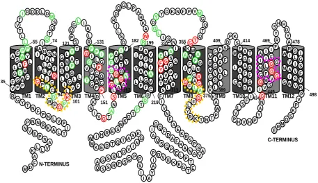
1.8.3 High affinity Nitrate Transporter Protein, NrtA
A. nidulans NrtA protein has a secondary structure consisting of 12 hydrophobic
TMs. This transport membrane protein has 507 residues and belongs to the NRT2
family, formally NNP family (See 1.8.1 sector), one of the 67 members of the MFS.
Homologue sequences from other organisms enable the comparison between proteins
consequently assisting the selection of certain conserved amino acids for mutational
studies. NrtA have 12TMs with two helix bundles containing a dual-topology
composed of six helices on each side. The two six-helix bundles are in the membrane
at opposite orientations and according to Huang et al. (2003). This suggests that a
motif duplication event could have occurred, in NrtA as TM1-TM6 is nearly identical