Original Article
Increased granulysin in the peripheral blood and
tissues of patients with oral lichen planus
Juanyong Xu1,2*, Lin Liu1,2*, Jing Shan1,2, Shan Li1,2, Chen Shen1,2, Chen Wang1,2, Yuan Fan1,2
1Jiangsu Key Laboratory of Oral Diseases, Nanjing Medical University, Nanjing, Jiangsu, China; 2Department of Oral Medicine, Affiliated Hospital of Stomatology, Nanjing Medical University, Nanjing, Jiangsu, China. *Equal con-tributors.
Received February 28, 2019; Accepted March 28, 2019; Epub May 1, 2019; Published May 15, 2019
Abstract: Oral lichen planus (OLP) is a chronic inflammatory disease of unclear etiology and pathogenesis. Granulysin (GNLY) participates in various immune responses and mediates various skin diseases. However, its expression in OLP has not been reported. This study was to investigate whether there was an abnormal expression of GNLY in the peripheral blood and tissues of patients with OLP. Twenty patients with non-erosive OLP, twenty patients with erosive OLP, and twenty healthy controls were enrolled. The mRNA expression of GNLY in the peripheral blood and tissues was detected using RT-qPCR. The protein expression of GNLY in the peripheral blood plasma was measured using ELISA. The GNLY in tissues was investigated using immunohistochemistry. The mRNA and protein expression of GNLY in non-erosive and erosive OLP patients, when compared with the controls, were upregulated both in the pe
-ripheral blood and tissue. Also, in non-erosive and erosive OLP lesion tissues, there was a weak positive expression of GNLY in the lymphocyte membrane of the lamina propria layer and was less expressed in the epithelial layer. In normal mucosa, GNLY was hardly expressed. Furthermore, the immunohistochemical levels of GNLY in non-erosive and erosive OLP lesion were significantly higher than that in the normal oral mucosa. In conclusions, the expression of GNLY in the peripheral blood and tissues of OLP patients were significantly higher than controls, suggesting that there exists an abnormality in the expression of GNLY in OLP which might be involved in the pathogenesis of OLP.
Keywords: Granulysin, oral lichen planus, peripheral blood, tissues, T lymphocytes
Introduction
Oral lichen planus (OLP) is a chronic inflamma -tory mucosal disease, which has been defined as a potentially precancerous condition, and the incidence is 0.1%-4% [1, 2]. However, little is currently known of the etiology and patho -genesis of OLP. Previous studies suggest that it may be an autoimmune disease caused by an abnormal immune response mediated by T lym-phocytes [1, 3].
Granulysin (GNLY) is a protein molecule in the cytoplasm of natural killer (NK) cells and cyto -toxic T lymphocytes (CTLs), located in the cyto-lytic granules of these two kinds of cells with perforin (PFP) and granzyme [4]. GNLY has the potential of killing microbes and tumor cells [5], and induces keratinocyte apoptosis through mitochondrial and receptor-mediated pathways [6]. At the same time, GNLY is also an immune
effector molecule with chemotactic and pro-inflammatory effects [7]. Sarwal thought that if the expression of GNLY could be used as a sign of acute rejection and steroid resistance in kid -ney transplantation, then GNLY may also serve as a sign of steroid resistance in polymyositis (PM) [8].
Increasing studies have revealed that GNLY is involved in the occurrence and development of some immune-mediated diseases. GNLY par -ticipates in multiple illnesses such as Stevens-Johnson syndrome/toxic epidermal necrolysis (SJS/TEN), psoriasis, and graft-versus-host dis -ease [9-11]. However, the involvement of GNLY in the immune response of OLP remains un-known.
detecting it in the peripheral blood and tissue of patients with OLP and healthy individuals.
Materials and methods
Patients and controls
Twenty patients with non-erosive oral lichen planus (OLP) (7 males and 13 females; age, 25-79 years; mean age, 56 years) and twenty patients with erosive OLP (6 males and 14 females; age, 26-74 years; mean age, 53 years) were enrolled. OLP patients in this study were from the Department of Oral Medicine, Affiliat-ed Hospital of Stomatology, Nanjing MAffiliat-edical University (Nanjing, China) diagnosed during the period of 2015-2018. All the patients were clinically and pathologically diagnosed with OLP according to the modified WHO diagnostic crite -ria [12, 13]. The erosive group’s symptoms were characterized by painful, ulcerated and erythematous areas, which reflected a more destructive phase of the disease [14]. The non-erosive group’s primary manifestations were asymptomatic Wickham striae on the oral mu-cosa with no painful, ulcerated and erythema -tous areas. Twenty healthy volunteers without systemic diseases who had undergone orthog-nathic surgery were recruited as the controls (7 males and 13 females; age, 27-68 years; mean
age, 48 years). There were no statistically sig-nificant differences in sex and age between the three groups. None of the subjects had received antibiotics or immunologic agents within at least 3 three months before the collection of specimens. The OLP patients also had no other oral mucosal diseases and autoimmune dis-eases. All the subjects gave written informed consent. This study complied with the guide-lines of the Declaration of Helsinki and was approved by the Ethical committee of Nanjing Medical University (Nanjing, China).
Peripheral blood T lymphocytes isolation
Peripheral blood samples (10 ml) from each subject were immediately isolated. The fresh blood was diluted with the phosphate buffered saline (PBS) solution and then collected in a centrifuge tube containing a lymphocyte isola -tion medium. Then the density gradient centrif -ugation was performed using Ficoll-Hypaque (Tianjin Haoyang Biology Manufacturing, China) to obtain peripheral blood mononuclear cells (PBMCs). We isolated T lymphocytes from pe-ripheral blood by Lymphoprep™ separation. The blood was diluted with PBS (Sigma-Aldrich, Munich, Germany) at 1:1 ratio. We put 5 ml diluted blood on 5 ml T lymphocytes separation medium to spin for 20 min at 800 g and col -lected the buffy coat after centrifugation. Then, we added five times more volume of PBS, washed the cells twice at 1500 rpm for 10 min -utes, and discard the supernatant. Next, we isolated T lymphocytes using EasySepTM human CD3positive selection kit (Stemcell Techno-logies, Canada) and counted the viability of T lymphocytes by the cytometer (> 95%). Finally, we used RPMI 1640 complete medium (Gibco, USA) to adjust the cell concentration and then placed T lymphocytes at an incubator with 37°C, 5% CO2.
Oral mucosa tissue
[image:2.612.91.288.66.298.2]Specimens from the oral mucosa of patients with OLP were collected, in which the area lesion was observed. All the biopsies were taken before an initial topical or systemic thera -py. The specimens of the control group were taken from the normal mucosa of healthy vol -unteers. This study divided all samples into two parts: one was used for immunohistochemistry whereas the other part was snap-frozen and stored at -80°C for RT-qPCR.
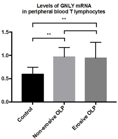
Figure 1. RT-qPCR results: the expression level of GNLY mRNA in non-erosive and erosive OLP patients was significantly higher than that in the control group
Quantitative real-time RT-polymerase chain reaction (RT-qPCR)
The expression levels of granulysin (GNLY) mRNA were detected both in the peripheral blood T lymphocytes and in oral mucosal tis-sues. According to the manufacturer’s instruc -tions, total cellular RNA was extracted with Total RNA kit I (OMEGA, USA). The RNA concen -tration was determined using spectrophotome-ter (GE, USA). Then, the RNA was reverse-tra-nscribed into cDNA using PrimeScriptTMRT Master Mix SYBR green (Takara, Japan). At the end of the PCR cycles, the melting curve analy -sis was performed to validate the specific gen -eration of the expected PCR product. The prim -er sequences w-ere designed in the laboratory and synthesized by General Biotech (General, PRC) based on the mRNA sequences obtained from the NCBI. For GNLY, the sequences of the primers used are: forward primer-CTCTCGTC-TGAGCCCTGAGT and reverse primer-TGGGC
T-TATCCACCATCTTC. The 2-ΔΔCt method was used to calculate the relative quantitative results to determine the mRNA expression of the target gene. Each RT-qPCR experiment was done in duplicates.
Enzyme-linked immunosorbent assay (ELISA)
To detect the protein expression of GNLY, we collected the peripheral blood plasma and cen-trifuged it at 5000 × g for 20 min. According to the ELISA kit instructions (Boster, USA), the O.D. values of GNLY were measured at 450 nm using a microplate reader at the end of 10 min of chromotherapy.
Immunohistochemistry
Immunohistochemistry was used to detect the expression of GNLY in the oral mucosa tissue specimens of the OLP group and the control group. The paraffin sections (4 μm) were dewaxed, hydrated, and then rinsed with tap water. The peroxidase blocking reagent was added to the slices and incubated for 10 min -utes. The primary antibody was added drop-wise to the slice and incubated at room tem-perature for 60 minutes,and then rinsed with PBS 3 times for 3 min each. The sections were incubated at room temperature with ready-to-use MaxVision™/HRP reagent for 15 mins. Colorimetric detection was developed with a freshly equipped diaminobenzidine (DAB) color reagent. Finally, the slides were counterstained with hematoxylin, dehydrated, and sealed. Antibody for GNLY (rabbit polyclonal antibody, ab204594, Abcam, UK) was used at a dilution 1:200 in PBS with 1% bovine serum albumin (BSA).
Histopathologic evaluation
A positive signal showed the brownish yellow staining in the cell nucleus or cytoplasm. All the slides were evaluated by the two senior oral pathologists independently. The result of stain -ing was semi-quantitatively evaluated, which was based on the ratio of staining intensity and proportion of positive cells. The intensity score was defined as 0, negative; 1, weak; 2, moder -ate; or 3, strong brown. The proportion score was defined as 0, negative; 1, < 10%; 2, 11-50%; 3, 51-80%; or 4, > 80% positive cells. The immunoreactive score was calculated as intensity score × proportion score, which was Table 1. mRNA expression levels of GNLY in the
peripheral blood T lymphocytes of the three groups (mean ± SD)
Control
group Non-erosive OLP Erosive OLP
n 20 20 20
[image:3.612.89.286.181.390.2]GNLY mRNA 0.59 ± 0.15 0.97 ± 0.20 0.94 ± 0.37
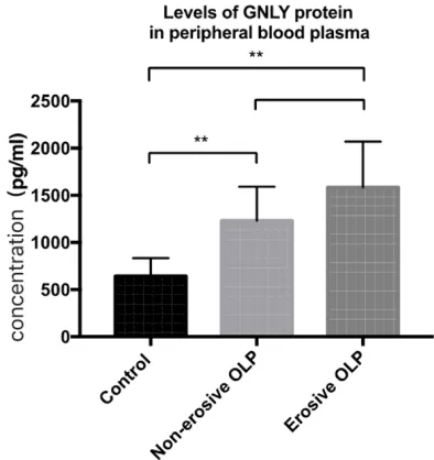
Figure 2. ELISA results: the protein expression level
of GNLY in non-erosive and erosive OLP patients was significantly higher than that in the control group
in the peripheral blood T lympho-cytes of non-erosive and erosive oral lichen planus (OLP) patients was significantly higher than that in the control group (*P < 0.05). The differences between non-erosive and erosive OLP were not significant
divided into three groups based on the final score: 0, negative; 1-4, weak positive expres -sion; > 4, strong positive expression [15, 16].
Statistics
All data were analyzed using a statistical soft -ware package (SPSS version 24.0; SPSS, Chi-cago, IL, USA). The results were expressed as mean ± standard deviation (SD). The differenc -es between the three groups differenc-es were evaluated using ANOVA analysis. Results were considered significant if P < 0.05.
Results
mRNA expression level of GNLY in the periph-eral blood T lymphocytes of non-erosive and erosive OLP patients and control group
The results of RT-qPCR indicated that the mRNA expression level of granulysin (GNLY)
(P > 0.05) (Figure 1). The data are shown in Table 1.
The protein expression level of GNLY in the peripheral blood plasma of non-erosive and erosive OLP patients and control group
The results of ELISA indicated that the protein expression level of GNLY in the peripheral blood plasma of non-erosive and erosive OLP patients was significantly higher than that in the control group (*P < 0.05, **P < 0.01). The differences between non-erosive and erosive OLP were not significant (P > 0.05) (Figure 2). The data are shown in Table 2.
mRNA expression level of GNLY in the oral mucosal tissues of OLP patients and control group
We detected the mRNA expression level of GNLY by RT-qPCR. The results indicated that the mRNA expression level of GNLY in the oral mucosal tissues of non-erosive and erosive OLP patients was significantly higher than that in the healthy controls (**P < 0.01). The differ -ences between non-erosive and erosive OLP were not significant (P > 0.05) (Figure 3). The data are shown in Table 3.
Immunohistochemical detection of GNLY in non-erosive and erosive OLP lesions and nor-mal oral mucosa
The results of immunohistochemistry showed that GNLY was hardly expressed in the normal oral mucosa (Figure 4A). However, in non-ero -sive and ero-sive OLP lesion tissues, GNLY showed weak positive expression mainly in the lymphocyte membrane of lamina propria layer and was less expressed in the epithelial layer (Figure 4B, 4C). The negative control group showed no staining (Figure 4D).
The immunoreactive score of GNLY
The results of immunohistochemistry showed that the immunoreactive score of GNLY in non-Table 2. Expression levels of GNLY in the plasma of the
three groups (pg/ml) (mean ± SD)
Control group Non-erosive OLP Erosive OLP
n 20 20 20
GNLY 645.12 ± 190.28 1231.46 ± 358.50 1585.93 ± 484.28
Figure 3. RT-qPCR results: the expression level of GNLY mRNA in non-erosive and erosive OLP patients was significantly higher than that in the control group
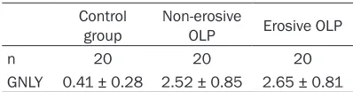
erosive and erosive OLP lesion was significantly higher than that in the normal oral mucosa (**P < 0.01) (Figure 5). The data of the immu -noreactive score (mean ± SD) of GNLY in non-erosive and non-erosive OLP lesion and normal oral mucosa are shown in Table 4.
Discussion
Oral lichen planus (OLP) is a common infla-mmatory disease of unknown etiology. A large body of evidence supports the role of T-cell-mediated immune dysregulation in the patho-genesis. The main pathological characteristics of OLP are hyperkeratosis of epithelium, hyper -plasia of stratum spinosum, colloid body, lique -faction degeneration of basal layer, and band -ed infiltration of T lymphocytes in lamina pro -pria, which are mainly activated T lymphocytes. It is hypothesized that the apoptosis of kerati -nocytes is associated with the pathogenesis of OLP and the different levels of apoptosis are involved in erosive and non-erosive switching of OLP to determine different clinical manifesta -tions [2, 17]. The colloid bodies has been iden-tified to be apoptotic bodies. However, it re-mains elusive how lymphocytes induce the apoptosis of keratinocytes and the infiltration into the oral submucosa [18].
Granulysin (GNLY), in the process of the body immunity, participates in various immune re- sponses including antibacterial, antiviral, and killing cancer cells, with granular components such as perforin and granzyme [6]. GNLY ex-pressed in cytotoxic T lymphocytes (CTLs) and natural killer (NK) cells [19, 20] can cause apoptosis and cytotoxic activity [21-23]. GNLY is the mediator of various skin diseases, includ -ing folliculitis [24], psoriasis [10, 11], acne, lichen planus, and viral vesicles [25]. It has been demonstrated that GNLY was involved of in the worsening of psoriatic arthritis and medi -ated the development of joint lesions [10]. In addition, the levels of serum GNLY are signifi -cantly associated with the severity of adverse
skin events [26]. It has been reported that GNLY is a key mediator for disseminated kerati -nocyte death in the skin lesions of SJS/TEN, suggesting that highly expressed GNLY might promote apoptosis in TEN lesions [9]. Deng et al. found that a low concentration of GNLY has a pro-inflammatory role through CCL5, while a high concentration of GNLY has a cytotoxic effect [7].
In this study, we found that the mRNA expres -sion level of GNLY in the peripheral blood T lym -phocytes and the concentration of GNLY in the plasma of OLP patients were significantly high -er than those in the control group, showing that GNLY might be involved in the occurrence and development of OLP. In addition, our findings showed that there was no significant difference between GNLY in non-erosive OLP and GNLY in erosive OLP. We suspected that this might be due to the simple division of the OLP patients into non-erosion and erosion groups simply according to their clinical manifestations. Ho-wever, OLP is a chronic disease, so the same patient may manifest the non-erosive or ero -sive type at different time periods, and the patient may be at various stages in the course of OLP. Although GNLY might participate in the immune pathogenesis of OLP, it might not be related to the severity of the disease.
Also, we found that the mRNA and protein expression levels of GNLY in non-erosive and erosive OLP tissues were significantly higher than those in the control group. The results were consistent with those in the peripheral blood of OLP, further demonstrating that GNLY might be involved in the pathogenesis of OLP process. The same outcomes were reported in the study of psoriatic arthritis: the percentage of GNLY cells in peripheral blood had no signifi -cant difference between patients of psoriatic arthritis in the acute phase and remittent st- age; but it was higher than in healthy examin-ees [10]. Moreover, GNLY was highly expressed in the skin lesions of SJS/TEN, which was also concordant with our findings [9]. Immunohis-tochemical results showed that GNLY was expressed positively in the lamina propria lym-phocytes and expressed weakly in the epitheli -al layer. However, GNLY scarcely existed in the normal mucosa. As CD8+ T cells and CD4+ T cells are densely infiltrated in OLP lesion, and GNLY is a small molecule found in CTL and NK cells, these indicate that with an increase in Table 3. mRNA expression levels of GNLY in the
oral mucosal tissues of the three groups (mean ± SD)
Control
group Non-erosive OLP Erosive OLP
n 20 20 20
the activated lymphocytes of an OLP lesion, more GNLY are secreted which may be associ -ated with the pathologic changes of OLP. OLP is a chronic disease of the mucous membranes, and colloid bodies is one crucial histologic fea -ture of OLP. Ultrastructural studies have con -firmed that the colloid bodies are apoptotic keratinocytes, but the specific pathway of kera -tinocyte apoptosis in OLP has not been clear. Our study found that GNLY had higher expres -sion in the oral mucosal tissue of OLP, which concurred with the result of skin lichen planus. Granzyme B and GNLY were significantly highly expressed in OLP lesions compared with nor-mal skin [27]. We hypothesized that the acti -Figure 4. Immunohistochemistry: (A) Non-erosive OLP lesion, (B) Erosive OLP lesion, (C) Normal oral mucosa, (D)
[image:6.612.91.524.71.398.2]Negative control (× 200).
Figure 5. Immunoreactive score: the expression level
of GNLY in non-erosive and erosive OLP lesion was significantly higher that than in the normal oral mu
-Table 4. Immunoreactive score of GNLY (mean ± SD)
Control
group Non-erosive OLP Erosive OLP
n 20 20 20
[image:6.612.89.287.447.685.2] [image:6.612.323.522.473.525.2]cytokines and mediate the apoptosis of kerati -nocytes, which might partly explain the patho-genesis of OLP.
In this paper, we studied the expression of GNLY in OLP. According to the experiments, we draw the following conclusions: the increasing GNLY in the peripheral blood and tissues of OLP suggested that GNLY might be involved in the pathogenesis of OLP. The microcirculation in the local microenvironment of OLP might cause the disease in a state of chronic inflam -mation, which resultsin a prolonged course and incurability of this disease.
Acknowledgements
This study was supported by the National Na- ture Science Foundation of China (Grant No. 81470748) and the Priority Academic Program Development of Jiangsu Higher Education Institutions (Grant No. PAPD 2018-87).
Disclosure of conflict of interest
None.
Address correspondence to: Dr. Yuan Fan, Jiangsu Key Laboratory of Oral Diseases, Nanjing Medical University, 136 Hanzhong Road, Nanjing 210029, Jiangsu, China. Tel: +86 25 85031817; Fax: +86 25 86516414; E-mail: [email protected]
References
[1] Kurago ZB. Etiology and pathogenesis of oral
lichen planus: an overview. Oral Surg Oral Med Oral Pathol Oral Radiol 2016; 122: 72-80. [2] Roopashree MR, Gondhalekar RV, Shashikanth
MC, George J, Thippeswamy SH and Shukla A. Pathogenesis of oral lichen planus--a review. J
Oral Pathol Med 2010; 39: 729-734.
[3] Goel S, Marwah A, Kaushik S, Garg VK and Gupta S. Role of serum interleukin-6 in decid
-ing therapy for multidrug resistant oral lichen planus. J Clin Exp Dent 2015; 7: e477-482.
[4] Krensky AM and Clayberger C. Granulysin: a novel host defense molecule. Am J Transplant
2005; 5: 1789-1792.
[5] Zhang H, Zhong C, Shi L, Guo Y and Fan Z. Granulysin induces cathepsin B release from lysosomes of target tumor cells to attack mito
-chondria through processing of bid leading to
Necroptosis. J Immunol 2009; 182: 6993-7000.
[6] Gamen S, Hanson DA, Kaspar A, Naval J, Kren-sky AM and Anel A. Granulysin-induced apop
-tosis. I. Involvement of at least two distinct
pathways. J Immunol 1998; 161: 1758-1764.
[7] Deng A, Chen S, Li Q, Lyu SC, Clayberger C and Krensky AM. Granulysin, a cytolytic molecule, is also a chemoattractant and proinflammatory
activator. J Immunol 2005; 174: 5243-5248. [8] Sarwal MM, Jani A, Chang S, Huie P, Wang Z,
Salvatierra O Jr, Clayberger C, Sibley R, Krensky AM and Pavlakis M. Granulysin expression is a marker for acute rejection and steroid resis
-tance in human renal transplantation. Hum
Immunol 2001; 62: 21-31.
[9] Chung WH, Hung SI, Yang JY, Su SC, Huang SP, Wei CY, Chin SW, Chiou CC, Chu SC, Ho HC, Yang CH, Lu CF, Wu JY, Liao YD and Chen YT. Granulysin is a key mediator for disseminated keratinocyte death in Stevens-Johnson syn -drome and toxic epidermal necrolysis. Nat Med 2008; 14: 1343-1350.
[10] Massari D, Prpic-Massari L, Kehler T, Kastelan M, Curkovic B, Persic V, Ruzic A and Laskarin G. Analysis of granulysin-mediated cytotoxicity in peripheral blood of patients with psoriatic ar -thritis. Rheumatol Int 2012; 32: 2777-2784. [11] Elgarhy LH, Shareef MM and Moustafa SM.
Granulysin expression increases with increas
-ing clinical severity of psoriasis. Clin Exp
De-rmatol 2015; 40: 361-366.
[12] Bardellini E, Amadori F, Flocchini P, Bonadeo S
and Majorana A. Clinicopathological features and malignant transformation of oral lichen
planus: a 12-years retrospective study. Acta Odontol Scand 2013; 71: 834-840.
[13] Rad M, Hashemipoor MA, Mojtahedi A, Zarei MR, Chamani G, Kakoei S and Izadi N.
Cor-relation between clinical and histopathologic
diagnoses of oral lichen planus based on mod
-ified WHO diagnostic criteria. Oral Surg Oral
Med Oral Pathol Oral Radiol Endod 2009; 107: 796-800.
[14] Rodriguez-Nunez I, Blanco-Carrion A, Garcia AG and Rey JG. Peripheral T-cell subsets in pa -tients with reticular and atrophic-erosive oral lichen planus. Oral Surg Oral Med Oral Pathol Oral Radiol Endod 2001; 91: 180-188. [15] Li Z, Wang Y, Qiu J, Li Q, Yuan C, Zhang W, Wang
D, Ye J, Jiang H, Yang J and Cheng J. The poly
-comb group protein EZH2 is a novel therapeu -tic target in tongue cancer. Oncotarget 2013; 4: 2532-2549.
[16] Liu LK, Jiang XY, Zhou XX, Wang DM, Song XL and Jiang HB. Upregulation of vimentin and ab
-errant expression of E-cadherin/beta-catenin
complex in oral squamous cell carcinomas:
correlation with the clinicopathological fea -tures and patient outcome. Mod Pathol 2010; 23: 213-224.
Eur Acad Dermatol Venereol 2017; 31:
880-886.
[18] Brant JM, Aguiar MC, Grandinetti HA, Rodrigues LV and Vasconcelos AC. A comparative study of
apoptosis in reticular and erosive oral lichen
planus. Braz Dent J 2012; 23: 564-569.
[19] Hanson DA, Kaspar AA, Poulain FR and Kren-sky AM. Biosynthesis of granulysin, a novel cy -tolytic molecule. Mol Immunol 1999; 36: 413-422.
[20] Ziesche E, Scheiermann P, Bachmann M, Sa-dik CD, Hofstetter C, Zwissler B, Pfeilschifter J and Muhl H. Dexamethasone suppresses in
-terleukin-22 associated with bacterial infec -tion in vitro and in vivo. Clin Exp Immunol 2009; 157: 370-376.
[21] Kaspar AA, Okada S, Kumar J, Poulain FR, Dr-ouvalakis KA, Kelekar A, Hanson DA, Kluck RM, Hitoshi Y, Johnson DE, Froelich CJ, Thom-pson CB, Newmeyer DD, Anel A, Clayberger C and Krensky AM. A distinct pathway of
cell-mediated apoptosis initiated by granulysin. J Immunol 2001; 167: 350-356.
[22] Semple PL, Watkins M, Davids V, Krensky AM, Hanekom WA, Kaplan G and Ress S. Induction of granulysin and perforin cytolytic mediator expression in 10-week-old infants vaccinated with BCG at birth. Clin Dev Immunol 2011;
2011: 438463.
[23] Won HK, Lee JW, Song WJ, Klaewsongkram J, Kang MG, Park HK, Lee HS, Kim MH, Chang YS, Cho SH and Min KU. Lamotrigine-induced toxic epidermal necrolysis confirmed by in vitro granulysin and cytokine assays. Asia Pac
All-ergy 2014; 4: 253-256.
[24] Oono T, Morizane S, Yamasaki O, Shirafuji Y, Huh WK, Akiyama H and Iwatsuki K. Invol-vement of granulysin-producing T cells in the development of superficial microbial folliculi
-tis. Br J Dermatol 2004; 150: 904-909.
[25] Morizane S, Suzuki D, Tsuji K, Oono T and Iw-atsuki K. The role of CD4 and CD8 cytotoxic T lymphocytes in the formation of viral vesicles. Br J Dermatol 2005; 153: 981-986.
[26] Suda G, Yamamoto Y, Nagasaka A, Furuya K, Kudo M, Chuganji Y, Tsukuda Y, Tsunematsu S, Sato F, Terasita K, Nakai M, Horimoto H, Sho T, Natsuizaka M, Ogawa K, Ohnishi S, Chuma M, Fujita Y, Abe R, Taniguchi M, Nakagawa M, Asahina Y and Sakamoto N. Serum granulysin levels as a predictor of serious telaprevir-in
-duced dermatological reactions. Hepatol Res
2015; 45: 837-845.