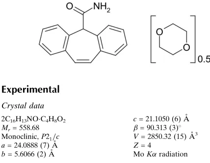
Cytenamide–1,4-dioxane (2/1)
Andrea Johnston,aAlastair J. Florence,a* Francesca J. A. Fabbiani,bKenneth Shanklandcand Colin T. Bedfordd
a
Solid-State Research Group, Strathclyde Institute of Pharmacy and Biomedical Sciences, The John Arbuthnott Building, University of Strathclyde, 27 Taylor Street, Glasgow G4 0NR, Scotland,b
University of Go¨ttingen, GZG, Department of Crystallography, Goldschmidtstrasse 1, D-37077 Go¨ttingen, Germany,cISIS Facility,
Rutherford Appleton Laboratory, Chilton, Didcot, Oxon OX11 0QX, England, and
dUniversity College London, Department of Chemistry, 20 Gordon Street, London
WC1H 0AJ, England
Correspondence e-mail: [email protected]
Received 13 June 2008; accepted 20 June 2008
Key indicators: single-crystal X-ray study;T= 160 K; mean(C–C) = 0.004 A˚;
Rfactor = 0.069;wRfactor = 0.122; data-to-parameter ratio = 13.5.
In the crystal structure of the title compound [systematic name: 5H-dibenzo[a,d ]cycloheptatriene-5-carboxamide–1,4-dioxane (2/1)], 2C16H13NOC4H8O2, the cytenamide
mol-ecules form a hydrogen-bonded R2 2
(8) dimer. The solvent molecule is located between two adjacent cytenamide dimers and forms N—H O hydrogen bonds with one cytenamide molecule from each dimer.
Related literature
For details on experimental methods used to obtain this form, see: Davis et al. (1964); Florence et al. (2003); Florence, Johnston, Fernandes et al. (2006). For related literature on cytenamide, see: Florence, Bedfordet al.(2008). For literature on related molecules, see: Cyret al.(1987); Fleischmanet al.
(2003); Florence, Johnston, Priceet al.(2006); Florence, Leech
et al.(2006); Bandoliet al.(1992); Harrisonet al.(2006); Leech
et al. (2007); Florence, Shankland et al. (2008). For other related literature, see: Etter (1990).
Experimental
Crystal data
2C16H13NOC4H8O2 Mr= 558.68 Monoclinic,P2=c
c= 21.1050 (6) A˚
= 90.313 (3)
V= 2850.32 (15) A˚3
= 0.09 mm1 T= 160 K
0.480.090.03 mm
Data collection
Oxford Diffraction Gemini S diffractometer
Absorption correction: multi-scan (ABSPACK; Oxford Diffraction, 2007)
Tmin= 0.84,Tmax= 1.00
(expected range = 0.838–0.997) 23004 measured reflections 5125 independent reflections 3677 reflections withI> 2(I)
Rint= 0.057
Refinement
R[F2> 2(F2)] = 0.068 wR(F2) = 0.121 S= 1.08 5125 reflections
380 parameters
H-atom parameters constrained
max= 0.47 e A˚3
[image:1.610.44.258.588.754.2]min=0.42 e A˚3
Table 1
Hydrogen-bond geometry (A˚ ,).
D—H A D—H H A D A D—H A
N1—H11 O2i
0.85 2.11 2.962 (3) 171 N1—H12 O4i
0.87 2.22 2.978 (3) 145 N2—H13 O1ii
0.87 1.95 2.823 (3) 177 N2—H14 O3ii
0.87 2.53 3.040 (3) 119
Symmetry codes: (i)x;yþ3 2;z
1
2; (ii)x;yþ 3 2;zþ
1 2.
Data collection: CrysAlis CCD (Oxford Diffraction, 2007); cell refinement: CrysAlis RED (Oxford Diffraction, 2007); data reduc-tion:CrysAlis REDandSORTAV(Blessing, 1997); program(s) used to solve structure:SIR92(Altomareet al., 1994); program(s) used to refine structure: CRYSTALS (Betteridge et al., 2003); molecular graphics: ORTEP-3 (Farrugia, 1997) and Mercury (Macrae et al., 2006); software used to prepare material for publication:PLATON
(Spek, 2003).
The authors thank the Basic Technology programme of the UK Research Councils for funding this work under the project Control and Prediction of the Organic Solid State (www.cposs.org.uk).
Supplementary data and figures for this paper are available from the IUCr electronic archives (Reference: TK2275).
References
Altomare, A., Cascarano, G., Giacovazzo, G., Guagliardi, A., Burla, M. C., Polidori, G. & Camalli, M. (1994).J. Appl. Cryst.27, 435.
Bandoli, G., Nicolini, M., Ongaro, A., Volpe, G. & Rubello, A. (1992).J. Chem. Crystallogr.22, 177–183.
Betteridge, P. W., Carruthers, J. R., Cooper, R. I., Prout, K. & Watkin, D. J. (2003).J. Appl. Cryst.36, 1487.
Blessing, R. H. (1997).J. Appl. Cryst.30, 421–426.
Cyr, T. D., Matsui, F., Sears, R. W., Curran, N. M. & Lovering, E. G. (1987).J. Assoc. Off. Anal. Chem.70, 836–840.
Davis, M. A., Winthrop, S. O., Thomas, R. A., Herr, F., Charest, M.-P. & Gaudry, R. (1964).J. Med. Chem.7, 88–94.
Etter, M. C. (1990).Acc. Chem. Res.23, 120–126. Farrugia, L. J. (1997).J. Appl. Cryst.30, 565.
Fleischman, S. G., Kuduva, S. S., McMahon, J. A., Moulton, B., Walsh, R. D. B., Rodriguez-Hornedo, N. & Zaworotko, M. J. (2003).Cryst. Growth Des.3, 909–919.
Florence, A. J., Baumgartner, B., Weston, C., Shankland, N., Kennedy, A. R., Shankland, K. & David, W. I. F. (2003).J. Pharm. Sci.92, 1930–1938. Acta Crystallographica Section E
Structure Reports Online
CrystEngComm, DOI: 10.1039/b719717a.
Florence, A. J., Johnston, A., Fernandes, P., Shankland, N. & Shankland, K. (2006).J. Appl. Cryst.39, 922–924.
Florence, A. J., Johnston, A., Price, S. L., Nowell, H., Kennedy, A. R. & Shankland, N. (2006).J. Pharm. Sci.95, 1918–1930.
Florence, A. J., Leech, C. K., Shankland, N., Shankland, K. & Johnston, A. (2006).CrystEngComm,8, 746–747.
Florence, A. J., Shankland, K., Gelbrich, T., Hursthouse, M. B., Shankland, N., Johnston, A., Fernandes, P. & Leech, C. K. (2008).CrystEngComm,10, 26– 28.
Harrison, W. T. A., Yathirajan, H. S. & Anilkumar, H. G. (2006).Acta Cryst.
C62, o240–o242.
Leech, C. K., Florence, A. J., Shankland, K., Shankland, N. & Johnston, A. (2007).Acta Cryst.E63, o675–o677.
Macrae, C. F., Edgington, P. R., McCabe, P., Pidcock, E., Shields, G. P., Taylor, R., Towler, M. & van de Streek, J. (2006).J. Appl. Cryst.39, 453–457. Oxford Diffraction (2007).CrysAlis CCD, CrysAlis RED andABSPACK.
Oxford Diffraction Ltd, Abingdon, Oxfordshire, England. Spek, A. L. (2003).J. Appl. Cryst.36, 7–13.
organic compounds
o1346
Johnstonet al. 2Csupporting information
Acta Cryst. (2008). E64, o1345–o1346 [doi:10.1107/S1600536808018709]
Cytenamide
–
1,4-dioxane (2/1)
Andrea Johnston, Alastair J. Florence, Francesca J. A. Fabbiani, Kenneth Shankland and Colin T.
Bedford
S1. Comment
Cytenamide (CYT) is an analogue of carbamazepine (CBZ), a dibenzazepine drug used to control seizures (Cyr et al.,
1987). CYT-dioxane hemisolvate was produced during an automated parallel crystallization study of CYT (Florence,
Johnston, Fernandes et al., 2006) as part of a wider investigation that couples automated parallel crystallization with
crystal structure prediction methodology to investigate the basic science underlying the solid-state diversity of CBZ
(Florence, Johnston, Price et al., 2006; Florence, Leech et al., 2006; Fleischman et al., 2003) and its closely related
analogues: CYT (Florence et al., 2008a),10,11-dihydrocarbamazepine (Bandoli et al., 1992; Harrison et al., 2006; Leech
et al., 2007) and cyheptamide (Florence, Shankland et al., 2008). The sample was identified as a new form using
multi-sample foil transmission X-ray powder diffraction analysis (Florence et al., 2003). Subsequent manual recrystallization
from a saturated 1,4-dioxane solution by slow evaporation at 298 K yielded a sample suitable for single-crystal X-ray
diffraction (Fig. 1).
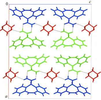
The reported crystal structure is essentially isostructural with that of CBZ-dioxane solvate (2/1) (Florence, Johnston,
Price et al., 2006) and accordingly displays very similar packing arrangements. Specifically, the molecules crystallize
with two CYT and one 1,4-dioxane molecules in the asymmetric unit. Pairs of CYT molecules form an R22(8) dimer
motif (Etter, 1990) via two N—H···O hydrogen bonds and a further two N—H···O contacts link CYT dimers with solvent
molecules to form an infinite chain that extends in the c-direction (Table 1 & Fig. 2).
S2. Experimental
A sample of cytenamide was synthesized according to a modification of the published method (Davis et al., 1964). A
single crystal of (I) was grown from a saturated solution of cytenamide in 1,4-dioxane by isothermal solvent evaporation
at 298 K.
S3. Refinement
Data were merged with SORTAV (Blessing, 1997) and a theta cut off of 25.0 ° was applied due to weak scattering.
H-atoms were found on a difference Fourier map and were initially refined with soft restraints on the bond lengths and
angles to regularize their geometry. The C-H distances are in the range 0.92 - 0.98 Å and Uiso(H) = 1.2-1.5Ueq(C). Atoms
C12 C13 C14 and to some extent C15 suffer from large and prolate thermal ellipsoids. During refinement, the crystal was
found to be twinned, according to the twin law expressed by the following matrix: 1 0 0.012, 0 - 1 0, 0 0 - 1 i.e.
approximately about the a axis, giving rise to a twin component of ca 10%. Inclusion of the twin resulted in a reduction
supporting information
sup-2
[image:4.610.129.480.75.285.2]Acta Cryst. (2008). E64, o1345–o1346
Figure 1
Figure 2
Packing diagram for (I) viewed down the b axis, showing the CYT R22(8) dimer motif further linked by N—H···O
hydrogen bonds between CYT and dioxane molecules to form an infinite chain in the [001] direction. Molecules are
coloured according to symmetry equivalence (CYT blue and green, dioxane molecules red) and hydrogen bonds are
represented by dashed lines.
5H-dibenzo[a,d]cycloheptatriene-5-carboxamide 1,4-dioxane hemisolvate
Crystal data
2C16H13NO·C4H8O2 Mr = 558.68
Monoclinic, P21/c
Hall symbol: -P 2ybc
a = 24.0888 (7) Å
b = 5.6066 (2) Å
c = 21.1050 (6) Å
β = 90.313 (3)°
V = 2850.32 (15) Å3 Z = 4
F(000) = 1184
Dx = 1.302 Mg m−3
Mo Kα radiation, λ = 0.71073 Å Cell parameters from 4484 reflections
θ = 3–27°
supporting information
sup-4
Acta Cryst. (2008). E64, o1345–o1346 Data collection
Area
diffractometer
Graphite monochromator
Detector resolution: 15.9745 pixels mm-1 ω scans
Absorption correction: multi-scan (ABSPACK; Oxford Diffraction, 2007)
Tmin = 0.84, Tmax = 1.00
23004 measured reflections 5125 independent reflections 3677 reflections with I > 2σ(I)
Rint = 0.057
θmax = 25.2°, θmin = 2.6° h = −28→28
k = 0→6
l = 0→25
Refinement
Refinement on F2
Least-squares matrix: full
R[F2 > 2σ(F2)] = 0.068 wR(F2) = 0.121 S = 1.08 5125 reflections 380 parameters 0 restraints
Primary atom site location: structure-invariant direct methods
Hydrogen site location: inferred from neighbouring sites
H-atom parameters constrained
Method = Modified Sheldrick w = 1/[σ2(F2) +
(0.03P)2 + 2.31P],
where P = [max(Fo2,0) + 2Fc2]/3
(Δ/σ)max < 0.001
Δρmax = 0.47 e Å−3
Δρmin = −0.42 e Å−3
Fractional atomic coordinates and isotropic or equivalent isotropic displacement parameters (Å2)
x y z Uiso*/Ueq
C27 0.08061 (11) 0.1544 (5) 0.64959 (12) 0.0235 C28 0.05571 (11) 0.1337 (6) 0.58956 (13) 0.0301 C29 0.06815 (12) −0.0532 (6) 0.54957 (13) 0.0341 C30 0.10567 (12) −0.2249 (6) 0.56844 (13) 0.0300 C31 0.13153 (12) −0.2035 (5) 0.62664 (12) 0.0262 C32 0.12027 (11) −0.0156 (5) 0.66747 (12) 0.0221 C33 0.24356 (17) 1.1206 (7) 0.52906 (16) 0.0547 C34 0.20870 (16) 1.1411 (8) 0.47152 (17) 0.0612 C35 0.26150 (18) 0.8437 (9) 0.42284 (16) 0.0654 C36 0.29643 (16) 0.8250 (8) 0.47959 (16) 0.0576 O2 0.20878 (8) 0.3034 (4) 0.67272 (9) 0.0319 O1 0.29456 (9) 0.8317 (4) 0.27135 (9) 0.0416 N1 0.28447 (10) 0.7810 (4) 0.16701 (10) 0.0276 O4 0.26727 (11) 0.8892 (5) 0.53470 (11) 0.0601 N2 0.21372 (10) 0.3049 (4) 0.77846 (10) 0.0301 O3 0.23813 (13) 1.0729 (6) 0.41616 (11) 0.0766
H12 0.2929 0.7005 0.1331 0.0337*
H13 0.2388 0.4167 0.7778 0.0346*
H14 0.2014 0.2492 0.8143 0.0341*
H11 0.2604 0.8928 0.1658 0.0335*
H21 0.3220 0.3680 0.2272 0.0227*
H41 0.3422 0.1772 0.1371 0.0304*
H51 0.3877 0.1358 0.0418 0.0373*
H61 0.4516 0.4256 0.0100 0.0391*
H71 0.4724 0.7421 0.0774 0.0366*
H91 0.4521 0.9769 0.1669 0.0320*
H101 0.4495 1.0070 0.2695 0.0308*
H121 0.4646 0.8312 0.3704 0.0366*
H131 0.4420 0.5553 0.4466 0.0428*
H141 0.3833 0.2408 0.4229 0.0431*
H151 0.3430 0.2207 0.3235 0.0324*
H181 0.1771 −0.1295 0.7304 0.0252*
H201 0.1550 −0.2818 0.8256 0.0321*
H211 0.1138 −0.2613 0.9244 0.0401*
H221 0.0540 0.0580 0.9478 0.0424*
H231 0.0330 0.3320 0.8699 0.0339*
H251 0.0491 0.5142 0.7700 0.0313*
H261 0.0473 0.4794 0.6683 0.0327*
H281 0.0308 0.2494 0.5766 0.0346*
H291 0.0505 −0.0629 0.5088 0.0391*
H301 0.1144 −0.3536 0.5422 0.0347*
H311 0.1579 −0.3182 0.6393 0.0303*
H331 0.2732 1.2396 0.5258 0.0635*
H332 0.2202 1.1544 0.5657 0.0653*
H341 0.1959 1.3056 0.4670 0.0711*
H342 0.1775 1.0333 0.4757 0.0712*
H351 0.2822 0.8002 0.3857 0.0752*
supporting information
sup-6
Acta Cryst. (2008). E64, o1345–o1346
H361 0.3290 0.9332 0.4750 0.0676*
H362 0.3100 0.6606 0.4851 0.0684*
Atomic displacement parameters (Å2)
U11 U22 U33 U12 U13 U23
Geometric parameters (Å, º)
C1—C2 1.539 (4) C20—H201 0.940
C1—O1 1.232 (3) C21—C22 1.382 (4)
C1—N1 1.320 (3) C21—H211 0.944
C2—C3 1.508 (4) C22—C23 1.375 (4)
C2—C16 1.511 (4) C22—H221 0.948
C2—H21 0.981 C23—C24 1.401 (4)
C3—C4 1.385 (4) C23—H231 0.944
C3—C8 1.408 (4) C24—C25 1.457 (4)
C4—C5 1.381 (4) C25—C26 1.337 (4)
C4—H41 0.946 C25—H251 0.955
C5—C6 1.382 (4) C26—C27 1.457 (4)
C5—H51 0.945 C26—H261 0.932
C6—C7 1.384 (4) C27—C28 1.404 (4)
C6—H61 0.940 C27—C32 1.400 (4)
C7—C8 1.397 (4) C28—C29 1.379 (4)
C7—H71 0.946 C28—H281 0.924
C8—C9 1.460 (4) C29—C30 1.378 (4)
C9—C10 1.341 (4) C29—H291 0.959
C9—H91 0.935 C30—C31 1.379 (4)
C10—C11 1.456 (4) C30—H301 0.934
C10—H101 0.929 C31—C32 1.389 (4)
C11—C12 1.397 (4) C31—H311 0.942
C11—C16 1.400 (4) C33—C34 1.477 (5)
C12—C13 1.378 (4) C33—O4 1.423 (4)
C12—H121 0.943 C33—H331 0.980
C13—C14 1.376 (4) C33—H332 0.977
C13—H131 0.924 C34—O3 1.422 (4)
C14—C15 1.379 (4) C34—H341 0.976
C14—H141 0.934 C34—H342 0.969
C15—C16 1.389 (4) C35—C36 1.464 (5)
C15—H151 0.928 C35—O3 1.410 (5)
C17—C18 1.542 (4) C35—H351 0.963
C17—O2 1.231 (3) C35—H352 0.979
C17—N2 1.319 (3) C36—O4 1.409 (4)
C18—C19 1.519 (4) C36—H361 0.997
C18—C32 1.511 (3) C36—H362 0.984
C18—H181 0.969 N1—H12 0.871
C19—C20 1.389 (4) N1—H11 0.855
C19—C24 1.401 (4) N2—H13 0.870
C20—C21 1.379 (4) N2—H14 0.873
C2—C1—O1 120.0 (2) C20—C21—H211 119.5
C2—C1—N1 117.9 (2) C22—C21—H211 120.8
O1—C1—N1 122.0 (3) C21—C22—C23 119.5 (3)
C1—C2—C3 113.0 (2) C21—C22—H221 120.9
supporting information
sup-8
Acta Cryst. (2008). E64, o1345–o1346
C3—C2—C16 115.6 (2) C22—C23—C24 122.0 (3)
C1—C2—H21 105.5 C22—C23—H231 119.2
C3—C2—H21 106.1 C24—C23—H231 118.9
C16—C2—H21 106.0 C19—C24—C23 117.9 (3)
C2—C3—C4 119.3 (3) C19—C24—C25 124.0 (2) C2—C3—C8 121.3 (2) C23—C24—C25 118.0 (3) C4—C3—C8 119.3 (2) C24—C25—C26 129.0 (3)
C3—C4—C5 121.5 (3) C24—C25—H251 115.1
C3—C4—H41 119.0 C26—C25—H251 115.8
C5—C4—H41 119.5 C25—C26—C27 129.7 (3)
C4—C5—C6 119.8 (3) C25—C26—H261 115.7
C4—C5—H51 119.2 C27—C26—H261 114.6
C6—C5—H51 121.0 C26—C27—C28 117.6 (3)
C5—C6—C7 119.3 (3) C26—C27—C32 124.0 (2)
C5—C6—H61 120.0 C28—C27—C32 118.3 (3)
C7—C6—H61 120.7 C27—C28—C29 121.5 (3)
C6—C7—C8 121.9 (3) C27—C28—H281 118.9
C6—C7—H71 119.4 C29—C28—H281 119.7
C8—C7—H71 118.8 C28—C29—C30 119.9 (3)
C3—C8—C7 118.1 (3) C28—C29—H291 119.7
C3—C8—C9 124.0 (2) C30—C29—H291 120.4
C7—C8—C9 118.0 (3) C29—C30—C31 119.3 (3)
C8—C9—C10 129.1 (3) C29—C30—H301 121.2
C8—C9—H91 113.3 C31—C30—H301 119.6
C10—C9—H91 117.6 C30—C31—C32 122.0 (3)
C9—C10—C11 129.7 (3) C30—C31—H311 119.5
C9—C10—H101 115.4 C32—C31—H311 118.5
C11—C10—H101 114.9 C18—C32—C27 121.4 (2) C10—C11—C12 117.9 (3) C18—C32—C31 119.5 (2) C10—C11—C16 123.7 (2) C27—C32—C31 119.0 (2) C12—C11—C16 118.4 (3) C34—C33—O4 111.5 (3) C11—C12—C13 121.5 (3) C34—C33—H331 107.5
C11—C12—H121 119.0 O4—C33—H331 109.5
C13—C12—H121 119.4 C34—C33—H332 107.9
C12—C13—C14 119.8 (3) O4—C33—H332 110.1
C12—C13—H131 119.7 H331—C33—H332 110.3
C14—C13—H131 120.5 C33—C34—O3 111.8 (3)
C13—C14—C15 119.5 (3) C33—C34—H341 109.3
C13—C14—H141 120.1 O3—C34—H341 109.4
C15—C14—H141 120.4 C33—C34—H342 108.3
C14—C15—C16 121.7 (3) O3—C34—H342 107.3
C14—C15—H151 119.5 H341—C34—H342 110.8
C16—C15—H151 118.8 C36—C35—O3 112.0 (3)
C2—C16—C11 121.4 (2) C36—C35—H351 110.5
C2—C16—C15 119.6 (3) O3—C35—H351 111.0
C11—C16—C15 118.9 (2) C36—C35—H352 108.2
C18—C17—O2 120.9 (2) O3—C35—H352 107.7
O2—C17—N2 122.3 (3) C35—C36—O4 111.7 (3) C17—C18—C19 112.4 (2) C35—C36—H361 109.1 C17—C18—C32 111.3 (2) O4—C36—H361 108.7 C19—C18—C32 115.1 (2) C35—C36—H362 110.6
C17—C18—H181 105.2 O4—C36—H362 108.0
C19—C18—H181 106.1 H361—C36—H362 108.7
C32—C18—H181 105.9 C1—N1—H12 120.5
C18—C19—C20 119.5 (3) C1—N1—H11 118.2
C18—C19—C24 120.9 (2) H12—N1—H11 121.2
C20—C19—C24 119.4 (2) C33—O4—C36 111.5 (3)
C19—C20—C21 121.4 (3) C17—N2—H13 118.9
C19—C20—H201 118.2 C17—N2—H14 120.4
C21—C20—H201 120.4 H13—N2—H14 120.7
C20—C21—C22 119.7 (3) C34—O3—C35 111.3 (3)
Hydrogen-bond geometry (Å, º)
D—H···A D—H H···A D···A D—H···A
N1—H11···O2i 0.85 2.11 2.962 (3) 171
N1—H12···O4i 0.87 2.22 2.978 (3) 145
N2—H13···O1ii 0.87 1.95 2.823 (3) 177
N2—H14···O3ii 0.87 2.53 3.040 (3) 119