
Aqua[4
000-(4-chlorophenyl)-2,2
000:6
000,2
000000-terpyridine]nitratocopper(II) nitrate [4
000-(4-chlorophenyl)-2,2
000:6
000,2
000000-terpyridine]-dinitratocopper(II) monohydrate
Juan Zhao, Shuo Shi,* Tianming Yao* and Zhongfei Tan
Department of Chemistry, Tongji University, Shanghai, People’s Republic of China Correspondence e-mail: [email protected], [email protected]
Received 30 December 2010; accepted 12 February 2011
Key indicators: single-crystal X-ray study;T= 293 K; mean(C–C) = 0.003 A˚; disorder in main residue;Rfactor = 0.044;wRfactor = 0.115; data-to-parameter ratio = 13.3.
The crystal structure of the title compound, [Cu(NO3
)-(C21H14ClN3)(H2O)]NO3[Cu(NO3)2(C21H14ClN3)]H2O,
con-sists of two crystallographically independent CuIIcomplexes, in which each copper cation is pentacoordinated by three N atoms of the chelating ligand and two O atoms of nitrate anions or water molecules. One of the coordinated nitrate anions is disordered over two set of sites in a 0.85:0.15 ratio.
Related literature
For the use of substituted terpyridine ligands in coordination chemistry due to their ability to form complexes with transi-tion metals, see: Chenet al.(2010); Fenget al.(2006); Houet al.
(2005); Mutai et al. (2001). For the synthesis of the title compound, see: Mutaiet al.(2001). For related structures, see: Granifoet al.(2004); Chenet al.(2010). For the biochemcial importance of terpyridine ligands, see: Bertrandet al.(2007); Maityet al.(2010).
Experimental
Crystal data
[Cu(NO3)2(C21H14ClN3 )]-[Cu(NO3)(C21H14ClN3)(H2 O)]-NO3H2O
Mr= 1098.76
Monoclinic,P21=c a= 14.7172 (2) A˚ b= 15.0680 (2) A˚ c= 20.4214 (3) A˚
= 105.377 (1) V= 4366.51 (10) A˚3 Z= 4
CuKradiation = 3.04 mm1 T= 293 K
0.300.200.20 mm
Data collection
Bruker SMART CCD area-detector diffractometer
Absorption correction: multi-scan (SADABS; Bruker, 2000) Tmin= 0.462,Tmax= 0.581
17732 measured reflections 8572 independent reflections 7142 reflections withI> 2(I) Rint= 0.027
Refinement
R[F2> 2(F2)] = 0.044 wR(F2) = 0.115 S= 1.00 8572 reflections 643 parameters
15 restraints
H-atom parameters constrained max= 0.56 e A˚
3 min=0.27 e A˚
3
Data collection:SMART(Bruker, 2000); cell refinement:SAINT
(Bruker, 2000); data reduction:SAINT; program(s) used to solve structure:SHELXS97(Sheldrick, 2008); program(s) used to refine structure:SHELXL97(Sheldrick, 2008); molecular graphics:XPin
SHELXTL(Sheldrick, 2008); software used to prepare material for publication:CIFTABinSHELXL97.
The authors thank the National Natural Science Foundation of China (20901060, 20871094) and the Program for Young Excellent Talents in Tongji University.
Supplementary data and figures for this paper are available from the IUCr electronic archives (Reference: NC2216).
References
Bertrand, H., Monchaud, D., Cian, A.-D., Guillot, R., Mergny, J.-L. & Teulade-Fichou, M.-P. (2007).Org. Biomol. Chem.5, 2555–2559.
Bruker (2000).SMART,SAINTandSADABS. Bruker AXS Inc., Madison, Wisconsin, USA.
Chen, F.-T. (2010).Acta Cryst.E66, m755.
Feng, H., Zhou, X.-P., Wu, T., Li, D., Yin, Y.-G. & Ng, S. W. (2006).Inorg. Chim. Acta,359, 4027–4035.
Granifo, J., Garland, M.-T. & Baggio, R. (2004).Inorg. Chem. Commun.7, 77– 81.
Hou, L., Li, D., Shi, W.-J., Yin, Y.-G. & Ng, S. W. (2005).Inorg. Chem.44, 7825– 7832.
Maity, B., Roy, M., Banik, B., Majumdar, R., Dighe, R. R., & Chakravarty, A. R. (2010).Organometallics,29, 3632–3641.
Mutai, T., Chen, J.-D., Arita, S. & Araki, K. (2001).J. Chem. Soc. Perkin Trans. 2, pp. 1045–1050.
Sheldrick, G. M. (2008).Acta Cryst.A64, 112–122. Acta Crystallographica Section E
Structure Reports
Online
supporting information
Acta Cryst. (2011). E67, m346 [doi:10.1107/S1600536811005290]
Aqua[4
′
-(4-chlorophenyl)-2,2
′
:6
′
,2
′′
-terpyridine]nitratocopper(II) nitrate [4
′
-(4-chlorophenyl)-2,2
′
:6
′
,2
′′
-terpyridine]dinitratocopper(II) monohydrate
Juan Zhao, Shuo Shi, Tianming Yao and Zhongfei Tan
S1. Comment
Substituted terpyridine ligands have attracted widespread attention in coordination chemistry due to their ability to form
complexes with transition metals, (Chen et al.,2010; Feng et al.,2006; Granifo et al.,2004; Hou et al.,2005; Mutai et
al.,2001). Recent studies reveal that under certain conditions, copper complexes with phenanthroline and polypyridine ligands have good DNA binding activity, which attracted great interest in biochemistry (Bertrand et al.,2007; Maity et
al.,2010). The crystal structure of the title compound was determined as part of an ongoing study of the properties of copper complex containing terpyridyl ligands.
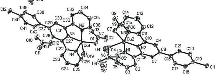
The crystal structure of the title compound consists of two crystallographically independent complexes, in which the
copper cations are always penta coordinated within a distorted square-pyramidal coordination (Fig.1). In one of these
complexes two copper atom is coordinated by three N atoms of the tridentrate chelating ligand and two O atoms of two
independent two nitrate anions.In the other complex the copper is coordinated by three N atoms of the chelating ligand,
one O atom of one nitrate anion and an O atom of one water molecule. The structure contains one additional nitrate anion
and one additional water molecule that are not connected to the copper cations. The central Cu–N bond lengths [Cu(1)—
N(5) = 1.9287 (16) Å and Cu(2)—N(2) = 1.9256 (18)Å] are slightly shorter than those to the outer N atoms of
2.0178 (18), 2.0277 (18), 2.0096 (19) and 2.014 (2)Å. The bond angles to the N atoms of the terpyridyl unit of 80.40 (8),
80.07 (8), 80.39 (7) and 79.97 (7) ° deviate from the ideal values which is a common characteristics for
terpyridyl-containing complexes (Granifo et al.,2004). The apical Cu–O bond lenghts (Cu(1)—O(1 W) 2.208 (2) Å and Cu(2)—
O(4) 2.163 (3) Å) are longer than the other Cu–O bonds.
S2. Experimental
4′-(4-Chlorophenyl)-2,2′:6′,2′′-terpyridine (Cl-ptp) has been synthesized by a procedure reported in the literature (Mutai
et al. 2001). A solution of Cl-ptp (42.35 mg, 0.124 mmol) in dichloromethane (3 ml) was mixed with anhydrous aceto-nitrile (3 ml) dissolving copper nitrate (29.96 mg, 0.124 mmol),then sealed and kept it in refrigerator. Several days later,
blue crystals were collected by filtration.
S3. Refinement
All H atom were positioned geometrically and refined as riding, with aromatic C—H =0.93 Å and with Uiso(H) =
Figure 1
ORTEP drawing of the title compound with labelling showing displacement ellipsoids at the 30% probability level. All
hydrogen atoms have been omitted for clarity.
Aqua[4′-(4-chlorophenyl)-2,2′:6′,2′′-terpyridine]nitratocopper(II) nitrate [4′-(4-chlorophenyl)-2,2′:6′,2′′
-terpyridine]dinitratocopper(II) monohydrate
Crystal data
[Cu(NO3)2(C21H14ClN3)][Cu(NO3)(C21H14ClN3) (H2O)]NO3·H2O
Mr = 1098.76
Monoclinic, P21/c a = 14.7172 (2) Å b = 15.0680 (2) Å c = 20.4214 (3) Å β = 105.377 (1)° V = 4366.51 (10) Å3
Z = 4
F(000) = 2232 Dx = 1.671 Mg m−3
Cu Kα radiation, λ = 1.54178 Å µ = 3.04 mm−1
T = 293 K Block, blue
0.30 × 0.20 × 0.20 mm
Data collection
Bruker SMART CCD area-detector diffractometer
Radiation source: fine-focus sealed tube Graphite monochromator
ω scans
Absorption correction: multi-scan (SADABS; Bruker, 2000) Tmin = 0.462, Tmax = 0.581
17732 measured reflections 8572 independent reflections 7142 reflections with I > 2σ(I) Rint = 0.027
θmax = 72.9°, θmin = 3.1° h = −18→17
k = −18→17 l = −24→25
Refinement Refinement on F2 Least-squares matrix: full R[F2 > 2σ(F2)] = 0.044 wR(F2) = 0.115 S = 1.00 8572 reflections 643 parameters 15 restraints
Primary atom site location: structure-invariant direct methods
Secondary atom site location: difference Fourier map
Hydrogen site location: inferred from neighbouring sites
H-atom parameters constrained w = 1/[σ2(F
o2) + (0.0792P)2] where P = (Fo2 + 2Fc2)/3 (Δ/σ)max = 0.001
Special details
Geometry. All e.s.d.'s (except the e.s.d. in the dihedral angle between two l.s. planes) are estimated using the full
covariance matrix. The cell e.s.d.'s are taken into account individually in the estimation of e.s.d.'s in distances, angles and torsion angles; correlations between e.s.d.'s in cell parameters are only used when they are defined by crystal symmetry. An approximate (isotropic) treatment of cell e.s.d.'s is used for estimating e.s.d.'s involving l.s. planes.
Refinement. Refinement of F2 against ALL reflections. The weighted R-factor wR and goodness of fit S are based on F2,
conventional R-factors R are based on F, with F set to zero for negative F2. The threshold expression of F2 > σ(F2) is used only for calculating R-factors(gt) etc. and is not relevant to the choice of reflections for refinement. R-factors based on F2 are statistically about twice as large as those based on F, and R- factors based on ALL data will be even larger.
Fractional atomic coordinates and isotropic or equivalent isotropic displacement parameters (Å2)
x y z Uiso*/Ueq Occ. (<1)
Cu2 0.37161 (3) 0.70822 (2) 0.243697 (17) 0.04814 (11) Cl1 −0.06351 (4) 0.37787 (4) −0.19024 (3) 0.05263 (14) N1 0.38825 (15) 0.58174 (12) 0.27589 (10) 0.0443 (4) N2 0.30021 (13) 0.64907 (11) 0.16278 (9) 0.0395 (3) N3 0.32876 (16) 0.81395 (13) 0.18301 (11) 0.0499 (4) N8 0.57336 (15) 0.69059 (13) 0.25635 (10) 0.0481 (4)
O4 0.5200 (2) 0.73693 (19) 0.28333 (17) 0.0547 (6) 0.85 O5 0.5407 (3) 0.6533 (3) 0.20330 (17) 0.0886 (10) 0.85 O6 0.6581 (2) 0.6877 (3) 0.28725 (18) 0.0811 (8) 0.85 O4′ 0.5462 (19) 0.7293 (19) 0.2997 (11) 0.085 (9)* 0.15 O5′ 0.5103 (12) 0.6818 (14) 0.2011 (8) 0.074 (5)* 0.15 O6′ 0.6530 (13) 0.6649 (19) 0.2589 (15) 0.107 (8)* 0.15 N9 0.28984 (17) 0.76429 (13) 0.34439 (11) 0.0520 (5)
H34A 0.5448 1.1112 0.4246 0.051* C35 0.52309 (16) 0.98441 (16) 0.39380 (11) 0.0465 (5) H35A 0.4834 0.9964 0.3510 0.056* C36 0.54549 (17) 0.89771 (15) 0.41408 (11) 0.0452 (4) H36A 0.5198 0.8517 0.3846 0.054* C37 0.86092 (15) 0.98066 (13) 0.75508 (10) 0.0376 (4) C38 0.87342 (17) 1.07184 (14) 0.75065 (11) 0.0447 (5) H38A 0.8495 1.1006 0.7094 0.054* C39 0.9212 (2) 1.12044 (14) 0.80709 (13) 0.0516 (5) H39A 0.9288 1.1814 0.8037 0.062* C40 0.95691 (18) 1.07797 (15) 0.86793 (11) 0.0473 (5) C41 0.9460 (2) 0.98785 (16) 0.87392 (12) 0.0600 (7) H41A 0.9706 0.9594 0.9152 0.072* C42 0.8979 (2) 0.94048 (14) 0.81760 (12) 0.0581 (7) H42A 0.8900 0.8797 0.8217 0.070* N10 0.89519 (16) 0.71939 (12) 0.86786 (10) 0.0501 (5) O10 0.9058 (2) 0.76082 (15) 0.92176 (12) 0.0773 (7) O11 0.95399 (15) 0.72583 (13) 0.83381 (10) 0.0598 (4) O12 0.82643 (18) 0.67101 (17) 0.84712 (13) 0.0789 (7) O2W 0.88021 (19) 1.31295 (18) 0.57881 (14) 0.0829 (7) H1O2 0.9248 1.2790 0.5993 0.124* H2O2 0.8795 1.3574 0.6043 0.124*
Atomic displacement parameters (Å2)
U11 U22 U33 U12 U13 U23
Geometric parameters (Å, º)
C16—C21 1.390 (3) C39—H39A 0.9300 C16—C17 1.399 (3) C40—C41 1.377 (3) C17—C18 1.383 (3) C41—C42 1.379 (3) C17—H17A 0.9300 C41—H41A 0.9300 C18—C19 1.378 (3) C42—H42A 0.9300 C18—H18A 0.9300 N10—O12 1.228 (3) C19—C20 1.383 (3) N10—O10 1.239 (3) C20—C21 1.386 (3) N10—O11 1.249 (3) C20—H20A 0.9300 O2W—H1O2 0.8500 C21—H21A 0.9300 O2W—H2O2 0.8501 Cu1—N5 1.9287 (16)
![Crystal structure of ethyl 2 {4 [(5 chloro 1 benzofuran 2 yl)methyl] 3 methyl 6 oxo 1,6 dihydropyridazin 1 yl}acetate](data:image/gif;base64,R0lGODlhAQABAIAAAP///wAAACH5BAEAAAAALAAAAAABAAEAAAICRAEAOw==)