organic papers
o1792
Yathirajanet al. C19H25N2S+C6H2N3O7 doi:10.1107/S1600536807011543 Acta Cryst.(2007). E63, o1792–o1794
Acta Crystallographica Section E
Structure Reports
Online
ISSN 1600-5368
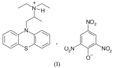
Ethopropazinium picrate
H. S. Yathirajan,aM. A. Ashok,a B. Narayana Acharaand Michael Bolteb*
aDepartment of Studies in Chemistry, University
of Mysore, Manasagangotri, Mysore 570 006, India, andb
Institut fu¨r Anorganische Chemie, J. W. Goethe-Universita¨t Frankfurt, Max-von-Laue-Strasse 7, 60438 Frankfurt/Main, Germany
Correspondence e-mail: [email protected]
Key indicators
Single-crystal X-ray study
T= 173 K
Mean(C–C) = 0.005 A˚
Rfactor = 0.077
wRfactor = 0.212
Data-to-parameter ratio = 12.7
For details of how these key indicators were automatically derived from the article, see http://journals.iucr.org/e.
Received 21 February 2007 Accepted 12 March 2007
#2007 International Union of Crystallography All rights reserved
The title compound [systematic name:
10-[2-(diethylamino)-propyl]phenothiazinium 2,4,6-trinitrophenolate], C19H25
-N2S +
C6H2N3O7
, is a pharmacologially active compound. The dihedral angle between the two outer aromatic rings of the phenothiazine unit is 38.64 (12). The crystal packing is
stabilized by N—H O hydrogen bonds and several weak
C—H O contacts. The molecular conformation of the cation
does not change significantly when it is crystallized with chloride or perrhenate as the anion.
Comment
Ethopropazine is an anticholinergic agent with some anti-histaminic and ganglionic blocking activity (Bratfos & Haug, 1979). The present work results in the formation of a salt by the interaction between ethopropazinium hydrochloride and 2,4,6-trinitrophenol in an aqueous medium.
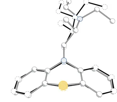
[image:1.610.234.430.388.506.2]A perspective view of the title compound, (I), is shown in Fig. 1. Bond lengths and angles can be regarded as normal (Cambridge Structural Database, Version 5.28, November
2006; MOGUL, Version 1.1; Allen, 2002; Brunoet al., 2004).
The dihedral angle between the two aromatic rings of the
phenothiazine unit is 38.64 (12). Crystallographic data for
ethopropazinium chloride, (Ia) (Marsau & Calas, 1971; Klein
et al., 1994), and ethopropazinium perrhenate, (Ib) (Gowdaet al., 1994), have been published, but since there is a coordinate error in the report by Marsau & Calas (1971), the structure given by Kleinet al.(1994) is employed for comparison. Least-squares overlays of the ethopropazinium cations of (I) and (Ia) (r.m.s. deviation 0.066 A˚ ) as well as of (I) and (Ib) (r.m.s. deviation 0.120 A˚ ), fitting only the phenothiazine units, are shown in Figs. 2 and 3. As can be seen, the molecular conformation is not significantly different in the compared
structures. The crystal packing is stabilized by N—H O
hydrogen bonds and several weak C—H O contacts
Experimental
Profenamine hydrochloride (0.7010 g, 0.02 M) and picric acid (0.4610 g, 0.02M) were dissolved in distilled water (100 ml), mixed and stirred in a beaker at room temperature. The separated yellow salt was washed well with distilled water, filtered and dried in a vacuum desiccator over phosphorus pentoxide. The complex was recrystallized from acetonitrile (m.p. 378 K).
Crystal data
C19H25N2S+C6H2N3O7
Mr= 541.58
Monoclinic,C2=c a= 36.876 (3) A˚
b= 8.4622 (4) A˚
c= 16.5727 (11) A˚
= 104.163 (6)
V= 5014.3 (6) A˚3
Z= 8
MoKradiation
= 0.19 mm1
T= 173 (2) K 0.290.260.25 mm
Data collection
Stoe IPDS-II two-circle diffractometer
Absorption correction: multi-scan (MULABS; Spek, 2003; Blessing, 1995)
Tmin= 0.938,Tmax= 0.945
22001 measured reflections 4421 independent reflections 3505 reflections withI> 2(I)
Rint= 0.081
Refinement
R[F2> 2(F2)] = 0.077
wR(F2) = 0.212
S= 1.06 4421 reflections 347 parameters
H atoms treated by a mixture of independent and constrained refinement
max= 0.46 e A˚ 3
min=0.55 e A˚ 3
Table 1
Hydrogen-bond geometry (A˚ ,).
D—H A D—H H A D A D—H A
N2—H2 O31i
0.95 (5) 1.92 (5) 2.841 (4) 161 (4) C1—H1A O31i
0.99 2.45 3.234 (4) 136 C23—H23 O31i 0.95 2.37 3.316 (4) 175
Symmetry code: (i)x;yþ1;z1 2.
H atoms were found in a difference map, but the C-bound H atoms were refined using a riding model, with C—H ranging from 0.95 to 0.99 A˚ and Uiso(H) = 1.2Ueq(C) or 1.5Ueq(Cmethyl). The H atom
bonded to N was freely refined.
Data collection: X-AREA (Stoe & Cie, 2001); cell refinement: X-AREA; data reduction: X-AREA; program(s) used to solve structure:SHELXS97(Sheldrick, 1997); program(s) used to refine structure:SHELXL97(Sheldrick, 1997); molecular graphics:XPin SHELXTL-Plus(Sheldrick, 1991); software used to prepare material for publication:SHELXL97.
MAA thanks the University of Mysore for research facil-ities.
References
Allen, F. H. (2002).Acta Cryst.B58, 380–388. Blessing, R. H. (1995).Acta Cryst.A51, 33–38.
Bratfos, O. & Haug, J. O. (1979).Acta Psychiatr. Scand.60, 1–9.
organic papers
Acta Cryst.(2007). E63, o1792–o1794 Yathirajanet al. C
[image:2.610.48.294.71.269.2]19H25N2S+C6H2N3O7
o1793
Figure 1The structure of the title compound, with the atom numbering. Displacement ellipsoids are drawn at the 30% probability level.
Figure 2
[image:2.610.326.515.72.273.2]Least-squares fit of the ethopropazinium cations in (I) (full bonds) and (Ia) (open bonds). H atoms have been omitted.
Figure 3
[image:2.610.323.516.318.487.2]Bruno, I. J., Cole, J. C., Kessler, M., Luo, J., Motherwell, W. D. S., Purkis, L. H., Smith, B. R., Taylor, R., Cooper, R. I., Harris, S. E. & Orpen, A. G. (2004).J. Chem. Inf. Comput. Sci.44, 2133–2144.
Gowda, N. M. M., Zhang, L. & Barnes, C. L. (1994).J. Chem. Crystallogr.24, 89–93.
Klein, C. L., Lear, J., O’Rourke, S., Williams, S. & Liang, L. (1994).J. Pharm. Sci.83, 1253–1256.
Marsau, P. & Calas, M.-R. (1971).Acta Cryst.B27, 2058–2062.
Sheldrick, G. M. (1991).SHELXTL-Plus. Release 4.1. Siemens Analytical X-ray Instruments Inc., Madison, Wisconsin, USA.
Sheldrick, G. M. (1997). SHELXS97 and SHELXL97. University of Go¨ttingen, Germany.
Spek, A. L. (2003).J. Appl. Cryst.36, 7–13.
Stoe & Cie (2001).X-AREA. Stoe & Cie, Darmstadt, Germany.
organic papers
o1794
Yathirajanet al. Csupporting information
sup-1
Acta Cryst. (2007). E63, o1792–o1794
supporting information
Acta Cryst. (2007). E63, o1792–o1794 [https://doi.org/10.1107/S1600536807011543]
Ethopropazinium picrate
H. S. Yathirajan, M. A. Ashok, B. Narayana Achar and Michael Bolte
10-[2-(diethylamino)propyl]phenothiazinium 2,4,6-trinitrophenolate
Crystal data
C19H25N2S+·C6H2N3O7− Mr = 541.58
Monoclinic, C2/c Hall symbol: -C 2yc a = 36.876 (3) Å b = 8.4622 (4) Å c = 16.5727 (11) Å β = 104.163 (6)° V = 5014.3 (6) Å3 Z = 8
F(000) = 2272 Dx = 1.435 Mg m−3
Mo Kα radiation, λ = 0.71073 Å Cell parameters from 17551 reflections θ = 1.7–24.9°
µ = 0.19 mm−1 T = 173 K Block, yellow
0.29 × 0.26 × 0.25 mm
Data collection
Stoe IPDS-II two-circle diffractometer
Radiation source: fine-focus sealed tube Graphite monochromator
ω scans
Absorption correction: multi-scan
(MULABS; Spek, 2003; Blessing, 1995) Tmin = 0.938, Tmax = 0.945
22001 measured reflections 4421 independent reflections 3505 reflections with I > 2σ(I) Rint = 0.081
θmax = 25.0°, θmin = 2.5° h = −43→41
k = −10→10 l = −19→19
Refinement
Refinement on F2
Least-squares matrix: full R[F2 > 2σ(F2)] = 0.077 wR(F2) = 0.212 S = 1.06 4421 reflections 347 parameters 0 restraints
Primary atom site location: structure-invariant direct methods
Secondary atom site location: difference Fourier map
Hydrogen site location: inferred from neighbouring sites
H atoms treated by a mixture of independent and constrained refinement
w = 1/[σ2(F
o2) + (0.1318P)2 + 4.3473P]
where P = (Fo2 + 2Fc2)/3
(Δ/σ)max = 0.003
Δρmax = 0.46 e Å−3
Δρmin = −0.55 e Å−3
Special details
Geometry. All e.s.d.'s (except the e.s.d. in the dihedral angle between two l.s. planes) are estimated using the full
supporting information
sup-2
Acta Cryst. (2007). E63, o1792–o1794
Refinement. Refinement of F2 against ALL reflections. The weighted R-factor wR and goodness of fit S are based on F2,
conventional R-factors R are based on F, with F set to zero for negative F2. The threshold expression of F2 > σ(F2) is used
only for calculating R-factors(gt) etc. and is not relevant to the choice of reflections for refinement. R-factors based on F2
are statistically about twice as large as those based on F, and R- factors based on ALL data will be even larger.
Fractional atomic coordinates and isotropic or equivalent isotropic displacement parameters (Å2)
x y z Uiso*/Ueq
supporting information
sup-3
Acta Cryst. (2007). E63, o1792–o1794
H23 0.6443 0.4650 0.1221 0.055* C24 0.68683 (10) 0.5534 (4) 0.0760 (2) 0.0525 (8) H24 0.6766 0.5208 0.0202 0.063* C25 0.72014 (11) 0.6338 (4) 0.0956 (2) 0.0593 (9) H25 0.7323 0.6616 0.0532 0.071* C26 0.73591 (10) 0.6742 (4) 0.1781 (2) 0.0568 (8) H26 0.7591 0.7292 0.1924 0.068* O31 0.58894 (6) 0.6879 (3) 0.58246 (13) 0.0500 (6) C31 0.56212 (9) 0.6614 (4) 0.52054 (18) 0.0479 (7) C32 0.53700 (10) 0.7777 (5) 0.4734 (2) 0.0581 (9) C33 0.50836 (10) 0.7423 (6) 0.4047 (2) 0.0682 (12) H33 0.4928 0.8240 0.3759 0.082* C34 0.50253 (10) 0.5873 (6) 0.3781 (2) 0.0654 (11) C35 0.52634 (10) 0.4675 (5) 0.41689 (19) 0.0601 (9) H35 0.5232 0.3620 0.3968 0.072* C36 0.55451 (9) 0.5054 (4) 0.48485 (18) 0.0501 (8) N32 0.54170 (9) 0.9432 (4) 0.4958 (2) 0.0689 (9) N34 0.47234 (10) 0.5499 (8) 0.3072 (2) 0.0889 (14) N36 0.57944 (8) 0.3774 (3) 0.52291 (16) 0.0521 (7) O321 0.55961 (9) 0.9794 (3) 0.5661 (2) 0.0787 (8) O322 0.52714 (8) 1.0442 (4) 0.4438 (2) 0.0908 (11) O341 0.45479 (9) 0.6596 (6) 0.26505 (18) 0.1063 (15) O342 0.46524 (9) 0.4110 (7) 0.2904 (2) 0.1079 (14) O361 0.59591 (8) 0.3034 (3) 0.47877 (16) 0.0707 (7) O362 0.58297 (7) 0.3499 (3) 0.59752 (14) 0.0557 (6)
Atomic displacement parameters (Å2)
U11 U22 U33 U12 U13 U23
supporting information
sup-4
Acta Cryst. (2007). E63, o1792–o1794
C24 0.066 (2) 0.0523 (18) 0.0402 (16) 0.0018 (16) 0.0147 (15) −0.0022 (14) C25 0.069 (2) 0.066 (2) 0.0502 (18) −0.0036 (18) 0.0288 (17) −0.0046 (16) C26 0.0534 (19) 0.067 (2) 0.0535 (19) −0.0064 (16) 0.0204 (16) −0.0075 (16) O31 0.0513 (13) 0.0556 (13) 0.0393 (11) −0.0017 (10) 0.0039 (10) 0.0045 (9) C31 0.0445 (17) 0.062 (2) 0.0364 (15) −0.0057 (14) 0.0085 (13) 0.0118 (13) C32 0.0493 (19) 0.072 (2) 0.0513 (18) −0.0073 (16) 0.0099 (15) 0.0249 (17) C33 0.0452 (19) 0.111 (3) 0.0456 (19) −0.010 (2) 0.0058 (15) 0.032 (2) C34 0.0467 (19) 0.114 (3) 0.0335 (16) −0.015 (2) 0.0067 (14) 0.0107 (19) C35 0.0517 (19) 0.095 (3) 0.0344 (15) −0.0158 (19) 0.0131 (14) −0.0032 (16) C36 0.0489 (17) 0.066 (2) 0.0356 (15) −0.0079 (15) 0.0111 (13) 0.0048 (14) N32 0.0534 (17) 0.071 (2) 0.077 (2) −0.0043 (15) 0.0071 (16) 0.0368 (18) N34 0.0479 (19) 0.185 (5) 0.0326 (16) −0.015 (3) 0.0079 (14) 0.002 (2) N36 0.0546 (16) 0.0591 (17) 0.0417 (14) −0.0094 (13) 0.0101 (12) −0.0048 (12) O321 0.0767 (19) 0.0561 (16) 0.090 (2) 0.0013 (13) −0.0049 (16) 0.0175 (14) O322 0.0670 (18) 0.093 (2) 0.105 (2) −0.0008 (15) 0.0071 (16) 0.0649 (19) O341 0.0587 (17) 0.212 (5) 0.0421 (15) −0.009 (2) 0.0003 (13) 0.032 (2) O342 0.0620 (19) 0.194 (4) 0.0610 (19) −0.015 (2) 0.0019 (15) −0.038 (2) O361 0.0749 (17) 0.0812 (19) 0.0587 (15) 0.0031 (14) 0.0218 (13) −0.0149 (13) O362 0.0672 (15) 0.0579 (14) 0.0405 (12) −0.0047 (11) 0.0104 (10) 0.0035 (10)
Geometric parameters (Å, º)
supporting information
sup-5
Acta Cryst. (2007). E63, o1792–o1794
C7—H7A 0.9800 C36—N36 1.460 (5) C7—H7B 0.9800 N32—O321 1.228 (5) C7—H7C 0.9800 N32—O322 1.239 (4) C11—C16 1.391 (4) N34—O342 1.222 (6) C11—C12 1.400 (5) N34—O341 1.244 (6) C12—C13 1.397 (4) N36—O361 1.231 (4) C13—C14 1.398 (4) N36—O362 1.233 (3) C13—H13 0.9500
supporting information
sup-6
Acta Cryst. (2007). E63, o1792–o1794
H5B—C5—H5C 109.5 C33—C34—C35 120.8 (3) N2—C6—C7 113.3 (3) C33—C34—N34 119.8 (4) N2—C6—H6A 108.9 C35—C34—N34 119.4 (5) C7—C6—H6A 108.9 C36—C35—C34 118.4 (4) N2—C6—H6B 108.9 C36—C35—H35 120.8 C7—C6—H6B 108.9 C34—C35—H35 120.8 H6A—C6—H6B 107.7 C35—C36—C31 125.6 (3) C6—C7—H7A 109.5 C35—C36—N36 116.8 (3) C6—C7—H7B 109.5 C31—C36—N36 117.5 (3) H7A—C7—H7B 109.5 O321—N32—O322 121.9 (4) C6—C7—H7C 109.5 O321—N32—C32 119.2 (3) H7A—C7—H7C 109.5 O322—N32—C32 119.0 (4) H7B—C7—H7C 109.5 O342—N34—O341 122.5 (4) C16—C11—C12 121.0 (3) O342—N34—C34 118.5 (5) C16—C11—S1 119.1 (2) O341—N34—C34 119.1 (5) C12—C11—S1 119.8 (2) O361—N36—O362 123.6 (3) C13—C12—C11 118.2 (3) O361—N36—C36 118.1 (3) C13—C12—N1 122.3 (3) O362—N36—C36 118.3 (3) C11—C12—N1 119.4 (3)
supporting information
sup-7
Acta Cryst. (2007). E63, o1792–o1794
S1—C11—C16—C15 −175.4 (2) C31—C32—N32—O322 161.0 (3) C11—S1—C21—C26 143.3 (3) C33—C34—N34—O342 −172.3 (3) C11—S1—C21—C22 −40.7 (3) C35—C34—N34—O342 10.2 (5) C26—C21—C22—C23 5.7 (5) C33—C34—N34—O341 8.5 (5) S1—C21—C22—C23 −170.2 (2) C35—C34—N34—O341 −169.0 (3) C26—C21—C22—N1 −174.4 (3) C35—C36—N36—O361 56.4 (4) S1—C21—C22—N1 9.7 (4) C31—C36—N36—O361 −122.1 (3) C12—N1—C22—C23 −143.9 (3) C35—C36—N36—O362 −123.9 (3) C1—N1—C22—C23 14.7 (4) C31—C36—N36—O362 57.6 (4)
Hydrogen-bond geometry (Å, º)
D—H···A D—H H···A D···A D—H···A
N2—H2···O31i 0.95 (5) 1.92 (5) 2.841 (4) 161 (4)
C1—H1A···O31i 0.99 2.45 3.234 (4) 136
C23—H23···O31i 0.95 2.37 3.316 (4) 175