With 7 text-figures Printed in Great Britain
MICROELECTRODE STUDY OF THE RESTING
AND ACTION POTENTIALS OF THE COCKROACH GIANT
AXON WITH SPECIAL REFERENCE TO THE ROLE
PLAYED BY THE NERVE SHEATH
BY Y. PICHON AND J. BOISTEL
Laboratoire de Physiologic Animate, Faculti des Sciences, Rennes, France
{Received 26 June 1967)
INTRODUCTION
Until recently the main contribution to the study of the insect nerve membrane has been made by the use of intracellular microelectrodes. Boistel & Coraboeuf (1954) were the first to adopt this technique to record resting and action potentials of the cockroach giant axon and were followed 3 years later by Yamasaki & Narahashi (1957). Fine-tipped micro-electrodes inserted into giant axons of Periplaneta americana have subsequently been extensively used for the study of electrical and ionic properties of insect nerve (Boistel & Coraboeuf, 1957; Boistel, 1959; Yamasaki & Narahashi, 1959 a,
b; Boistel, i960; and others). More recently, the same technique has also been
em-ployed for the study of the electrical activity of cockroach giant axons induced either by electrical stimulation of cereal nerves (Pichon & Boistel, 1965 b), by electrical polarization of the last abdominal ganglion (Pichon & Boistel, 1965 a, 1966a), or by the application, at the level of this ganglion, of high potassium salines (Pichon & Boistel, 1965 a, c). A similar technique has also been adopted by Callec & Boistel (1965 a, b, 1966a, b, 1967) for the electrophysiological study of the sixth abdominal ganglion of the cockroach.
across the fat-body sheath of Carausius. The above explanation was however not the only possible one; Treherne (1962a, b) has shown, for example, that the ionic composition of the extracellular fluid surrounding the nerve cells of Periplaneta differs from that of the haemolymph, a Donnan equilibrium maintaining an excess of in-organic cations in the extracellular fluid. In sheathed nerve cords this high concentra-tion of caconcentra-tions might result in some modificaconcentra-tions of the electrical behaviour of the giant axons. So, according to the curves given by Yamasaki & Narahashi (1959a), the resting potential would be reduced to approximately 54 mV. (Treherne, 1962 A). More-over, some modifications of the shape and the height of the action potential were also to be expected. It is clear, therefore, that further investigations are needed in this direction.
The aim of the present experiments was to determine more accurately these modi-fications of the membrane potentials in cockroach nerve cords studied in vitro. A subsequent paper will describe experiments on intact nerve cords bathed in the insect's own haemolymph.
METHODS
In this study membrane potentials were recorded under various experimental con-ditions. Male adult cockroaches (Periplaneta americana) reared at room temperature were used.
Recording apparatus. Microelectrodes were drawn from Pyrex tubing (1*5 mm.
external diameter) and filled with 3 M-KC1. They had a resistance ranging from 15 to 25 MQ. Since good mechanical strength of the tip is required for proper impalement of the giant fibres through the nerve sheath, only fresh-filled microelectrodes were used. Experiments have shown, indeed, that, whereas tip diameter does not change when the microelectrode is kept for many days in KC1 solution (Boisseau & Boistel,
1965), mechanical strength greatly decreases with time.
The microelectrode was connected to the input of a high inpedance and low grid-current amplifier (Bioelectric Instruments) by means of an agar-Ringer bridge and an Ag-AgCl wire. The reference electrode consisted of an Ag-AgCl wire embedded in agar-Ringer and was immersed in the bathing fluid. The output potential of the amplifier was recorded on one beam of an oscilloscope (Tektronix type 502 A) and by an ink-writer (Beckman Instruments). This potential was differentiated in a passive RC network with a time constant of about i-^fisec. and recorded on the other beam of the oscilloscope.
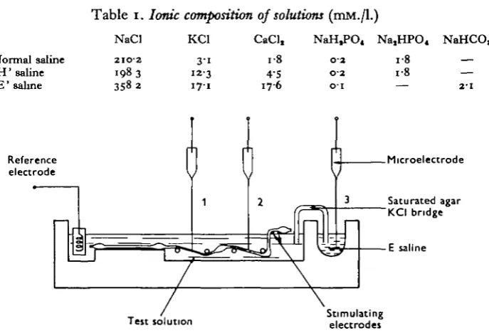
Solutions. The ionic composition of the normal and modified salines are given in
Table 1. Ionic composition of solutions (mM./l.)
NaCl KCI CaCl, NaH,PO4 Na,HPO« NaHCO,
Normal saline 'H'saline 'E'saline
2102 1983 3582
3-1 12-3 17-1
4-5 176
01 02 01
i-8 i-8
2 - 1
Reference electrode
Microelectrode
Saturated agar KCI bridge
E saline
\
Test solution Stimulatingelectrodes
Fig. i. Schematic diagram of the experimental arrangement The microelectrode iB shown in three different positions: position, 1, impalement of the desheathed part of the nerve cord (between 4th and 5th ganglia); position 2, impalement of a sheathed part of the nerve cord (between 3rd and 4th ganglia); position 3, measurement of the microelectrode tip potential in ' E' saline. The nerve cord was stimulated electrically between and and 3rd ganglia by means of a pair of Ag-AgCl electrodes.
Experimental procedure. The abdominal nerve cord, including the five last abdominal
ganglia, was removed from the animal. In some experiments it was desheathed between the 4th and the 5th ganglia by means of fine needles; in others, the nerve sheath was kept intact. The preparation was then mounted in the nerve chamber shown in Fig. 1. Antidromic electrical stimulation was applied by means of a pair of Ag-AgCl electrodes located in the right part of the chamber and at the surface of the saline. Microelectrodes were inserted either into the desheathed region (position 1 of the electrode), or into a sheathed region (position 2 of the electrode). Electrode tip potentials were measured, when needed, as the difference between the potentials recorded with the microelectrode tip dipping on one hand in the test solution and on the other hand in the ' E' saline (position 3 of the microelectrode). If the saturated KCI bridge is assumed to abolish junction potentials, this should give correct measurements of the tip potential between the test saline and ' E' salines. Measurements of the membrane potentials of the giant axons were made after an equilibration time of more than 30 min. in the test solution. DC potentials were recorded on the ink-writer. Measurements of resting potential were made 2-3 min. after the impalement. Measurements of 'sheath potential' were made when the microelectrode was slowly withdrawn from the nerve cord.
RESULTS
(1) Introduction of a microelectrode into a sheathed nerve cord
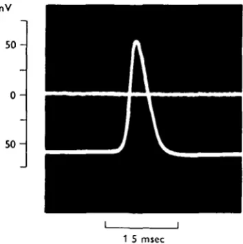
The nerve cord was mounted in the nerve chamber and slightly stretched. The microelectrode was then advanced until its tip caused a small depression in the connective. A small vibration of the holder led to a rapid penetration of the micro-electrode through the sheath into an underlying giant axon. Under these conditions a negative potential was recorded and adequate electrical stimulation gave rise to a large action potential (Fig. 2). If the microelectrode was kept in this position no modification of the resting or action potential was noticed.
mV
5 0
0
5 0
[image:4.451.136.313.211.389.2]-1 5 msec
Fig. 3. Superimposed tracings of the potential at the tip of the microelectrode before and after impalement of a giant axon in a sheathed nerve cord. An action potential was elicited by electrical stimulation of the nerve cord. Notice that a low resting potential (55 mV.) was associated with a large action potential ( n o mV.). 190 C. Normal saline.
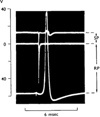
When the microelectrode was removed from the preparation the potential returned to its original zero level. However, when the movement was carried out slowly enough, it was possible, in most cases, to record two successive changes of the microelectrode tip potential: a positive deflexion (70-75 mV.) followed by a rapid negative deflexion (10-15 mV.) to the original zero potential (Fig. 3). It was agreed to call 'sheath potential' this last negative deflexion and resting potential the potential difference between inside of a giant axon and external solution. The meaning of this 'sheath potential' will be discussed in further detail in the last section of this paper.
A schematic drawing of the DC potential changes associated with vertical move-ments of the microelectrode tip together with photographic recordings of the electrical events following electrical stimulation of the preparation are shown in Fig. 4.
(2) Membrane potentials in sheathed and desheathed nerve cords bathed in normal saline
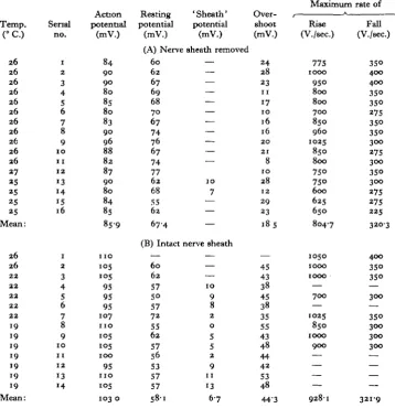
(Table 2). Typical tracings of action potentials and their derivatives are given in Fig. 5 (A and B). It is clearly seen that, whereas action potentials were higher in sheathed nerve cords (103-0! 5-9 mV.) than in desheathed preparations (85-9 ±4-6 mV.), rest-ing potentials were lower in sheathed preparations (58-1 + 5-4 mV.) than in desheathed ones (67-4 + 6-2 mV.); thus, the mean value of the overshoot was of 44-3 + 5-8 mV. in the first case instead of 18-5 ± 7* 1 mV. in the last case. This very important difference
mV
• 4 0 - 1
0
40
-i SP
T
RP
[image:5.451.145.314.152.350.2]6 msec
Fig. 3. Superimposed tracings showing changes in the tip potential of the microelectrode as it was withdrawn from a giant axon in a sheathed nerve cord. Before being withdrawn, the microelectrode was inside a giant axon and a full-sized action potential ( n o mV.) was elicited by electrical stimulation (lower tracing). After the microelectrode has been partly withdrawn, a positive deflexion was recorded (upper tracing). After complete withdrawal of the micro-electrode its tip potential agam reached the initial zero potential (middle tracing). The intracellular action potential is truncated. 190 C. Normal saline.
Nerve sheath
Extracellular ^ - = space"*" r—-Axon
40 mV
6 msec
Fig. 4. Potential changes associated with vertical movements of the microelectrode. The sobd line represents the DC potential variations of the microelectrode tip. Photographic recordings show electrical events following electrical stimulation of the preparation for postulated positions of the microelectrode tip (upper drawings). The intracellular action potential is truncated. 190 C. Normal saline.
[image:5.451.64.389.434.587.2]in the overshoots (25-8 mV.) may be attributed to the 'sheath potential' only with difficulty, the mean recorded value of this potential being only of 6-7 mV. It is not quite impossible, however, that the true value of this 'sheath potential' has been higher than the recorded one. Maximum rates of rise and rates of fall of the action potential were slightly higher in sheathed nerve cords (928-11122-0 and 321-9 ± 38-0 V./sec.) than in desheathed ones (804-7 ± 128-9 a n ci 320-3 ± 49-4 V./sec.) but no significant change in the shape of the action potential has been noted.
Table 2. Membrane potentials of cockroach giant axons in normal saline
Temp. (°C.) 26 26 26 26 26 26 26 26 26 26 26 27 25 25 25 25 Mean: 26 26 22 22 22 22 22 19 19 19 19 19 19 19 Mean: Serial no. 1 2 3 4 5 6 7 8 9 1 0 11 1 2 13 14 15 16 1 2 3 4 5 6 7 8 9 1 0 11 1 2 13 14 Action potential (mV.) 84 90 90 80 85 80 83 90 96 88 82 87 90 80 84 85 859 n o 105 •°5 95 95 95 107 n o 105 105
1 0 0
95 n o 105 103 0 Resting potential (mV.) (A) Nerve 60 62 67 69 68 70 67 74 76 67 74 77 62 68 55 62 674 (B) Intact — 60 62 57 5° 57 72 55 62 57 56 53 57 57 581 'Sheath' potential (mV.) sheath removed — — — — — — — — — — — 10 7 — — — nerve sheath — — •— 1 0 9 8 2 0 5 5 2 9 11 13 6-7 Over-shoot (mV.) 24 28 23 11 17 10 16 16 2 0 21 8 10 28 12 29 23 185 — 45 43 38 45 38 35 55 43 48 44 42 53 48 443
M a x i m u m
K Rise (V./sec.) 775 1000 95O 800 800 700 850 960 1025 850 800 750 750 600 625 650 804-7 1050 1000 1000 — 700 — 1025 850 1000 900 — — — — 928-1 rate of Fall (V./sec.) 35° 400 400 35O 35O 275 35O 35° 300 275 300 35° 300 275 275 225 3203 400 350 35° — 300 — 35° 300 300 300 — — — — 3219
(3) Effects of 'H' saline on membrane potentials
The differences between sheathed and desheathed nerve cords were more pro-nounced than in preceding experiments (Table 3, Fig. 5 C, D). Whereas resting po-tentials were only slightly higher in desheathed preparations (57-3 + 5-3 mV. instead of 55-6 ± 4-2 mV.), action potentials were three times smaller in desheathed prepara-tions (36-5 + 7-6 mV.) than in sheathed ones (107-9 ± °'° mV.); m the first case there was an undershoot of 20-7 + 8-4 mV., whereas in the second there was an overshoot of 52-2 + 7-1 mV. It should be noted that the action potentials recorded in desheathed preparations were often in two phases (Fig. 5 C). The mean values for the maximum rate of rise were 244-2+ 106-4 V./sec. in desheathed nerve cords for 1071-61159-5 V./ sec. in sheathed ones; the maximum rates of fall were ioo-o± 3-6 V./sec. in the first case and 398-7 + 44-0 V./sec. in the second. The mean value of the 'sheath potential' was ii"5 + 8-2 mV.
0
0
-1400 V./sec
50 mV.
[image:7.451.96.358.226.440.2]1 msec
Fig. 5. Effects of changing ionic composition of the external solution on the action potential (lower tracings) and its derivative (upper tracings) of sheathed (B and D) and desheathed (A and C) nerve cords. A and B, normal saline; C and D , ' H ' saline. Interrupted line indicates zero potential. A, B and D: 260 C.; C: 270 C.
(4) Effects of desheathing procedure upon electrical properties of giant axons
The preceding results raised the question of the extent to which local desheathing of the nerve cord altered the electrical properties of the giant axons along their whole length. Experiments were performed in which measurements of membrane potential were made in sheathed and desheathed regions of the same nerve cords. Similar differ-ences to those mentioned above were observed, despite the fact that recordings in the sheathed part were made more than 2 hr. after the desheathing had been carried out (Fig. 6, Table 4): in the sheathed portion the action potentials were 62 mV. higher and the resting potentials 18-5 mV. lower than in desheathed portions of the nerve cord. It will therefore be seen that, whereas a solution having the same ionic composi-tion as the haemolymph in respect of K+ and Ca2+ produced a drastic decrease in the
magnitude of the action potential in desheathed preparations, removal of the nerve sheath led only to local changes, thus demonstrating that diffusion processes along the extracellular spaces were very slow.
Table 3. Membrane potentials of cockroach giant axons in'H' saline
Temp.
P C )
2 7 2 7 2 7 2 7 27 2 7 2 7 2 7 2 7 2 7 2 7 2 7 2 7 2 7 2 7 Mean: 25 25 2 6 2 4 2 6 2 6 2 6 2 6 2 6 2 6 2 6 2 6 2 6 2 6 2 6 2 6 Mean: Serial no. 1 2 3 4 5 6 7 8 9 1 0 11 1 2 13 14 15 1 2 3 4 5 6 7 8 9 1 0 11 1 2 13 14 15 16 Action potential (mV.) 35 3° 3 0 34 3 0 35 35 58 30 4 0 3° 4 2 4 0 4 4 35 36-5 " 5 1 0 4
105
1 0 4 1 0 2 98 n o 1 0 4 " 5 1 1 7 1 1 8 n o n o 1 0 7
i ° 5
1 0 2
1079 Resting potential (mV.) (A) Nerve SO 58 6 2 5 9 56 55 55 67 61 64 6 2 51 56 53 5° 57 3 'Sheath potential' (mV.) sheath removed —. 15 7 — — — — — — — — — — — —
(B) Intact nerve sheath 5 0 57 58 4 9 58 58 55 5 1 58 52 6 0 55 6 2 6 2 53 52 55-6 2 6 16 9 14 — 7 5 9 2 0 2 0 2 0 8 0 0 17 2 " • 5
Over-shoot (mV.)
- 1 5 - 2 8 - 3 2 - 2 5 - 2 6 — 2 0 — 20 - 9 - 3 1 - 2 4 - 3 2 - 9 - 1 6 - 9 - 1 5
- 2 0 7
65 47 47 55 4 4 4 0 55 53 57 65 58 55 48 45 52 5°
5 2 2
Maximum
Rise
(V./sec.)
53° — 2 0 0
2 2 s
2 0 0
35O
1 5 0 2 3 0 1 5 0 2 0 0 — 3 0 0
275
3 0 0 —
244-2
1250
9 0 0
925 1025 1050 1050 1300 1200 1200 1350 1125 1050 875 775 1050 1020 io7i'6 rate of Fall (V./sec.)
1 2 s — 5 0 125 5 0 IOO 50 150 IOO IOO — IOO IOO 1 5 0 — IOO 375 35° 35° 375
4 2 5 4 2 5
475
4 0 0 4 2 5
475 4 0 0 45° 375 4 0 0 34° 34O
398-7
(5) Effects of' E' saline on membrane potentials
365
0 — 0 — 1400 V/sec. 50 mV 1 msec [image:9.451.121.343.78.366.2]Fig. 6. Effects of haemolymph ions on the action potential and its derivative in a desheathed part (A) and in a sheathed part (B) of the same nerve cord. Same preparation as in fig 5 C. 27° C
Table 4. Membrane potentials of giant axons in a partly
desheathed nerve cord bathed in 'H' saline
Temp. (°C) 27 27 2 7 27 27 2 7 27 Mean: 27 2 7 27 27 2 7 Mean: Serial no. 1 2 3 4 5 6 7 1 2 3 4 5 Action potential (mV.) 58 3° 4 0 3° 42 4 0 4 4 40-6 95 95 " 3 n o
1 0 0
IO2'6 Resting potential (mV.) 'Sheath potential' (mV.) (A) Desheathed part 67 61 64 62 5 1 56 53 5 9 1
(B) 32 4 0 45 4 4 4 2 40-6 — — — — — — — — Sheathed part 35 33 1 0 28 27 2 6 6
Over-shoot (mV.)
- 9 - 3 1 - 2 4 - 3 2 - 9 - 1 6 - 9
- i 8 - 6
63 55 68 66 58 62 0 Maximum Rise (V./sec.)
2 3 0 1 5 0 2 0 0 — 3 0 0
275
3 0 0
2425 1130 1250 1600 1560 1420 13920 rate of Fall (V./sec.)
1 5 0 1 0 0 1 0 0 —
1 0 0 1 0 0 1 5 0
1166
4 2 0 34° 4 1 0
4 8 0
4 1 0
26-3 ± 7-2 mV. The maximum rates of rise (830-9 ± 116-8 V./sec.) and rates of fall (343*6 + 35*3 V./sec.) were of the same order of magnitude as those recorded for desheathed preparations in normal saline.
Table 5. Membrane potentials of cockroach giant axons in'E' saline
Temp. (°C.) 2 7 2 7 2 7 2 7 2 7 2 7 2 7 2 7 2 7 2 7 2 7 Mean: Serial no. 1 2 3 4 S 6 7 8 9 1 0 1 1 Action potential (mV.) 85 90 95 8 0 87 9 0 9 0 98 83
1 0 2 1 0 0
9 0 9
Resting potential (mV.) 'Sheath potential' (mV.) Nerve sheath removed
67 6 0 69 67 62 64 65 67 6 0 62 68 64-6 — — — — — — — — — — — — Over-shoot (mV.) 18 30 2 6 13 25 2 6 25 31 2 3 4 0 32
2 6 3
Maximum Rise (V./sec.) 775 875 1020 73O 73° 7 7 0 7 7 0 8 4 0 7 0 0
1050
8 8 0
8309
rate of Fall (V./sec.)
3 6 0
37o
4 1 0 34°
34°
34O 34O 34O 2 6 0
34° 34O 343-6 0 — 1400 V/sec 50 mV 1 msec
Fig. 7. Effects of extracellular ions on the action potential and its derivative of a giant axon in a desheathed nerve cord. 270 C.
From these results, it was therefore concluded that the ionic composition of the extracellular fluid might be at least partly responsible for maintaining normal resting and action potentials despite the relatively high potassium concentrations. It is not impossible, however, that osmotic pressure itself might play some rdle in avoiding the enlargement of extracellular spaces and in concentrating ions (by water loss) in the extracellular fluid.
(6) Errors in measurements of resting potential and 'sheath potential'
Part of this difference might be attributed to the ' sheath potential', the origin of which needed to be studied in more detail.
We have seen that the inside surface of the nerve sheath is positive with respect to outside. This sign is not the one which should be expected if the sheath acted as a semi-permeable barrier with a Donnan equilibrium operating between the extracellular fluid and the bathing fluid. The recorded values of this 'sheath potential' were
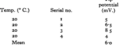
ex-Table 6. Tip-potential differences between 'E' and 'H' salines
Tip potential Temp. (° C.) Serial no. (mV.)
20 1 5 20 2 6-5 20 3 85 20 4 4
[image:11.451.126.323.184.258.2]Mean 6fo
Table 7. Membrane potentials and 'sheath potentials' in a sheathed nerve cord in
'H' saline and microelectrode tip potentials between 'E' and 'H' salines
5. (° C.)
2 1 2 1 2 1 2 1 2 1 2 1 2 1 2 1 2 1 2 1 2 1
Mean
Serial no.
1
2
3 4 5 6 7 8 9
1 0 1 1
Action potential
(mV.) 1 0 7 1 0 5
95 95
1 0 0
8 0 92
1 0 0 1 0 0
96 99 9 7 2
Resting potential
(mV.) 50
54
5°
49 55 55
50
57 53
5° 52 5»'3
' Sheath potential (mV.)
19
1 0 0 1 1
3
0 0 1 0
9 9
14
7 7
T i p potential
(mV.)
1 0
3
0 1 1 1
3 3 4 9 6 3 4 8
[image:11.451.45.403.256.445.2]The possible error in measurements of resting potential in sheathed nerve cords brought about by this ' sheath potential' will be discussed later.
DISCUSSION
The above results have shown that whereas action potentials recorded in sheathed nerve cords are of greater magnitude than those recorded in desheathed preparations, resting potentials are always lower in the first case (Table 8). It has also been demon-strated that diffusion processes along the extracellular spaces are very slow and that the potential difference existing across the nerve sheath seems to be extremely variable from one impalement to another. Moreover, it has been pointed out that the use of
Table 8. Effects of ions on sheathed and desheathed nerve cords
Maximum rate of Action
potential (mV.)
(a) Deaheathed nerve
cords
(6) Sheathed nerve cords
(a) Desheathed nerve
cords
(6) Sheathed nerve cord
Desheathed nerve
Resting 'Sheath Over-potential Over-potential' shoot (mV.) (mV.) (mV.)
(A) Normal galine 67-416-2 —
Rise (V./sec.)
Fall (V./sec.)
— I 8 - 5 1 7 - I 804-71128-9 320-3149-4 103-015-9 58-115-4 6-714-2 44-315-8 928-1 Ii22-o 321-9138-0
(B) ' H ' Saline
36-517-6 57-3±53 — - 2 0 7 1 8 - 4 244-21106-4 IOO- + 3-6 107-916-0 55-614* 11-518-2 53-217-1 1071-61159-5 398-71440
(C) 'E'saline
90-917-2 64613-3 — 26-317-2 830-91116-8343-61353 a saline containing cations at the extracellular level results in quite normal resting and action potentials in desheathed preparations despite of a relatively high potassium concentration. The maximum rates of rise and rate of fall recorded in different con-ditions show that there is no significant difference in the shape of the action potentials except for desheathed nerve cords bathed in ' H ' saline.
fluid might correspond to the measured resting potential plus the measured 'sheath potential'. In that case the true resting potential of the axons would be higher than the measured one. This possibility has been provisionally discarded for two reasons. First, it has been shown that measured resting potentials were rather constant for the same preparation whereas' sheath potentials' varied widely from one impalement to another. Secondly, it was observed that potentials of the same sign as ' sheath potentials' have been recorded in desheathed cords whose axons had a quite normal resting potential. One could suspect therefore the so-called 'sheath potential' to be an artifact deter-mined by variations of the microelectrode tip potential between two different ionic solutions, similar to those recorded between ' H' and ' E' salines. Moreover, one could imagine the ' sheath potential' as originating not from the nerve sheath itself but from the underlying perineurial and glial cells. It has, in fact been suggested that these cells might be involved in the extra-axonal sodium regulation in Carausius nerve cords (Treherne & Maddrell, 19676). The suggested movement of sodium ions across these cells would result in a potential difference relative to the outside medium. The polarity of this potential difference would depend upon the mobility of sodium ions relative to that of the anion which would be secreted with sodium in the channels of the mesaxon folds. Experiments on the isolated nerve sheath, being carried out at the present time, should permit us to decide whether the ' sheath potential' exists or not. It must be noted here that possible errors in measurements of resting potential may arise from microelectrode tip potentials, the importance of which cannot be well appreciated. Proper elimination of tip potentials using thorium chloride (Agin & Holtzman, 1966) would perhaps permit more reliable results.
Experiments with a saline containing Na+, K+ and Ca2+ at the extracellular level have clearly shown that quite normal action potentials can be obtained, despite a relatively high potassium concentration. This finding is in good agreement with results of Treherne (1962 c) who has shown that elevation of the cation concentration to the extracellular levels resulted in delayed conduction block in desheathed cockroach nerve cords irrigated with high potassium saline.* Great care must be exercised, however, in interpreting our results, for the relatively high resting potential in extra-cellular saline may be responsible for the production of these action potentials, whereas in sheathed nerves, large action potentials were associated with much lower resting potentials. The mean resting potential recorded in extracellular saline (64-6 mV.) cannot be interpreted in terms of external potassium concentration. Moreover, it has been demonstrated (Yamasaki & Narahashi, 1959a) that in the case of cockroach axons the magnitude of the resting potential undergoes little change when the external sodium concentration is varied over a wide range. It must be noted here, however, that an increase in the resting potential in high-sodium salines has been reported by Seyama & Irisawa (1967). These authors have shown that, with constant external potassium concentrations, the resting potential of the skate heart was 14 mV. higher in 200% sodium solutions than in 50% ones. The major difference might be due, in that case, to the variations of the microelectrode tip potential and to an increase of potassium equilibrium potential, EK, caused by an increase of intracellular potassium in hypertonic solutions. Such an explanation might be valuable in the case of the giant axon of the cockroach bathed in extracellular saline. On the other hand, Narahashi (1966) has shown that there was little or no change in resting potential when external calcium concentration was varied from 0-18 to 54 mM./l. It is possible, however, that high calcium concentrations may play some role in preventing membrane depolariza-tion at high potassium concentradepolariza-tions (cf. Stampfli & Nishie, 1956). Osmotic pressure may also play a similar role (Stampfli & Nishie, 1955). On the other hand, it does not seem unlikely that chloride ions may be partly responsible for the increased resting potential; in that case, an excess of chloride ions in the extracellular saline would result in a higher chloride equilibrium potential, Ed, and consequently to an increased resting potential. Further investigations concerning these problems will be carried out using the voltage-clamp technique.
When the nerve sheath is removed from the cockroach nerve cord, the resting poten-tial of the giant axons is increased whereas the amplitude of the action potenpoten-tial is decreased. This phenomenon seems different from that reported by Treherne & Maddrell (19676). These authors have shown, indeed, that in Carausius nerve cord removal of the fat-body sheath does not affect the amplitude of the action potential whereas a positive potential of about 15 mV., probably associated with this fat-body sheath, is responsible for an apparent reduction of the resting potential in intact nerve cords (Maddrell & Treherne, 1966; Treherne & Maddrell, 1967 a). A lower resting potential in the intact nerve cord of the cockroach might be associated with a positive
potential across the nerve sheath, the 'sheath potential', reported in a previous paper (Pichon & Biostel, 1966a). In that case, however, the magnitude of the action potential must be unaffected when the nerve sheath is removed. The possibility remains that some damage to the giant axons in removing the nerve sheath or a greater injury to these axons when they are impaled with a microelectrode in a desheathed nerve cord (as pointed out earlier) may be responsible for such a reduction of the action potential. This would agree with the fact that removal of the nerve sheath made the impalement of the axons of Carausius a virtual impossibility (Treherne & Maddrell, 19676). It is logical to conclude, however, from the present experiments, that the elevated concen-trations of cations in the extracellular fluid is sufficient to explain the differences between sheathed and desheathed preparations, the so-called 'sheath potential' being related to variations of the microelectrode tip potential. The mechanism responsible for the maintenance of this specialized ionic composition inside the extra-cellular spaces is not clear. This differential ionic distribution between external solution and extracellular fluid has been shown probably to result from a Donnan-like equilibrium (Treherne, 1962a, b). The nature of the anion causing this effect is not known, but if we assume this anion to be a large molecule in solution in extra-cellular spaces then the nerve sheath acting as a semi-permeable barrier would be polarized, the inside being negative with respect to the outside. Such a potential has never been recorded. On the other hand, if an extracellular fixed-charge system is involved, the activity coefficient of extracellular cations might be lower than that in a free solution and would be equivalent to that in the bathing medium as pointed out by Treherne & Maddrell (19676). A possible local regulation by the glial cells at the region of the axon surfaces, also suggested by these authors for sodium ions, is not to be excluded.
We are indebted to Dr J. E. Treherne for helpful discussions and for reading and correcting this manuscript.
SUMMARY
1. The use of very fine-tipped and mechanically strong microelectrodes has allowed reliable recordings of resting and action potentials to be made in cockroach giant axons in sheathed and desheathed nerve cords.
2. When the microelectrode was withdrawn from a giant axon in an intact connec-tive the first posiconnec-tive change in the potential from the resting level, was in most cases followed by a negative deflexion to the original zero level, the 'sheath potential'. The values of this' sheath potential' together with the resting potential, the action potential, the maximum rate of rise and maximum rate of fall of the action potential have been measured in three different salines.
3. In normal saline, resting potentials were lower in sheathed preparations (58-1 ± 5*4 mV.) than in desheathed ones (67-4 + 6*2 mV.), whereas action potentials were higher in the former (103 + 5-9 mV.) than in the latter (85-9 + 4-6 mV.).
being 55-6 ± 4-2 mV. and the mean action potential 107-9 ± 6'° m^ - Local desheathing of the nerve cord led only to local disturbance of the resting and action potentials, thus indicating that diffusion processes along the extracellular spaces were very slow. 5. The use of a saline in which cation concentrations have been elevated to the extracellular level resulted in normal resting potentials (64-6+ 3-3 mV.) and action potentials (90-9 + 7-2 mV.) in desheathed cords, despite the relatively high potassium concentration (17-1 mM./L).
6. Recordings of the maximum rates of rise and rates of fall showed that there was no significant modification in the shape of the action potential in these different experimental conditions.
7. The values of the ' sheath potential' were very variable from one impalement to another and it is suggested that this potential might be related to variations of the microelectrode tip potential bathed in different ionic solutions.
8. The low resting potentials and high action potentials of giant axons in intact nerve cords may result from an excess of inorganic cations in the extracellular fluid.
REFERENCES
ADRIAN, R. H. (1956). The effect of internal and external potassium concentration on the membrane potential of frog muscle, J. Pkystol. London, 133, 631—58.
AGIN, D. & HOLTZMAN, D. (1966). Glass microelectrodes: the origin and elimination of tip potentials.
Nature, Lend, a n , 1194.
BOISSEAU, J. & BOISTEL, J. (1965). Controle au microscope electronique de microelectrodes de verre de difterentes longueurs vides ou remplies de solutions conductrices. J. Phytiol. Paris. 57, 529-35. BOISTEL, J. (1959). Quelques caractenstiques electnques de la membrane de la fibre nerveuse au repos
d'un Insecte (Periplaneta americana). C. r. Seanc. Soc. Biol. 153, 1009-13.
BOISTEL, J. (i960). Caracttristiques FonctionneUes des Fibres Nerveuses et des Ricepteurs Tactdes et
Olfactifs des Insectes, 14.7 pp. Pans: Librairie Arnette.
BOISTKL, J. & CORABOEUF, E. (1954). Potenoels de membrane et potentiels d'action de nerf d'Insecte recueillis a l'aide de microelectrodes intracellulaires. C. r. hebd. Seanc. Acad. Sci., Paris 338, 2116-18. BOISTKL, J. & CORABOEUF, E. (1958). R61e joue par les ions sodium dans la genese de l'activite electrique
du tissu nerveux d'Insecte. C. r. hebd. Seanc. Acad Sci., Parts X47, 1781-3.
CALLKC, J. J. & BOISTHL, J. (1965a). Essai de localisation, a l'aide de microelectrodes, de zones synap-tiques au niveau de 6eme ganglion abdominal d'un Insecte (cas particulier de Periplaneta americana).
J. Pkysiol., Pans 57, 233.
CALLEC, J. J. & BOISTEL, J. (19656). Analysis with microelectrodes of the synaptic transmission at the level of the sixth abdominal ganglion of a cockroach, Periplaneta americana. In The Physiology of the
Insect Central Nervous System pp. 59-65 (Eds. J. E. Treheme and J. W. L. Beament). London and
New York: Academic Press.
CALLEC, J. J. & BOISTEL, J. (1966a). Etude de divers types d'activites electnques enregistrees par micro-electrodes capillaires au niveau du dernier (6eme) ganglion abdominal de Periplaneta americana L.
C. r. Sianc. Soc. Biol. 160, 1943-7.
CALLEC, J. J. & BOISTEL, J. (19666). Phenomenes d'excitation et d'inhibition au niveau du dernier (6ime) ganglion abdominal de la blatte, Periplaneta americana L. C. r. Sianc. Soc. Biol. 160, 2418-24.
CALLEC, J. J. & BOISTEL, J. (1967). Les effets de l'ac&ylcholine aux niveaux synaptiques et somatiques dans le cas du dernier ganglion abdominal de la blatte, Periplaneta amertcana L. C. r. Seanc. Soc. Biol.
161, 442—6.
MADDRELL, S. H. P. & TREHERNE, J. E. (1966). A neural fat-body sheath in a phytophagous insect
(Carausius morosus). Nature, Lond. 211, 215-16.
NARAHASHI, T. (1966). Dependence of excitability of cockroach giant axons on external divalent cations.
Comp., Biochem. Phystol. 19, 759—74.
NARAHASHI, T. & YAMASAKI, T. (i960). Mechanism of the after-potential production in giant axons of the cockroach. J. Physwl. Lond. 151, 75-88.
PICHON, Y. & BOISTEL, J. (1965a). Etude comparative des effets du potassium et de ceux de la polarisa-tion electnque sur le 6«me ganglion abdominal de la blatte, Periplaneta americana L. J. Phystol.,
PICHON, Y. & BOISTKL, J. (19656). Complements a l'etude electrophysiologique de la transmission synaptique au niveau du 6eme ganglion abdominal d'une blatte, Penplaneta americana L. $hne Congris
International de I'U.I.E.I.S., Touioute (in the Press).
PICHON, Y. & BOISTEL, J. (1965 c). Effets des ions K+sur l'activite ' spontanee' du 6eme ganglion abdomi-nal de la blatte, Penplaneta americana. C.r. Stanc. Soc. Biol. 160, 380-5.
PICHON, Y. & BOISTKL, J. (1966a). Application aux fibres geantes de blattes (Penplaneta americana L.
Blabera craniifer Burm.) d'une technique permettant 1'introduction d'une microelectrode dans le
tissue nerveux sans resection pr^alable de la game. J. Pkyriol., Paris, 58, 592.
PICHON, Y. & BOISTEL, J. (19666). Effets de la polarisation electnque du sixieme ganglion abdominal de la blatte, Penplaneta americana, sur l'activiti des fibres geantes. C.r. Sianc. Soc. Biol. 160, 1728-33. SBYAMA, I. & IRISAWA, H. (1967). The effect of high sodium concentration on the action potential of
the skate heart. J. gen. Pkystol. 50, 505-17.
STAMPFLI, R. & NISHIE, K. (1955). Effects of aniso-osmotic solutions on resting and action potentials of myelinated nerve. Helv. physiol. pharmac. Ada 13, 33—4.
STAMPFLJ, R. & NISHIE, K. (1956). Effects of calcium free solutions on membrane-potential of myelinated nerve fibres of the brazilian frog, Leptodactyhu ocellatus. Helv. Physiol. pharmac. Acta. 14, 93-104. TBEHEHNE, J. E. (1962)0. Distribution of water and inorganic ions in the central nervous system of an
insect (Penplaneta americana L.) Nature, Lond. 193, 750—2.
TREHERNE, J. E. (19626). The distribution and exchange of some ions and molecules in the central nervous system of Penplaneta americana L. J. Exp. Biol. 39, 193-217.
TREHERNE, J. E. (1962 c). The effects of the iomc composition of the extracellular fluid on the electrical activity of the cockroach abdominal nerve cord. J. Exp. Biol. 39, 631-41.
TREHERNE, J. E. & MADDRELL, S. H. P. (1967a). Membrane potentials in the central nervous system of a phytophagous insect (Caraustus morosus). J. Exp. Biol. 46, 413-21.
TREHERNE, J. E. & MADDRELL, S. H. P. (19676). Axonal function and ionic regulation in the central nervous system of a phytophagous insect (Caraustus morosus). J. Exp. Biol. 47, 235—47.
YAMASAKI, T. & NARAHASHI, T. (1957). Intracellular microelectrode recordings of resting and action potentials from the insect axon and the effects of DDT on the action potential. Studies on the mechanism of action of insecticides. Botyu-Kagaku aa, 305-13.
YAMASAKI, T. & NARAHASHI, T. (1959a). The effects of potassium and sodium ions on the resting and action potentials of the cockroach giant axon. J. Insect. Physiol. 3, 146-58.
YAMASAKI, T. & NARAHASHI, T. (19596). Electrical properties of the cockroach giant axon. J. Insect.