E-M. Larsson1
s.
Holtas1 0. Nilsson2Received November 4, 1988; revision requested
December 29, 1988; revision received February 8,
1989; accepted February 21, 1989.
Presented at the congress of the European So-ciety of Neuroradiology, Wurzburg, W. Germany,
September 1988.
This work was supported by grants from the Fbrenade Liv Mutual Group Life Insurance Co., Stockholm, Sweden, and from the Swedish Medical Research Council (project No.
B89-39X-08164-03A).
' Department of Diagnostic Radiology, University
Hospital, S-221 85 Lund, Sweden. Address reprint requests to E-M. Larsson.
2 Department of Neurology, University Hospital,
S-221 85 Lund, Sweden.
0195-6108/89/1005-1071
© American Society of Neuroradiology
Gd-DTPA-Enhanced MR of
Suspected Spinal Multiple
Sclerosis
1071
A prospective study was undertaken to evaluate the potential of Gd-DTPA-enhanced MR to differentiate active from inactive demyelinating lesions of the cervical spinal cord. Five patients with elongated high-signal-intensity lesions in the cervical cord on long TR/TE spin-echo MR images and a clinical suspicion of demyelinating disease had MR before and after IV Gd-DTPA. Delayed contrast enhancement (after 45-60 min) of the lesions was seen on short TR/TE images in two patients with clinically active disease, but no enhancement could be detected in three patients with stable disease. The patients with active disease underwent repeated MR examinations until the enhance-ment disappeared. The decrease in Gd-DTPA enhanceenhance-ment paralleled a decrease in clinical signs and symptoms of cervical myelopathy.
MR is useful in evaluating patients suspected of having demyelinating disease. The MR finding of asymptomatic lesions in the brain lends support to the diagnosis of multiple sclerosis. Other possible causes of myelopathy, such as spinal cord compres-sion and intramedullary tumor, can be excluded with the use of MR.
AJNR 10:1071-1076, September/October 1989
MR is the only technique that allows direct visualization of demyelinating lesions in the spinal cord [1-3]. Such lesions are usually elongated and have a high signal intensity on long TRJTE spin-echo images. No abnormality is seen on short TRJTE images unless the cord is enlarged because of focal edema accompanying an acute lesion or is atrophic in chronic disease [2, 3]. Acute demyelinating lesions are indistinguishable from older, chronic lesions on long TRJTE images [4, 5). In pa-tients with clinically definite or suspected multiple sclerosis (MS), a correlation has been described between recent clinical exacerbation of the disease and contrast enhancement of lesions in the brain on CT [6-8] as well as on MR [4, 9]. A similar correlation might be expected for demyelinating lesions in the spinal cord, but none has, to our knowledge, been reported. The aim of this prospective investigation was to evaluate the potential of Gd-DTPA-enhanced MR in differentiating acute, active demyelinating lesions from inactive lesions in the cervical spinal cord.
Subjects and Methods
Five patients with elongated high-signal-intensity lesions in the cervical spinal cord on long TR/TE images and a clinical suspicion of demyelinating disease were included in the study. Clinical data on the patients are summarized in Table 1. None of the patients had a definite MS diagnosis since all had isolated myelopathy without clinical evidence of lesions in any other part of the CNS.
In all patients, visual evoked potentials (VEPs) were recorded and CSF was examined for oligoclonal bands and immunoglobulin G (lgG)jalbumin ratio.
1072 LARSSON ET AL. AJNR: 1 0, September/October 1989
TABLE 1: Clinical, Laboratory, and MR Findings in Patients with Suspected Demyelinating Disease
Type of Finding Case 1 Case 2 Case 3 Case 4 Case 5
Clinical
Age (years) 43 40 44 49 44
Gender F M F F M
Duration of disease 1 mo 1.5 mo 4 mo 9 yr 21 yr
Clinical course One attack One attack One attack Progressive Attacks,
pro-gressive Laboratory
Intrathecal lgG synthesis Pathologic Normal Pathologic Pathologic Pathologic
Visual evoked potentials Pathologic Pathologic Normal Pathologic Pathologic
MR
Clinically active or stable Active Active Stable Stable Stable
Cervical cord (long TR/TE) Lesion Lesion Lesion Lesion Lesion
Cord enlargement or atrophy (short TRfTE) Enlargement Normal Normal Atrophy Atrophy
Contrast enhancement of cervical cord (short Yes Yes No No No
TR/TE)
Brain (long TR/TE) One lesion Normal Multiple le- Normal Multiple lesions
sions
Note.-F = female; M = male; mo = month(s); yr = year(s); lgG = immunoglobulin G.
cervical spine. Sagittal images were obtained using spin-echo pulse sequences with a short TR/TE, 500/16/3 (TRITE/excitations) in four patients and 300/16/5 in one, as well as a long TR/TE, 2000/84/1. A 256 x 256 matrix was used and the pixel size was 1.0 mm. The slice thickness was 5 mm with a 2.1-mm gap between slices.
Gd-DTPA was administered IV at a dose of 0.1 mmoljkg body weight.
Imaging protocols consisted of short TR/TE images before and 10, 45, and 60 min and in one case also 24 hr after the patient received
Gd-DTPA. Long TR/TE images were obtained before and 30 min
after Gd-DTPA injection. In two patients with contrast enhancement on the primary MR examination (cases 1 and 2), MR before and after Gd-DTPA was repeated 3 months later. In case 1 a second follow-up examination was performed 5V2 months after the first MR study. Signal-intensity measurements were performed to estimate the degree of contrast between lesion and normal cord on short TRfTE
images before and after IV administration of Gd-DTPA. The signal intensity was determined in a region of interest in the lesion and was
compared with the signal intensity in the cord cranial and caudad to the lesion.
MR of the brain without Gd-DTPA was performed using a head coil. Axial long TRfTE images (2000/60/1) with a 7 -mm slice thickness and 3-mm interslice gap were obtained.
Three patients (cases 1-3) who had had their first attack of cervical myelopathy (Table 1) underwent a neurologic examination within 1
week before the primary MR study. One patient (case 1) was
exam-ined with MR 4 weeks and another (case 2) 6 weeks after the onset
of the attack; at that time none of the patients showed clinical signs
of remission of the attack. In case 3, MR was performed 4 months after the onset of the attack when the patient was in a clinically stable phase of the disease. Cases 4 and 5, with a slowly progressing myelopathy (Table 1 ), did not show any notable worsening of disease
during the weeks before and after the MR examination. Cases 1 and 2 were categorized as clinically active and cases 3-5 as clinically
stable at the time of the primary MR examination.
In cases 1 and 2, repeated neurologic examinations were per-formed during the course of the attack and one neurologic examina-tion was performed after the last follow-up MR study.
Results
The laboratory tests for intrathecal synthesis of lgG were positive in all patients except case 2. VEPs showed a
patho-logic response in all except case 3 (Table 1). All patients had
elongated high-signal-intensity lesions in the cervical spinal
cord on long TR/TE images before injection of Gd-DTPA (Figs. 1 A, 2A, and 3A).
Clinically Stable Patients
On short TR/TE images, general atrophy of the cervical cord was seen in case 5 and local atrophy at the level of the
lesion in case 4 (Fig. 1 B). These two patients had a long duration of disease (Table 1 ). In case 3, with a shorter duration
of the disease, the cord had a normal sagittal diameter. _No
Gd-DTPA enhancement was observed in these three patients.
Two of the patients had multiple asymptomatic
high-signal-intensity lesions compatible with MS plaques in the brain on long TR/TE images, whereas MR of the brain was normal in the third patient.
Clinically Active Patients
The cervical cord was enlarged at the level of the lesion on short TRfTE images in case 1 (Fig. 2B) and had a normal
configuration in case 2 (Fig. 3B).
Contrast enhancement of the lesion was observed in both patients on short TR/TE images. The enhancement was best seen on images obtained 45 and 60 min after Gd-DTPA
injection (Figs. 2 and 3). Signal-intensity measurements re
-vealed a 25% contrast enhancement 60 min after Gd-DTPA in both cases, but the two studies could not be fully compared quantitatively owing to differences in TR (300 msec in case 1 and 500 msec in case 2). A slight enhancement was already present 1 0 min after injection but was not as obvious as after 45 and 60 min (Figs. 2 and 3). No remaining enhancement was detected after 24 hr (case 1) (Fig. 2). A slight enhance-ment was also discerned on long TRfTE images 30 min after Gd-DTPA injection.
[image:2.612.56.568.99.277.2]AJNR:1 0, SeptemberfOctober 1989 Gd-DTPA MR IN MULTIPLE SCLEROSIS 1073
Fig. 1.-Case 4: 49-year-old woman with
slowly progressing myelopathy, clinically stable at time of MR.
A, Long TR/TE image (2000/84) shows
elon-gated high-signal-intensity lesion in cord at C2-C3.
8, Short TR/TE image (500/16) after Gd-DTPA
shows local atrophy of cervical cord at C2-C3 and no contrast enhancement. Area of increased signal in cord at level of vertebral body of C4 is considered an artifact since it was present also
on corresponding image before Gd-DTPA
injec-tion.
A
decreased and the contrast enhancement on short TRfTE images appeared less intense 1 0 as well as 60 min after Gd-DTPA (Fig. 2G). In a third MR examination 51/z months after the first, the width of the cord was normal and no contrast enhancement was present (Fig. 2H). The lesion could not be identified on long TR/TE images without Gd-DTPA after 3 and 51/z months. In case 2 the contrast enhancement on short
TR/TE images had disappeared completely after 3 months
and the lesion could not be detected on long TR/TE images without Gd-DTPA. In both patients the decrease in contrast enhancement paralleled a decrease in clinical signs and symp-toms of cervical myelopathy.
MR of the brain showed one small high-signal-intensity lesion in case 1 and was normal in case 2 (Table 1 ).
No adverse reactions to IV administration of Gd-DTPA were observed in the five patients.
Discussion
In this investigation we found delayed Gd-DTPA enhance-ment of suspected demyelinating lesions in the cervical cord in two patients with active disease but no enhancement in three patients with clinically stable disease (Table 1; Figs.
1-3). The two patients with active disease underwent repeated
MR examinations until the enhancement had disappeared (Fig. 2), and the decrease in enhancement could be correlated with a decrease in clinical signs and symptoms.
The possibility of differentiating active demyelinating lesions from inactive lesions in the brain has been investigated by iodinated contrast-enhanced CT [6-8] and Gd-DTPA-en-hanced MR [4, 5, 9, 10]. The inflammatory process of an active demyelinating lesion is associated with a transient breakdown of the blood-brain barrier, which is responsible for the contrast enhancement seen on CT and MR [4, 7]. The enhancement of active but not of inactive lesions in the cervical cord in our study is in agreement with the previous CT and MR investigations of lesions in the brain, in which a correlation between contrast enhancement and clinical activity
B
has been found in most patients [4-9]. Recently, suspected Gd-DTPA enhancement of a clinically active lesion in the cervical cord on MR was reported in a patient with definite MS, whereas another patient with clinically active suspected MS did not exhibit any enhancement of a lesion in the cord [11]. In the same study, a third patient with definite MS
without acute symptoms showed no enhancement of a cer
-vical lesion. Because inactive lesions are not depicted on Gd-DTPA-enhanced short TRfTE images, a long TR/TE scan without Gd-DTPA should always be obtained first to detect both active and inactive lesions. Most acute demyelinating lesions are accompanied by edema as the increased perme-ability of the blood-brain barrier allows a leakage of water [3, 6]. This may cause transient local enlargement of the cord at the level of the lesion [2, 3], as was seen in one of our patients. With resolution of edema, the T2 relaxation time
frequently shortens and the lesion diminishes, which may
make detection more difficult on long TR/TE images [3].
The delayed enhancement, best seen about 45-60 min after IV injection of Gd-DTPA in our study, correlates with previously reported delayed iodinated contrast enhancement of MS plaques in the brain on CT [6-8]. Gd-DTPA enhance-ment of MS plaques in the brain was reported to be visualized on short TR/TE images obtained only 3 min after contrast injection in most patients, whereas a few lesions were better seen on delayed (55-min) images [4]. In the same investiga-tion, initial enhancement displayed on a 3-min image was absent on the 55-min image in two patients [4]. Enhancement of intracranial and intraspinal tumors usually is most marked immediately after injection of contrast material [11-15], but delayed scans have recently been reported to show a further increase in signal intensity in some tumors, especially small ones [15, 16]. A delay in enhancement has been observed also in necrotic tumor tissue [11, 13]. It is presumed that demyelinating lesions produce a minor blood-brain barrier
disruption compared with the disruption associated with ma
[image:3.612.59.555.85.290.2]ra-1074 LARSSON ET AL.
A
8
c
D
F
G
AJNR:10, September/October 1989
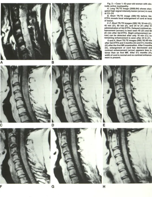
Fig. 2.-Case 1: 43-year-old woman with clin-ically active myelopathy.
A, Long TR/TE image (2000/84) shows elon
-gated high-signal-intensity lesion (arrow) in cord at C3-C4.
B, Short TR/TE image (300/16) before
Gd-DTPA reveals local enlargement of cord at level of lesion.
C-F, ShortTR/TE images (300/16) 10 min (C),
45 min (0), 60 min (E), and 24 hr (F) after IV
administration of Gd-DTPA. Delayed contrast en
-hancement (arrows) is best seen 45 (0) and 60 (E) min after Gd-DTPA. Slight enhancement (ar· row) can be detected after only 10 min (C); no remaining enhancement is seen after 24 hr (F).
G and H, Short TR/TE images (500/16) 60 min
after Gd-DTPA and 3 months (G) and 5V. months
(H) alter the first MR examination. Alter 3 months
(G), enlargement of cord has decreased and
contrast enhancement (arrow) appears less in
-tense than on first MR. Alter 5'1• months (H), width of cord is normal and no contrast enhance-ment is present.
E
[image:4.618.55.563.76.731.2]AJNR:1 0, September/October 1989 Gd-DTPA MR IN MULTIPLE SCLEROSIS 1075
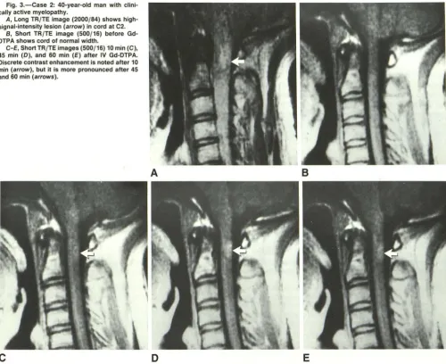
Fig. 3.-Case 2: 40-year-old man with clini-cally active myelopathy.
A, Long TR/TE image (2000/84) shows
high-signal-intensity lesion (arrow) in cord at C2. B, Short TR/TE image (500/16) before Gd -DTPA shows cord of normal width.
C-E, Short TR/TE images (500/16) 10 min (C),
45 min (D), and 60 min (E) after IV Gd-DTPA. Discrete contrast enhancement is noted after 10 min (arrow), but it is more pronounced after 45
and 60 min (arrows).
c
A
D
cellular space and thereby improve the detection of demyeli-nating lesions in the brain [7, 8]. The same mechanism is probably responsible for the marked increase in signal
inten-sity on delayed scans compared with the early postinjection
scans of the spinal cord in our study. Because the Gd-DTPA
enhancement pattern of lesions in the brain does not seem
to be identical in all patients [ 4] and our myelopathy material
is small, we suggest that early as well as delayed scans of
suspected demyelinating lesions should be obtained after
Gd-DTPA. If delayed scans had not been obtained in our
investi-gation, the discrete contrast enhancement 10 min after
Gd-DTPA would probably have been interpreted as equivocal. The evolution of acute Gd-DTPA-enhancing cerebral
de-myelinating lesions to inactive nonenhancing lesions over time
has been demonstrated on MR in a dog with experimental
allergic encephalomyelitis [5). The decrease in the Gd-DTPA
enhancement of the lesions in the spinal cord over the course
of the disease in two of our patients is in agreement with
these findings. Presumably, the duration of the contrast
en-hancement is variable, correlating with the variable course of
the disease in different patients.
8
E
MR is not required for confirmation when a definite diag-nosis of MS has been made on clinical grounds. Clinically,
dissemination of the lesions in space and time is included in
the diagnostic criteria of MS [17]. In patients with isolated
myelopathy and a clinical suspicion of demyelinating disease,
as in our investigation, MR is useful for demonstrating
asymp-tomatic lesions in the brain, lending further support to the
diagnosis of MS. In patients with a normal MR scan of the
brain, an MR examination of the spinal cord can provide
support for the diagnosis of MS if elongated
high-signal-intensity lesions are detected on long TR/TE images, espe
-cially if they are associated with local or general atrophy of
the cord on short TRfTE images. In addition, other potential
causes of myelopathy, such as spinal cord compression and
intramedullary tumor, can be excluded [2, 3). It should be
emphasized that no MR findings are specific for MS and that
neither CSF examination nor evoked potential recordings can
be used as specific tests for MS [2, 18, 19). MR can be used
to support the diagnosis of MS by revealing dissemination in
space, as mentioned above. In addition, Gd-DTPA-enhanced
[image:5.614.59.559.83.491.2]1076 LARSSON ET AL. AJNR:1 0, September/October 1989
of dissemination in time when contrast-enhancing active le -sions and nonenhancing inactive lesions are observed in the same examination. This requires, however, further studies of the enhancement pattern of demyelinating lesions and a de-velopment of the technique to reliably detect enhancement of
all active lesions.
Another potential use of MR in demyelinating disease is to
evaluate the effect of treatment [20]. This is often difficult because of the great variability in the natural course of the disease. The use of serial Gd-DTPA-enhanced MR may be of additional value for this purpose when the technique has been improved.
REFERENCES
1. Maravilla KR, Weinreb JC, Suss R, Nunnally RL. Magnetic resonance
demonstration of multiple sclerosis plaques in the cervical cord. AJNR
1984;5: 685-689
2. Nilsson 0, Larsson E-M, Holtas S. Myelopathy patients studied with
magnetic resonance for multiple sclerosis plaques. Acta Neurol Scand
1987;76:272-277
3. Miller DH, McDonald WI, Blumhardt LD, et al. Magnetic resonance imaging
in isolated noncompressive spinal cord syndromes. Ann Neurol 1987;
22:714-723
4. Grossman Rl, Gonzalez-Scarano F, Atlas SW, Galetta S, Silberberg DH.
Multiple sclerosis: gadolinium enhancement in MR imaging. Radiology
1986;161 :721-725
5. Kuharik MA, Edwards MK, Farlow MR, et al. Gd-enhanced MR imaging of
acute and chronic experimental demyelinating lesions. AJNR 1988;9:
643-648
6. Barrett L, Drayer B, Shin C. High-resolution computed tomography in
multiple sclerosis. Ann Neurol 1985; 17:33-38
7. Sears ES, McCammon A, Bigelow R, Hayman LA. Maximizing the harvest
of contrast enhancing lesions in multiple sclerosis. Neurology 1982;
32:815-820
8. Vifiuela F, Fox AJ, Debrun GM, Feasby TE, Ebers GC. New perspectives
in computed tomography of multiple sclerosis. AJR 1982;139: 123-127 9. Gonzalez-Scarano F, Grossman Rl, Galetta S, Atlas SW, Silberberg DH.
Multiple sclerosis disease activity correlates with gadolinium-enhanced
magnetic resonance imaging. Ann Neurol1987;21 :300-306
10. Gowin W, Weihe W, Appel C, Mariss G. Die magnetische Reso
nanzto-mographie bei der nichtakuten Multiplen Sklerose vor und nach
Kontrastmittelgabe (Gd-DTPA). Rontgenpraxis 1986;39:367-377
11. Sze G, Krol G, Zimmerman RD, Deck MD. Intramedullary disease of the
spine: diagnosis using gadolinium-DTPA-enhanced MR imaging. AJNR 1988;9:847-858
12. Bydder GM, Brown J, Niendorf HP, Young IR. Enhancement of cervical
intraspinal tumors in MR imaging with intravenous gadolinium-DTPA. J Comput Assist Tomogr 1985;9:847-851
13. Schbrner W, Laniado M, Niendorf HP, Schubert C, Felix R. Time-dependent
changes in image contrast in brain tumors after gadolinium-DTPA. AJNR
1986;7: 1013-1020
14. Sze G, Krol G, Zimmerman RD, Deck MD. Malignant extradural spinal
tumors: MR imaging with Gd-DTPA. Radiology 1988;167:217-223 15. Valk J. Gd-DTPA in MR of spinal lesions. AJNR 1988;9:345-350 16. Russell EJ, Geremia GK, Johnson CE, et al. Multiple cerebral metastases:
delectability with Gd-DTPA-enhanced MR imaging. Radiology 1987;
165:609-617
17. Schumacher GA, Beebe G, Kibler RF, et al. Problems of experimental trials
of therapy in multiple sclerosis: report by the panel on the evaluation of
experimental trials of therapy in multiple sclerosis. Ann NY Acad Sci
1965;122:552-568
18. Poser CM, Paty DW, Scheinberg L, et al. New diagnostic criteria for
multiple sclerosis: guidelines for research protocols. Ann Neurol 1983;
13:227-231
19. Paty DW, Asbury AK, Herndon RM, et al. Use of magnetic resonance
imaging in the diagnosis of multiple sclerosis. Policy statement. Neurology
1986;36: 1575
20. Kappos L, Stadt D, Ratzka M, et al. Magnetic resonance imaging in the
evaluation of treatment in multiple sclerosis. Neuroradiology 1988;30: