A Thesis Submitted for the Degree of PhD at the University of Warwick
http://go.warwick.ac.uk/wrap/52292
This thesis is made available online and is protected by original copyright. Please scroll down to view the document itself.
Organometallic Iridium Anticancer
Complexes
A Thesis Submitted for the Degree of
Doctor of Philosophy
by
Zhe Liu
Acknowledgements i
Declaration iii
Abstract iv
Abbreviations v
Chapter 1 Introduction 1
1.1 Metal-Based Anticancer Agents 2
1.2 Organometallic Anticancer Complexes 7
1.2.1 CyclopentadienylAnticancer Complexes 8
1.2.2 Ruthenium and Osmium Arene Anticancer Complexes 10
1.3 Iridium Anticancer Agents 15
1.3.1 Iridium(I) Anticancer Complexes 16
1.3.2 Iridium(III) Anticancer Complexes 16
1.4 IrIII Pentamethylcyclopentadienyl Complexes as Catalytic Agents 21
1.5 Aims 25
1.6 References 26
Chapter 2 Experimental Methods and Materials 34
2.1 Instrumentation and Methods 35
2.1.1.1 Experimental 37
2.1.1.2 Water Suppression 37
2.1.2 pH Measurements 38
2.1.3 Determination of pKa Values 38
2.1.4 X-ray Crystallography 39
2.1.5 Elemental Analysis 39
2.1.6 Electrospray Ionisation Mass Spectrometry (ESI-MS) 40
2.1.7 Inductively Coupled Plasma Mass Spectroscopy (ICP-MS) 40
2.1.8 UV-Vis Absorption Spectroscopy 41
2.1.9 Computational Methods 42
2.1.10 Determination of Partition Coefficient, Log P 42
2.1.11 Cancer Cell Growth Inhibition 43
2.1.11.1 Cytotoxicity against A2780 Human Ovarian Cancer Cells 43
2.1.11.1.1 Materials and Maintenance 44
2.1.11.1.2 In Vitro Growth Inhibition Assay 44
2.1.11.2 NCI/DTP Cytotoxicity 46
2.2 Synthesis and Characterisation of Starting Materials 47
2.2.1 Materials 48
2.2.2 Syntheses 48
2.2.3 X-ray Crystal Structure 49
3.1 Introduction 57
3.2 Experimental Section 59
3.2.1 Materials 59
3.2.2 Syntheses 60
3.2.3 Methods 70
3.2.3.1 X-ray Crystallography 70
3.2.3.2 Kinetics of Hydrolysis 71
3.2.3.3 Determination of pKa Values 71
3.2.3.4 Computation 71
3.2.3.5 Interactions with Nucleobases 72
3.2.3.6 Cytotoxicity 72
3.2.3.7 log P Determination 73
3.2.3.8 Cellular Accumulation, Cellular Distribution, and DNA
Binding in A2780 Human Ovarian Cancer Cells 73
3.2.3.9 Sequence Preference of DNA Adducts 74
3.2.3.10 Fluorescence Measurements 75
3.2.3.11 Viscometry 75
3.2.3.12 ICP-MS Analysis 76
3.3 Results 76
3.3.2 Hydrolysis Studies 88
3.3.3 pKa Determination 94
3.3.4 Interactions with Nucleobases 96
3.3.5 Cytotoxicity 102
3.3.6 Hydrophobicity (log P) 109
3.3.7 Cell Accumulation and DNA Binding 110
3.3.8 Distribution of Iridium in Cell Fractions 111
3.3.9 Replication Mapping of Ir−DNA Adducts 113
3.3.10 Ethidium Bromide (EtBr) Displacement 115
3.3.11 Viscometry 116
3.4 Discussion 117
3.4.1 X-ray Crystal Structures 117
3.4.2 Hydrolysis and pKa of Aqua Adducts 118
3.4.3 Interactions with Nucleobases 121
3.4.4 Hydrophobicity (log P) and Cell Accumulation 123
3.4.5 Distribution of Iridium in Cells 124
3.4.6 DNA Binding in A2780 Human Ovarian Cancer Cells 125
3.4.7 EtBr Displacement and Viscometry 125
3.4.8 Cytotoxicity 126
3.5 Conclusions 129
4.1 Introduction 143
4.2 Experimental Section 146
4.2.1 Materials 146
4.2.2 Syntheses 147
4.2.3 Methods 152
4.2.3.1 X-ray Crystallography 152
4.2.3.2 Determination of pKa Values 152
4.2.3.3 Computation 153
4.2.3.4 Interactions with Nucleobases 153
4.2.3.5 log P Determination 154
4.2.3.6 Cytotoxicity 154
4.2.3.7 ICP-MS Analysis 154
4.3 Results 155
4.3.1 Synthesis and Characterisation 155
4.3.2 Structural and Electronic Differences between Complexes
32 and 7 158
4.3.3 Hydrolysis Studies 161
4.3.4 pKa Determination 162
4.3.5 Interactions with Nucleobases 165
4.3.7 Hydrophobicity (log P) 179
4.4 Discussion 180
4.4.1 X-ray Crystal Structures 180
4.4.2 Hydrolysis and pKa of Aqua Adducts 181
4.4.3 Interaction with Nucleobases 184
4.4.4 Hydrophobicity (log P) 187
4.4.5 Cytotoxicity 188
4.5 Conclusions 190
4.6 References 192
Chapter 5 Hydride-Transfer Reactions of Cyclopentadienyl Iridium
Aqua Complexes 198
5.1 Introduction 199
5.2 Experimental Section 201
5.2.1 Materials 201
5.2.2 Methods 202
5.2.2.1 NMR Spectroscopy 202
5.2.2.2 UV-Vis Absorption Spectroscopy 202
5.2.2.3 pH Measurements 202
5.2.2.4 Reduction of NAD+ by 4A Using Formate as a Hydride
Source 203
5.2.2.8 Catalytic Conversion of 1,4-NADH to NAD+ by 5A 204
5.3 Results 205
5.3.1 Reduction of NAD+ Using Formate as a Hydride Source 205
5.3.2 NMR Spectroscopy of Reactions with 1,4-NADH 208
5.3.3 Hydrogenation of Pyruvate 211
5.3.4 Reaction with Enzymatically Produced 1,4-NADH 212
5.3.5 Detection of H2 214
5.3.6 Catalytic Studies on Reactions with 1,4-NADH 216
5.3.6.1 Effect of 1,4-NADH on the Catalytic Reaction 216
5.3.6.2 Effect of 5A and Temperature on the Catalytic Reaction 219
5.4 Discussion 220
5.4.1 Reduction of NAD+ Using Formate as a Hydride Source 220
5.4.2 Hydride-Transfer from 1,4-NADH 223
5.5 Conclusions 226
5.6 References 228
Chapter 6 Future Work 231
6.1 Cyclopentadienyl Ligand, Chelating Ligand and Leaving Group 232
6.2 Intercalation into DNA 233
6.4 Other Biological Targets 236
6.5 Bio-inspired Hydride-Transfer Reactions 236
6.6 References 238
Chemical Structures of Iridium Complexes Studied in This Thesis 240
Courses Attended 242
I would like to thank Professor Peter John Sadler for his supervision and
encouragement throughout the project. I am very grateful for everything that he has
taught me and for all help and opportunities he has given me to explore different
areas of research. Without him, I would not have come this far. The help he gave
me are too much to be detailed here, but they are all in my heart. I have thoroughly
enjoyed working in the PJS group and the BBQ every year at his home.
A big and special thanks goes to my dearest friend Dr. Abraha Habtemariam for
sharing his knowledge, professional supervision, and all the help he gave me.
Thanks a lot for your infinite patience, guidance, and advice throughout the three
years. Cannot forget your help to get into the 'iridium world', and to set up and deal
with every dangerous experiment. I will miss our chats in the lab and in the office.
A big and special thanks goes to Dr. Ana M. Pizarro and Captain Dr. Luca
Salassa. Thanks Ana for all the perfect biological works and infinite patience in
helping me accessing the cell-testing data. Her serious attitude to research
impressed me deeply. Thanks captain Luca for his awesome calculations, graphics
for TOC in our publications and helpful discussions. Without you our office is quiet.
With you, our office is full of smiles. You are No.1, Captain!
A big and special thanks goes to all PJS members, both past and present, with
whom I formed many friendships. Thanks to Dr. Pieter C. A. Bruijnincx for his help
Bhayat, Claire Booyjzsen, Hui-chung Tai, Dr. Jun Du, and Dr. Maria J.
Romero-Castro for spending a happy time with me at Warwick. I am so very grateful of
having had the chance to have you as a friend. I am going to miss you guys a lot.
Thanks also to Sally A. Fletcher for her help with log P and cell accumulation
studies.
Thanks to Professor Viktor Brabec, Dr. Anna Kisova and Dr. Oldrich Vrana of
the Institute of Biophysics at Academy of Sciences of the Czech Republic for their
collaboration on the phenanthroline complexes. Thanks to Dr. Guy Clarkson for
solving the X-ray crystal structures. Thanks to Philip Aston and Dr. Lijiang Song
for their help with mass spectrometry, and Dr. Ivan Prokes for NMR training.
Thanks to Professor Colin Murrell and Dr. Andrew Crombie for assistance with gas
chromatography. I would also like to thank National Cancer Institute
Developmental Therapeutics Program (NCI/DTP, U.S.A.) for in vitro cytotoxic test.
I would like to thank the University of Warwick Research Scholarship for
financial support. Thanks to EU COST Action D39 for giving me the opportunity to
attend and present at their meeting.
Finally I would like to thank my parents and my friends for their support and
encouragement. In particular a very big special thank to my dearest wife and my
mother-in-law for their dedication, patience, supports and staying with me over the
I hereby declare that except where specific reference is made to other sources, the
work contained in this thesis is the original work of the author. It has been
composed by myself and has not been submitted, in whole or in part, for any other
degree, diploma, or other qualification.
Some of the work presented in this thesis has been published:
1. Liu, Z.; Habtemariam, A.; Pizarro, A. M.; Fletcher, S. A.; Kisova, A.; Vrana, O.; Salassa, L.; Bruijnincx, P. C. A.; Clarkson, G. J.; Brabec, V.; Sadler, P. J.
Organometallic Half-Sandwich Iridium Anticancer Complexes. J. Med. Chem.2011,
54, 3011-3026.
2. Liu, Z.; Salassa, L.; Habtemariam, A.; Pizarro, A. M.; Clarkson, G. J.; Sadler, P. J. Contrasting Reactivity and Cancer Cell Cytotoxicity of Isoelectronic
Organometallic Iridium(III) Complexes. Inorg. Chem.2011, 50, 5777-5783.
3. Liu, Z.; Habtemariam, A.; Pizarro, A. M.; Clarkson, G. J.; Sadler, P. J. Organometallic Iridium(III) Cyclopentadienyl Anticancer Complexes Containing
C,N-Chelating Ligands. Organometallics2011, 30, 4702-4710.
4. Liu, Z.; Habtemariam, A.; Sadler, P. J.; Soldevila, J. Novel Iridium/Rhodium Anti-Cancer Compounds. 2010, International Patent Application No. PCT/GB2011/ 000776.
Zhe Liu
Abstract
Cisplatin has been used to treat various types of cancers for over 30 years, however, a number of serious side-effects of cisplatin have stimulated the quest for other metal-based anticancer agents. Iridium complexes are generally thought to be too inert to possess high reactivity, and therefore, there are only a few previous reports of the antitumour activity of iridium complexes.
In this thesis a wide range of organometallic IrIII cyclopentadienyl complexes of the type [(η5-Cpx)Ir(XY)Cl]0/+ (where Cpx = pentamethylcyclopentadienyl (Cp*), tetramethyl(phenyl)cyclopentadienyl (Cpxph) or tetramethyl(biphenyl)cyclopenta-dienyl (Cpxbiph), XY = N,N-, N,O- or C^N-chelating ligand) has been synthesised and characterised. All the complexes hydrolyse rapidly in aqueous solution. Complexes with N,N-chelating ligands readily form adducts with 9-ethylguanine but not 9-ethyladenine; C^N- or N,O-chelated complexes bind to both purines. Guanine residues are preferential binding sites for 1,10-phenanthroline complexes on plasmid DNA. Replacement of the neutral N,N-bound chelating ligand by the negatively-charged C,N-bound analogues can improve biological activity. In addition, cytotoxic potency towards A2780 human ovarian cancer cells increases with phenyl substitution on Cp*: Cpxbiph > Cpxph > Cp*. This can be rationalised by increased hydrophobicity with more extended phenyl ring, resulting in increased cellular uptake and increased intercalative ability. Notably, several complexes exhibited submicromolar anticancer activity.
The interconversion of 1,4-NADH and NAD+ through hydride-transfer reactions in the presence of cyclopentadienyl IrIII aqua complexes was studied. It is shown that the IrIII aqua complexes not only converts NAD+ to 1,4-NADH using formate as the hydride source, but can also catalyse the reverse reaction with hydride donation from 1,4-NADH to a iridium centre, recovered by protonation of bound hydride with generation of H2.
A adenine
A absorbance
acac acetylacetonate
ADH alcohol dehydrogenase
azpy-NMe2 4-(2-pyridylazo)-N,N-dimethylaniline
bip biphenyl
bpy 2,2'-bipyridine
bpy-Me2 4,4′-dimethyl-2,2′-bipyridine
bpy(OH)2 bipyridine-3,3′-diol
bq benzo[h]quinoline
CCDC Cambridge Crystallographic Data Centre
CDDP cis-[PtCl2(NH3)2]
COD cis,cis-1,5-cyclo-octadiene
Cp cyclopentadienyl
Cp* pentamethylcyclopentadienyl
Cpxph tetramethyl(phenyl)cyclopentadienyl
Cpxbiph tetramethyl(biphenyl)cyclopentadienyl
CT calf thymus
DDW double deionised water
dha dihydroanthracene
dpq dipyrido[3,2-f:2′,3′-h]quinoxaline
dppz dipyrido[3,2-a:2′,3′-c]phenazine
en ethylenediamine
EPS electrostatic potential surface
9-EtA 9-ethyladenine
9-EtG 9-ethylguanine
G guanine
GI50 50% growth inhibition concentration
hmb hexamethylbenzene
IC50 50% growth inhibition concentration
impy-NMe2 p-dimethylaminophenyliminopyridine
KP1019 [trans-RuCl4(Ind)2][IndH]
LC50 50% cell number decrease concentration
LDH lactate dehydrogenase
9-MeA 9-methyladenine
MG-MID mean graph midpoint
NAD+ β-nicotinamide adenine dinucleotide
NADH reduced β-nicotinamide adenine dinucleotide
NAMI-A [trans-RuCl4(DMSO)(Im)][ImH]
NCI/DTP National Cancer Institute Developmental
Therapeutics Program
phen 1,10-phenanthroline
phpy 2-phenylpyridine
phq 2-phenylquinoline
pico picolinate
SD standard deviation
SRB sulforhodamine B
TGI 100% growth inhibition concentration
tha tetrahydroanthracene
TOF turnover frequency
TON turnover number
tpy 2-(p-tolyl)pyridine
transPt trans-[PtCl2(NH3)2]
Chapter 1: Introduction
Chapter 1
This Chapter introduces metallodrugs as anticancer agents, especially focusing on
the previous studies on ruthenium, osmium and iridium organometallic anticancer
complexes. Finally, the involvement of IrIII complexes in biologically relevant
hydride-transfer reactions is described.
1.1
Metal-Based Anticancer Agents
Cancer is caused when genetic damage to cells prevents them from being
responsive to normal tissue controls. Each year, approximately 12.7 million people
worldwide are diagnosed with cancer. In 2008 it caused about 13% of all human
deaths worldwide (7.6 million), and is becoming the leading cause of death.1
Different therapies, such as chemotherapy, radiotherapy and surgery, can be used to
treat cancer, depending on the type of cancer and the extent of the disease.2,3 Most
commonly, chemotherapy is an effective way to kill cancer cells.
Metal complexes have played key roles in the development of pharmacy and
modern chemotherapy.4,5 Medicinal applications of metal complexes as therapeutic
agents can be traced back almost 5000 years.6 In modern medicinal inorganic
chemistry, the most successful metal-based anticancer drug is cisplatin, cis
-[PtCl2(NH3)2] (Figure 1.1), discovered about 40 years ago.7,8 Today, cisplatin is still
one of the world’s best selling anticancer drugs. It is mainly used in the treatment of
ovarian, head and neck, bladder, cervical and lymphomas cancers. Over the past
decades, more than 3000 platinum complexes have been synthesised and tested for
Chapter 1: Introduction
At present, four platinum drugs have entered world-wide clinical use: cisplatin,
carboplatin, oxaliplatin and nedaplatin (Figure 1.1). It is estimated that 50%–70% of
cancer patients are treated with a platinum drug.11
Nuclear DNA is now widely accepted to be the ultimate target of cisplatin and
related platinum therapeutics. The main mechanism of action of cisplatin is that it
becomes activated by aquation to form [Pt(NH3)2Cl(H2O)]+ and [Pt(NH3)2(H2O)2]2+
once inside the cell, and subsequently coordinatively bind to DNA via N7 positions
of purines bases to afford primarily 1,2- or 1,3-intrastrand crosslinks and a lower
number of interstrand crossslinks, Figure 1.2.12 Although the activated (aquated)
cisplatin can interact with other biomolecules, its antitumour activity derives from
its capability to form bifunctional DNA cross-links.13 The coordination of platinum
to two adjacent bases causes the DNA to bend (kink) by around 45°, Figure 1.3.14
The cisplatin–DNA adducts cause various cellular responses, such as replication
arrest, transcription inhibition, cell-cycle arrest, DNA repair and apoptosis.12
Pt Cl
Cl
H3N H3N
Pt NH3 NH3 O O O O Pt N H2 H2 N O O O O Pt NH3 NH3 O O O Cisplatin Carboplatin Oxaliplatin Nedaplatin
Figure 1.2. Formation and effects of cisplatin adducts.12
Figure 1.3. X-ray structure of cis-[Pt(NH3)2(d(pGpG))] showing the kinking of
[image:21.595.205.356.392.646.2]Chapter 1: Introduction
Regardless of the achievements of current platinum drugs, there are some major
drawbacks which are restricting their clinical use: they are efficient only in a limited
range of cancers, some tumours can have acquired or intrinsic resistance and they
often cause severe side-effects like nausea, bone marrow suppression and kidney
toxicity. The clinical success and drawbacks of platinum-based anticancer drugs,
therefore, have stimulated the exploration for other metal-based anticancer
complexes (circled metals in Figure 1.4),15 which may address the problems
associated with platinum drugs, produce significantly different lesions on DNA, and
exhibit more potent cytotoxicity towards tumours which are non-responsive to
current chemotherapy.
Figure 1.4. Metals (red circled) with known anticancer compounds.
In this frame, ruthenium compounds are considered to be suitable candidates for
anticancer drug design since they have a rich redox chemistry (RuII and RuIII), and
exhibit a similar spectrum of kinetics to platinum(II).16 A number of ruthenium
ruthenium(III) complexes have entered clinical trials, [trans-RuCl4(Ind)2][IndH]
(KP1019, Ind = indazole) and [trans-RuCl4(DMSO)(Im)][ImH] (NAMI-A, DMSO
= dimethylsulfoxide, Im = imidazole), Figure 1.5. NAMI-A showed marked
efficacy against metastases,17,18 whereas the structurally-similar KP1019 exhibited
activity against colon carcinoma and some primary explanted human tumours.19,20
Previous work has shown that RuIII complexes are activated by reduction in vivo to
more reactive RuII due to the reduced stability of RuII–Cl bonds.21,22 This has
resulted in increased interest in the anticancer RuII complexes, especially
organometallic RuII arene complexes. These complexes are discussed in section
1.2.2. HN N Ru NH N Cl Cl Cl Cl NH H N HN N Ir NH N Cl Cl Cl Cl NH H N SOMe2 Ru Cl Cl Cl Cl HN NH N HN
Figure 1.5. Chemical structures of KP1019 (left) and NAMI-A (right).
An anticancer gold phosphine compound has been reported (Figure 1.6), which
shows a different mechanism of action compared to cisplatin and targets
Chapter 1: Introduction
exhibit anticancer activity, and Ga(III) maltolate (Figure 1.6) has recently entered
clinical trials for the treatment of bone disease and related conditions.24
Figure 1.6. Chemical structures of the gold(I) phosphine and gallium(III) maltolate anticancer compounds.
1.2
Organometallic Anticancer Complexes
Organometallic chemistry evolved rapidly during the second half of the 20th
century.25 Organometallic complexes have at least one metal-carbon bond,
according to most definitions. Organometallics have a wide choice of metals and
ligands, varying oxidation states and coordination numbers, great structural variety
(linear, octahedral, etc.), and kinetic stability. Therefore, organometallic complexes
provide a platform for the development of anticancer agents.26 Au
Ph2P
Ph2P PPh2 PPh2
Ga
O
O O
O O
O
1.2.1 Cyclopentadienyl Anticancer Complexes
A range of organometallic complexes, containing one or more six-electron-donor
η5
-cyclopentadienyl ligand (C5H5 −
, abbreviated as Cp), exhibit promising anticancer
activity. The Cp ligands are generally bound via all 5 carbon atoms to a metal centre
(π-bonded, η5-coordination).
Sandwich ferrocenium salts [(C5H5)2FeIII]+ were the first antineoplastic iron
complexes,27 which sparked the development of organometallic anticancer
complexes.28,29 Ferrocene [(C5H5)2FeII] does not possess anticancer activity,
however, large number of modified ferrocene complexes showed promising
anticancer activity owing to their redox activity. A notable example is ferrocifen,
Figure 1.7, which consists of ferrocene modified tamoxifen (an organic drug for
breast cancer treatment). Ferrocifen has a very significant effect on breast cancers
which are not treatable by tamoxifen. This appears benefit from oxidative damage
to DNA after oxidation of the ferrocenyl group to ferrocenium in the cells.30
O(CH2)2NMe2 O(CH2)4NMe2
Fe
Tamoxifen Ferrocifen
Chapter 1: Introduction
Titanocene dichloride Cp2TiCl2, Figure 1.8 A, entered clinical trials as an
anticancer agent in the 1990s. It was designed because of the possibility of forming
bifunctional cross-links on DNA in a similar manner to cisplatin, which might
induce apoptosis and cancer cell death. However, the complex binds more strongly
to phosphate backbone than to DNA.31 Also it is difficult to identify the active
species and formulate for administration due to complicated hydrolysis, loss of the
Cp ligand and formation of hydroxo species. Clinical results for titanocene
dichloride were not satisfactory and the trials have now been abandoned.32,33
Nonetheless, there is continuing interest in substituted titanocenes which might
offer greater aqueous stability or cytotoxicity (Figure 1.8 B and C).34-36 For example,
titanocene Y (Figure 1.8C) containing methoxyphenyl substitutions on Cp rings
showed more potent cytotoxicity.35 Some other metallocenes, like Cp2VCl2 and
Cp2NbCl2, also possess anticancer activity.37
Ti Cl
Cl
Ti Cl
Cl N
Ti Cl
Cl MeO
MeO
(A) (B) (C)
The increasing use of pentamethylcyclopentadienyl (Cp*) compounds in recent
years is a significant development of organometallic chemistry. Not only are such
compounds usually more soluble and more readily crystallised than their
unsubstituted cyclopentadienyl (Cp) analogs, but they are generally more stable as a
result of the steric and electron-donation effects of the five methyl groups. This is
particularly so for the Cp* iridium complexes, where the η5-C5Me5 acts as an
excellent stabilising ligand toward Ir(III).38 The [Cp*Ir] fragment is often used to
stabilise the elusive molecule 1,4-dithiobenzoquinone.39,40 In addition, the formed
iridium complexes showed promising anticancer activity against A2780 human
ovarian cancer cell line. The pentamethylcyclopentadienyl iridium anticancer
complexes are discussed in section 1.3.2.
1.2.2 Ruthenium and Osmium Arene Anticancer Complexes
This section is focused on recent data concerned with the chemistry and
anticancer activity of RuII and OsII arene complexes. It is believed that the activity
of the RuIII compounds is dependent on in vivo reduction to the more reactive
RuII.21,22 Neutral η6-coordinated (π-bound) arenes can stabilise ruthenium and
osmium in their +2 oxidation state. With these in mind, work in the Sadler
laboratory has focused on the anticancer potential of half-sandwich RuII and OsII
arene complexes of the type, [(η6-arene)(Ru/Os)(YZ)(X)], where YZ is a bidentate
chelating ligand and X is a good leaving group (e. g. Cl), Figure 1.9. The structure
Chapter 1: Introduction
building blocks, the monodentate ligand X, the bidentate ligand YZ and the arene,
to design and tune the kinetic parameters and pharmacological properties of these
complexes.
M
X
Y Z
R
M = Ru/Os
Figure 1.9. General structure of RuII/OsII arene compounds.
Ru R
H2N
NH2
Cl
+
tetrahydroanthracene (tha) dihydroanthracene (dha)
biphenyl (bip) p-cymene (p-cym)
[image:28.595.269.341.210.333.2]benzene (bz)
Figure 1.10. [(η6-arene)Ru(en)Cl]+ anticancer complexes (arene shown in right).
Ruthenium arene complexes containing the chelating ligand ethylenediamine,
(en), [(η6-arene)Ru(en)Cl]+, showed promising anticancer activity both in vitro and
in vivo, Figure 1.10.41,42 The mechanism of action for this type of complexes first
Importantly, this step is suppressed in the blood because of the high chloride
concentration (ca. 100 mM). Once the complexes cross the cell and nuclear
membranes, the complexes are largely hydrolysed to give the reactive species [(η6
-arene)Ru(en)(H2O)]2+ because of the much lower chloride concentration (4 mM).43
The aqua species are then thought to bind to nuclear DNA with a high affinity to the
N7 position of guanine bases.44-46 Despite these similarities with cisplatin, [(η6
-arene)Ru(en)Cl]+ complexes also showed some obviously different mechanism of
action with cisplatin. For example, the Ru arene complexes only form
monofunctional adducts while cisplatin form bifunctional adducts. In addition, the
[(η6-arene)Ru(en)Cl]+ complexes is active against cisplatin-resistant cancer cell
lines.42
For this type of complex, it also has been found that the cytotoxicity against
human ovarian A2780 cancer cell line increases with the size of the coordinated
arene in the order benzene (IC50 = 17 µM, where IC50 is the concentration that
inhibits cell growth by 50%) < p-cymene (IC50 = 10 µM) < biphenyl (IC50 = 5 µM)
< dihydroanthracene (IC50 = 2 µM) < tetrahydroanthracene (IC50 = 0.5 µM).42 This
might indicate that the hydrophobic arene can be involved in increased cellular
uptake, and in addition might be involved in the increased ability of intercalation
into the main target DNA. Indeed, the X-ray crystal structure of 9-ethylguanine
(9-EtG) adduct [(η6-dha)Ru(en)(9EtG-N7)]2+ has shown the base-stacks between the
Chapter 1: Introduction
Figure 1.11. (A) X-ray structure for [(η6-dha)Ru(en)(9EtG-N7)]2+; (B) A space-filling model of the ruthenium complex bound to B-DNA, illustrating how the
extended dha arene can intercalate between guanine base pairs.46
Compared with the lighter congener ruthenium, osmium organometallics have
been little explored as therapeutic agents, probably due to its reputation of being
relatively substitution inert and highly toxic. However the Sadler group have
recently explored a new class of potential osmium(II) arene anticancer agents.
The osmium analogue of the active ethylenediamine (en) RuII complex, [(η6
-bip)Os(en)Cl]+, Figure 1.12A, also exhibited promising activity against the human
ovarian cancer A2780 cell line (IC50 = 9 µM). However, the hydrolysis rate of the
osmium en compound is ca. 40 times slower than that of the RuII analogue,
highlighting the lower reactivity of OsII.47 In addition, aqua adducts of osmium
complexes were significantly more acidic by 1-2 units than their ruthenium
Os
H2N
NH2 Cl + Os Cl O O Os Cl N O O
(A) (B) (C)
Figure 1.12. Osmium (II) arene complexes with N,N-, O,O- and N,O-chelating ligands. (A) [(η6-bip)Os(en)Cl]+; (B) [(η6-p-cym)Os(acac)Cl]; (C) [(η6-p
-cym)Os(pico)Cl].
Changing the chelating ligand from the neutral N,N-bound en to the anionic
O,O-chelator acetylacetonate (acac) (Figure 1.12B), significantly increases the extent
and rate of hydrolysis (too fast to monitor by NMR at 298 K). Density functional
theory (DFT) calculations have shown that the hydrolysis barrier of the OsII acac
complex is significantly lower than that of the en complex by nearly 30 kJ·mol–1.49
However, hydrolysis of the acac compounds is complicated by the formation of the
hydroxo-bridged dimer {[(η6-arene)Os]2(µ-OH)3}+, with loss of the acac ligand.
This hydroxo-bridged dimer is the only observed species at micromolar
concentrations in solutions similar to those used in biological cell culture tests,47
which resulted in the inactivity toward the human ovarian (A2780) and human lung
(A549) cancer cell lines.
Intermediate behaviour to that of the N,N- and O,O-chelated osmium arene
complexes is observed for complexes containing anionic N,O-chelators. The
Chapter 1: Introduction
ligand (Figure 1.12C). This complex hydrolyses at an intermediate rate, is stable in
aqueous solutions at micromolar concentrations, and is active towards both A549
and A2780 cell lines.50 A series of osmium(II) arene anticancer complexes
containing picolinate derivatives have also been studied.51
The organometallic osmium arene azopyridine complex [(η6-p
-cym)Os(4-(2-pyridylazo)-N,N-dimethylaniline)I]PF6, shows nanomolar activity in vitro in a few
of human cancer cell lines,52 and exhibits activity in vivo against HCT116 human
colon cancer xenografts in mice.53 Its activity may involve redox mechanisms.
1.3
Iridium Anticancer Agents
Iridium is a transition metal of the platinum-group family, which is located in the
middle of osmium and platinum in the periodic table. Like osmium, iridium
complexes are also generally thought to be too inert to possess high reactivity. This
tenet seems to be proved by the fact that [Ir(acac)(COD)] (COD = cis,cis
-1,5-cyclo-octadiene) showed much less antitumour activity than the rhodium analogue,54,55
and that trans-[IrCl4(DMSO)(Im)][ImH]56 and trans-[IrCl4(Im)2][ImH] (ImH =
imidazole),57 (analogues of NAMI-A and the imidazole analogue of indazole
complex KP1019, respectively) have all been found to be biologically inactive. In
comparison with ruthenium and platinum complexes, the group 9 iridium
1.3.1 Iridium(I) Anticancer Complexes
The majority of the complexes of iridium(I) are 4-coordinate and have
square-planar geometry.
[Ir(acac)(COD)] (COD = cis,cis-1,5-cyclo-octadiene) is one of the earliest
iridium anticancer complexes (Figure 1.13). It was found to give 100% cures in
mice bearing Ehrlich ascites carcinoma without toxic deaths at dose levels of 50 and
100 mg/kg/day in 1978.55 In contrast to [Rh(acac)(COD)], the iridium(I) analogue
had no effect on lung metastases.54
Interestingly, the dinuclear iridium(I) compound, [IrCl(COD)]2, Figure 1.13, was
found to have antimetastatic activity in the Lewis lung model, although it did not
inhibit primary tumours.58
Ir O
O
Figure 1.13. Early IrI anticancer complexes [Ir(acac)(COD)] and [IrCl(COD)]2.
1.3.2 Iridium(III) Anticancer Complexes
Iridium complexes with a d6 (IrIII) electron configuration are
coordinatively-saturated when the coordination number is 6. Iridium(III) complexes generally have
Chapter 1: Introduction
In one of the earliest studies, no activity was found for several iridium(III)
ammine complexes when tested against the solid Sarcoma 180 tumour.59 Compound
mer-[Ir(NH3)3Cl3] also showed inactivity against the Sarcoma 180 and the
ADJ/PC6A systems while its rhodium(III) analogue, mer-[Rh(NH3)3Cl3], inhibited
both tumours.
Although the anticancer properties of iridium compounds have been explored
almost 40 years, IrIII anticancer agents started to attract attentions just a few years
ago. From 2007, Sheldrick and coworkers reported a series of cytotoxic
pentamethylcyclopentadienyl and trichlorido IrIII complexes containing
N,N-chelating polypyridyl ligands, Figure 1.14.60-63
Ir Cl Cl Cl S O H3C H3C
N N N N bpy phen dpq dppz dppn Ir L N N N N dpq dppz dppn n+
L = Cl (NH2)2CS (NMe2)2CS
Table 1.1. IC50 (μM) of IrIII Anticancer Complexes Containing N,N-chelating
Polypyridyl Ligands.60-62
Compound IC50 (μM)
MCF-7 HT-29 [(η5-C5Me5)Ir(phen)Cl]+ >100 >100
[(η5-C5Me5)Ir(dppz)Cl]+ 2.3(0.4) 7.4(0.9)
[(η5-C5Me5)Ir(dppn)(NMe2)2CS)]2+ 0.17(0.02) 0.41(0.16) fac-[IrCl3(DMSO)(bpy)] >100 >100 fac-[IrCl3(DMSO)(phen)] 4.6(0.5) 4.6(0.2) fac-[IrCl3(DMSO)(dpq)] 5.5(0.9) 6.1(0.7) fac-[IrCl3(DMSO)(dppz)] 0.8(0.3) 1.5(0.2) fac-[IrCl3(DMSO)(dppn)] 0.21(0.11) 1.3(0.4)
In general, the antiproliferative effects of this type of N,N-bound polypyridyl IrIII
complexes are governed by the size of the polypyridyl ligands in the order of bpy <
phen, dpq < dppz < dppn, Table 1.1. Increasing the surface area of the polypyridyl
ligand generally results in a significant increase in the intercalative binding strength.
A side-on intercalation mode was established by 2D NOESY for the interaction of
[(η5-C5Me5)Ir(dppz)(H2metOMe)]3+ (HmetOH = L-methionine) with the
hexanucleotide d(GTCGAC)2.64 The polypyridyl IrIII complexes interact with DNA
via coordination, intercalation or a combination of both. For example, complexes
[(η5-C5Me5)Ir(pp)Cl]+ (pp = dpq, dppz, dppn) interact with DNA initially through
kinetically preferred intercalation, followed by thermodynamically preferred
Chapter 1: Introduction
Increasing the size of polypyridyl ligands also resulted in higher cellular uptake,
correlated well with their cytotoxicity.61
Lo and co-workers have reported a series of luminescent cyclometalated Ir(III)
complexes containing polypyridine as biological labels and probes.65,66 Some of
these complexes were also found to be highly cytotoxic toward HeLa cells.
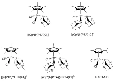
The Dyson group has explored a series of pentamethylcyclopentadienyl IrIII
complexes containing monodentate ligands PTA
(1,3,5-triaza-7-phosphatricyclo-[3.3.1.1]decane) and mPTA (1-methyl-1,3,5-triaza-7-phosphaadamantane), Figure
1.15. In contrast to Ru p-cymene analogue RAPTA-C, which showed effective
inhibition against bovine cat B with IC50 value of 2.5 ± 0.5 μM,67 all these IrIII
complexes were inactive in vitro against A2780 human ovarian cancer cell line and
bovine cat B cells (IC50 > 300 μM).68 Complex [(Cp*)Ir(PTA)Cl2] is considered to
undergo rapid hydrolysis in water to give mono hydroxo species,
[(Cp*)Ir(PTA)(OH)Cl], similar to [(Cp*)Ir(PTA)2Cl]+, followed by further
substitution by sulphur atom of the cysteine residue in the active site of cat B.
However, IrIII complex forms the weakest M-S bond compared to RuII and OsII
analogues by DFT calculation, which may caused the inactivity against bovine cat
Ir Cl Cl P N N N Ir
Cl P N
N N
P
N N
N
[(Cp*)Ir(PTA)Cl2] [(Cp*)Ir(PTA)2Cl]+
Ir
Cl
Cl P
N N N+
Ir
Cl P
N N N+ P N N N Ru Cl Cl P N N N
[image:37.595.122.491.84.349.2][(Cp*)Ir(mPTA)Cl2]+ [(Cp*)Ir(PTA)(mPTA)Cl]2+ RAPTA-C
Figure 1.15. IrIII and RuII monodentate PTA and mPTA complexes.
For [(η6-arene)Ru(Y)(Z)(X)]n+ complexes, replacing the monodentate Y and Z
ligands by a bidentate chelating group YZ generally tends to improve anticancer
activity and stability in aqueous solution.42 Several pentamethylcyclopentadienyl
IrIII anticancer complexes containing N,O- or N,N-chelating ligands have been
studied. N,O-bound 1,2-naphthoquinone-1-oximate complex [(η5-C5Me5)Ir(η2
-C10H6N2O)Cl], Figure 1.16A, exerted a more potent cytotoxicity against HeLa
(cervical carcinoma) and HL60 (leukemia) cancer cells than cisplatin.69 However,
no interaction with double-stranded DNA was observed for the cytotoxic complex.
Another N,O-bound quinolin-8-ol complex [(η5-C5Me5)Ir(qol)Cl], Figure 1.16B,
showed reasonable effectiveness against C-32, SNB-19 and SK-Mel tumour cells.70
Chapter 1: Introduction
unknown. N,N-bound 2-(pyridine-2-yl)thiazole complex [(η5-C5Me5)Ir(pyTz)Cl],
Figure 1.16C, is inactive toward A2780 human ovarian cancer cell line and
cisplatin-resistant variant (A2780cisR) with IC50 value > 300 μM.71 The inactivity
may be due to the absence of interaction with DNA since the complex lacks of
either intercalating group or strong hydrogen bond donors on the N,N-chelating
ligand.
Ir O
N Cl
O
Ir Cl N
O
Ir Cl N
S N
(A) (B) (C)
Figure 1.16. Iridium(III) anticancer complexes containing N,O- or N,N-chelating ligands.69-71
1.4
Ir
IIIPentamethylcyclopentadienyl Complexes as Catalytic
Agents
The global demand for iridium in 2007 was 3700 kg, out of which 750 kg was for
catalysis.72 Recently, remarkable efforts have been devoted towards the
development of aqueous-phase organometallic catalysis due to the relatively easy
mixtures.73 Iridium(III) pentamethylcyclopentadienyl (Cp*) complexes are
extremely useful in synthesis due to their ability to catalyse an array of synthetic
transformations, often with unique selectivity, such as in C=O hydrogenation,
hydroamination, and asymmetric allylic substitutions.74 The iridium Cp* dimer
[(Cp*)IrCl2]2 is an effective catalyst for conversion of alcohols into amides via
oximes.75 Some iridium aqua complexes are reported to catalyse CO2 hydrogenation
due to formation of active hydride complexes.76 IrIII Cp* compounds are also used
as catalysts in a range of other reactions. For example, a number of IrIII Cp*
complexes containing C^N, or N,N-chelating ligands are highly active for
water-oxidation when driven with a chemical oxidant.77
Transfer hydrogenation reactions provide an alternative to direct hydrogenation
for the reduction of a range of substrates. Catalytic iridium complexes are the most
important catalyst for transfer hydrogenation reactions.78 In the reduction of ketones
and aldehydes (one of the fundamental applications of transfer hydrogenation),
iridium hydride species are often formed as actually effective agents in the reaction
pathway. Typically, formic acid, formate, or alcohol can be used as the source of
hydride. A water-soluble iridium hydride complex [(Cp*)Ir(bpy)H]+ (bpy =
2,2'-bipyridine) has been reported as a robust catalyst for acid-catalysed transfer
hydrogenation of carbonyl compounds, Eq. 1.1.79 HCOOH and HCOONa are used
as hydrogen donors to form the hydride complex and the crystal structure is shown
Chapter 1: Introduction
C R2 R1
O
+ [Ir-H]+ H
+
H2O R1 C R2 H OH
+ [Ir-OH
[image:40.595.111.505.85.347.2]2]2+ (Eq. 1.1)
Figure 1.17. Crystal structure of hydride complex [(Cp*)Ir(bpy)H]+.79
For another example, a Cp* IrIII catalyst containing a diamine ligand (HCOONa
as hydrogen donor) showed a higher activity in transfer hydrogenation of aldehydes
than its ruthenium and rhodium analogues.80
In numerous biological hydrogen transfer reactions, an important example is the
reduction of coenzyme NAD+ (β-nicotinamide adenine dinucleotide) to its reduced
form 1,4-NADH (Figure 1.18), by accepting two electrons and a proton from a
substrate in the presence of an enzyme. NAD+/NADH have been known as classic
molecules involving in energy metabolism, reductive biosynthesis and
antioxidation.81-83 Various transition metal hydrides have been studied as catalysts
for the reduction of NAD+ and NAD+-models to the corresponding 1,4-NADH and
shown to catalyse the reduction of NAD+ in the presence of formate as the hydride
source.87 This reduction is regioselective, giving the biologically relevant
1,4-NADH isomer. N N N N NH2 O OH OH H H H H O P O O N H + NH2 O O O P O O O OH OH H H H H ― ― 1 2 3 4 5 6
NAD
+ N N N N NH2 O OH OH H H H H O P O O N NH2 O O O P O O O OH OH H H H H ― ―1,4-NADH
1 2 3 4 5 6 H H [image:41.595.181.454.203.496.2]Chapter 1: Introduction
1.5
Aims
The general aim of this thesis is to investigate the design and reactivity of
organometallic anticancer iridium(III) complexes containing different substituted
cyclopentadienyl and chelating ligands. The studies are focused on systematic
variations in the design of the complexes and studying the relationships between
their physical, chemical and biological properties. More specific aims are as follows.
1. To synthesise and characterise organometallic iridium(III) complexes
with variations to the cyclopentadienyl and chelating ligands with a view
to tune their reactivity and improve their biological properties.
2. To develop structure-activity relationships, investigate the influences of
changing cyclopentadienyl ligands with different substitution and
chelating ligands on their aqueous chemistry and biological activity,
including rates and extent of hydrolysis, acidity of bound water,
nucleobase binding, cytotoxicity, distribution in cellular fractions,
cellular uptake and DNA binding.
3. To investigate the conversion between NAD+ and NADH using
organometallic iridium complexes as catalysts, explore the possibility of
1.6
References
(1) Jemal, A.; Bray, F.; Center, M. M.; Ferlay, J.; Ward, E.; Forman, D. CA-Cancer
J. Clin.2011, 61, 69-90.
(2) Hooning, M. J.; Aleman, B. M. P.; Hauptmann, M.; Baaijens, M. H. A.; Klijn, J.
G. M.; Noyon, R.; Stovall, M.; van Leeuwen, F. E. J. Clin. Oncol. 2008, 26, 5561-5568.
(3) Albain, K. S.; Swann, R. S.; Rusch, V. W.; Turrisi Iii, A. T.; Shepherd, F. A.;
Smith, C.; Chen, Y.; Livingston, R. B.; Feins, R. H.; Gandara, D. R.; Fry, W. A.;
Darling, G.; Johnson, D. H.; Green, M. R.; Miller, R. C.; Ley, J.; Sause, W. T.; Cox,
J. D. The Lancet2009, 374, 379-386.
(4) Metallotherapeutic Drugs and Metal-Based Diagnostic Agents: The Use of
Metals in Medicine; Gielen, M.; Tiekink, E. R. T., Eds.; Wiley: Chichester, 2005.
(5) Thompson, K. H.; Orvig, C. Science2003, 300, 936-939. (6) Orvig, C.; Abrams, M. J. Chem. Rev.1999, 99, 2201-2204.
(7) Rosenberg, B.; Van Camp, L.; Krigas, T. Nature1965, 205, 698-699.
(8) Rosenberg, B.; Vancamp, L.; Trosko, J. E.; Mansour, V. H. Nature 1969, 222, 385-386.
(9) Weiss, R. B.; Christian, M. C. Drugs1993, 46, 360-377.
Chapter 1: Introduction
(13) Jamieson, E. R.; Lippard, S. J. Chem. Rev.1999, 99, 2467-2498.
(14) Sherman, S.; Gibson, D.; Wang, A.; Lippard, S. Science1985, 230, 412-417. (15) Köpf-Maier, P. Eur. J. Clin. Pharmacol.1994, 47, 1-16.
(16) Reedijk, J. Platinum Met. Rev.2008, 52, 2-11.
(17) Alessio, E.; Mestroni, G.; Bergamo, A.; Sava, G. Curr. Top. Med. Chem.2004,
4, 1525-1535.
(18) Sava, G.; Pacor, S.; Mestroni, G.; Alessio, E. Clin. Exp. Metastasis 1992, 10, 273-280.
(19) Berger, M. R.; Garzon, F. T.; Keppler, B. K.; Schmahl, D. Anticancer Res.
1989, 9, 761-766.
(20) Hartinger, C. G.; Zorbas-Seifried, S.; Jakupec, M. A.; Kynast, B.; Zorbas, H.;
Keppler, B. K. J. Inorg. Biochem.2006, 100, 891-904.
(21) Clarke, M. J.; Zhu, F.; Frasca, D. R. Chem. Rev.1999, 99, 2511-2534.
(22) Kelman, A. D.; Clarke, M. J.; Edmonds, S. D.; Peresie, H. J. J. Clin. Hematol.
Onc.1977, 7, 274-288.
(23) Berners-Price, S. J.; Bowen, R. J.; Galettis, P.; Healy, P. C.; McKeage, M. J.
Coord. Chem. Rev.1999, 185-186, 823-836.
(24) Jakupec, M. A.; Keppler, B. K. Curr. Top. Med. Chem.2004, 4, 1575-1583. (25) Halpern, J. Pure Appl. Chem.2001, 73, 209-220.
(26) Gasser, G.; Ott, I.; Metzler-Nolte, N. J. Med. Chem.2011, 54, 3-25.
(28) Köpf-Maier, P.; Köpf, H. Chem. Rev.1987, 87, 1137-1152.
(29) Köpf-Maier, P.; Köpf, H. In Bioinorganic Chemistry; Springer
Berlin/Heidelberg: 1988; 70, pp 103-185.
(30) Top, S.; Vessières, A.; Leclercq, G.; Quivy, J.; Tang, J.; Vaissermann, J.;
Huché, M.; Jaouen, G. Chem.-Eur. J.2003, 9, 5223-5236.
(31) Guo, M.; Guo, Z.; Sadler, P. J. Biol. Inorg. Chem.2001, 6, 698-707.
(32) Kröger, N.; Kleeberg, U. R.; Mross, K.; Edler, L.; Hossfeld, D. K. Onkologie
2000, 23, 60-62.
(33) Mross, K.; Robben-Bathe, P.; Edler, L.; Baumgart, J.; Berdel, W. E.; Fiebig, H.;
Unger, C. Onkologie2000, 23, 576-579.
(34) Allen, O. R.; Gott, A. L.; Hartley, J. A.; Hartley, J. M.; Knox, R. J.; McGowan,
P. C. Dalton Trans.2007, 5082-5090.
(35) Oberschmidt, O.; Hanauske, A. R.; Pampillón, C.; Sweeney, N. J.; Strohfeldt,
K.; Tacke, M. Anti-Cancer Drugs2007, 18, 317-321
(36) Allen, O. R.; Croll, L.; Gott, A. L.; Knox, R. J.; McGowan, P. C.
Organometallics2004, 23, 288-292.
(37) Fricker, S. P. Metal compounds in cancer therapy; Chapman & Hall: London,
1994.
(38) White, C. Y., A.; Maitlis, P. M. Inorg. Chem.1992, 29, 228-234.
(39) Amouri, H.; Moussa, J.; Renfrew, A. K.; Dyson, P. J.; Rager, M. N.;
Chapter 1: Introduction
(41) Morris, R. E.; Aird, R. E.; del Socorro Murdoch, P.; Chen, H.; Cummings, J.;
Hughes, N. D.; Parsons, S.; Parkin, A.; Boyd, G.; Jodrell, D. I.; Sadler, P. J. J. Med.
Chem.2001, 44, 3616-3621.
(42) Aird, R. E.; Cummings, J.; Ritchie, A. A.; Muir, M.; Morris, R. E.; Chen, H.;
Sadler, P. J.; Jodrell, D. I. Br. J. Cancer2002, 86, 1652-1657.
(43) Wang, F.; Chen, H.; Parsons, S.; Oswald, I. D. H.; Davidson, J. E.; Sadler, P. J.
Chem.-Eur. J.2003, 9, 5810-5820.
(44) Novakova, O.; Chen, H.; Vrana, O.; Rodger, A.; Sadler, P. J.; Brabec, V.
Biochemistry2003, 42, 11544-11554.
(45) Liu, H.-K.; Wang, F.; Parkinson, J. A.; Bella, J.; Sadler, P. J. Chem.-Eur. J.
2006, 12, 6151-6165.
(46) Chen, H.; Parkinson, J. A.; Parsons, S.; Coxall, R. A.; Gould, R. O.; Sadler, P.
J. J. Am. Chem. Soc.2002, 124, 3064-3082.
(47) Peacock, A. F. A.; Habtemariam, A.; Fernández, R.; Walland, V.; Fabbiani, F.
P. A.; Parsons, S.; Aird, R. E.; Jodrell, D. I.; Sadler, P. J. J. Am. Chem. Soc.2006,
128, 1739-1748.
(48) Peacock, A. F. A.; Habtemariam, A.; Moggach, S. A.; Prescimone, A.; Parsons,
S.; Sadler, P. J. Inorg. Chem.2007, 46, 4049-4059.
(49) Peacock, A. F. A.; Melchart, M.; Deeth, R. J.; Habtemariam, A.; Parsons, S.;
Sadler, P. J. Chem.-Eur. J.2007, 13, 2601-2613.
(51) van Rijt, S. H.; Peacock, A. F. A.; Johnstone, R. D. L.; Parsons, S.; Sadler, P. J.
Inorg. Chem.2009, 48, 1753-1762.
(52) Fu, Y.; Habtemariam, A.; Pizarro, A. M.; van Rijt, S. H.; Healey, D. J.; Cooper,
P. A.; Shnyder, S. D.; Clarkson, G. J.; Sadler, P. J. J. Med. Chem. 2010, 53, 8192-8196.
(53) Shnyder, S. D.; Fu, Y.; Habtemariam, A.; van Rijt, S. H.; Cooper, P. A.;
Loadman, P. M.; Sadler, P. J. Med. Chem. Comm.2011, 2, 666-668.
(54) Sava, G.; Giraldi, T.; Mestroni, G.; Zassinovich, G. Chem. Biol. Interact.1983,
45, 1-6.
(55) Giraldi, T.; Sava, G.; Mestroni, G.; Zassinovich, G.; Stolfa, D. Chem. Biol.
Interact.1978, 22, 231-238.
(56) Messori, L.; Marcon, G.; Orioli, P.; Fontani, M.; Zanello, P.; Bergamo, A.;
Sava, G.; Mura, P. J. Inorg. Biochem.2003, 95, 37-46.
(57) Marcon, G.; Casini, A.; Mura, P.; Messori, L.; Bergamo, A.; Orioli, P.
Metal-Based Drugs2000, 7, 195-200.
(58) Sava, G.; Zorzet, S.; Perissin, L.; Mestroni, G.; Zassinovich, G.; Bontempi, A.
Inorg. Chim. Acta1987, 137, 69-71.
(59) Cleare, M. J. Coord. Chem. Rev.1974, 12, 349-405.
(60) Schäfer, S.; Sheldrick, W. S. J. Organomet. Chem.2007, 692, 1300-1309. (61) Scharwitz, M. A.; Ott, I.; Gust, R.; Kromm, A.; Sheldrick, W. S. J. Inorg.
Chapter 1: Introduction
(62) Schäfer, S.; Ott, I.; Gust, R.; Sheldrick, W. S. Eur. J. Inorg. Chem.2007, 3034-3046.
(63) Geldmacher, Y.; Kitanovic, I.; Alborzinia, H.; Bergerhoff, K.; Rubbiani, R.;
Wefelmeier, P.; Prokop, A.; Gust, R.; Ott, I.; Wölfl, S.; Sheldrick, W. S. Chem. Med.
Chem.2011, 6, 429-439.
(64) Frodl, A.; Herebian, D.; Sheldrick, W. S. J. Chem. Soc., Dalton Trans. 2002, 3664-3673.
(65) Leung, S.-K.; Liu, H.-W.; Lo, K. K.-W. Chem. Commun. 2011, DOI: 10.1039/c1031cc11423a.
(66) Lo, K. K.-W.; Zhang, K. Y.; Li, S. P.-Y. Pure Appl. Chem.2011, 83, 823-840. (67) Casini, A.; Gabbiani, C.; Sorrentino, F.; Rigobello, M. P.; Bindoli, A.;
Geldbach, T. J.; Marrone, A.; Re, N.; Hartinger, C. G.; Dyson, P. J.; Messori, L. J.
Med. Chem.2008, 51, 6773-6781.
(68) Casini, A.; Edafe, F.; Erlandsson, M.; Gonsalvi, L.; Ciancetta, A.; Re, N.;
Ienco, A.; Messori, L.; Peruzzini, M.; Dyson, P. J. Dalton Trans. 2010, 39, 5556-5563.
(69) Wirth, S.; Rohbogner, C.; Cieslak, M.; Kazmierczak-Baranska, J.; Donevski, S.;
Nawrot, B.; Lorenz, I.-P. J. Biol. Inorg. Chem.2010, 15, 429-440.
(70) Sliwinska, U.; Pruchnik, F. P.; Ulaszewski, S.; Latocha, M.; Nawrocka-Musial,
D. Polyhedron2010, 29, 1653-1659.
(71) Gras, M.; Therrien, B.; Süss-Fink, G.; Casini, A.; Edafe, F.; Dyson, P. J. J.
(72) Jollie, D. Platinum 20082008, 42-43
(73) Aqueous-Phase Organometallic Catalysis, 2 ed.; Cornils, B.; Herrmann, W. A.,
Eds.; Wiley-VCH: Weinheim, Germany, 2004.
(74) Iridium Complexes in Organic Synthesis; Oro, L. A.; Claver, C., Eds.;
WILEY-VCH: Weinheim, Germany, 2008.
(75) Owston, N. A.; Parker, A. J.; Williams, J. M. J. Org. Lett.2006, 9, 73-75. (76) Ogo, S.; Kabe, R.; Hayashi, H.; Harada, R.; Fukuzumi, S. Dalton Trans.2006, 4657-4663.
(77) Blakemore, J. D.; Schley, N. D.; Balcells, D.; Hull, J. F.; Olack, G. W.;
Incarvito, C. D.; Eisenstein, O.; Brudvig, G. W.; Crabtree, R. H. J. Am. Chem. Soc.
2010, 132, 16017-16029.
(78) Saidi, O.; Williams, J. In Iridium Catalysis; Andersson, P. G., Ed.; Springer
Berlin / Heidelberg: 2011; 34, pp 77-106.
(79) Abura, T.; Ogo, S.; Watanabe, Y.; Fukuzumi, S. J. Am. Chem. Soc.2003, 125, 4149-4154.
(80) Wu, X.; Liu, J.; Li, X.; Zanotti-Gerosa, A.; Hancock, F.; Vinci, D.; Ruan, J.;
Xiao, J. Angew. Chem., Int. Ed.2006, 45, 6718-6722.
(81) Belenky, P.; Bogan, K. L.; Brenner, C. Trends Biochem. Sci.2007, 32, 12-19. (82) Berger, F.; Ramírez-Hernández, M. H.; Ziegler, M. Trends Biochem. Sci.2004,
29, 111-118.
Chapter 1: Introduction
(84) Lo, H. C.; Leiva, C.; Buriez, O.; Kerr, J. B.; Olmstead, M. M.; Fish, R. H.
Inorg. Chem.2001, 40, 6705-6716.
(85) Lo, H. C.; Buriez, O.; Kerr, J. B.; Fish, R. H. Angew. Chem., Int. Ed.1999, 38, 1429-1432.
(86) Yan, Y.; Melchart, M.; Habtemariam, A.; Peacock, A.; Sadler, P. J. Biol. Inorg.
Chem.2006, 11, 483-488.
Chapter 2
Experimental Methods and Materials
Chapter 2: Experimental Methods and Materials
This Chapter describes the main experimental techniques used in this work. More
specific methods relating to individual experiments are described in the appropriate
Chapters. The synthesis and characterisation of the cyclopentadienyl ligands and
dimers used for the synthesis of the iridium complexes in subsequent Chapters is
also covered here.
2.1
Instrumentation and Methods
2.1.1 Nuclear Magnetic Resonance Spectroscopy (NMR)1-3
Nuclear magnetic resonance (NMR) spectroscopy is a powerful technique which
exploits the magnetic properties of nuclei to determine physical and chemical
properties of atoms or molecules. All chemical elements that possess a nuclear spin
of ½ or greater are in principle observable by NMR. Most studied nuclei with NMR
are 1H, 13C and 15N. When placed in a magnetic field (B0), nuclei with a spin ½ will
distribute over two states with different energies (α and β), and process around B0 at
a characteristic frequency (Larmor frequency). The slight population difference
between the α and β states results in a bulk magnetization vector (M0) along the
axes of B0. The application of a second magnetic field (B1) at the Larmor frequency
of the nuclei will cause the nuclei to resonate. In most NMR experiments, the nuclei
are irradiated with brief pulses of B1, allowing the spins to return to their
equilibrium between the pulses referred to as relaxation. The relaxation of the spins
causes the NMR signal to decay with time, producing the observed Free Induction
where signals are plotted as a function of frequency. Depending on the local
chemical environment, different nuclides in a molecule resonate at slightly different
frequencies and this gives rise to the different chemical shifts observed in the NMR
spectra. The chemical shift can be used to obtain structural information about the
molecule by understanding the different chemical environments of the atoms in the
molecule.
2D NMR spectra provide more information about a molecule than 1D NMR
spectra and are especially useful in determining the structure of a molecule,
particularly for molecules that are too complicated to work with using 1D NMR. In
the COSY (correlation spectroscopy), TOCSY (total correlation spectroscopy) or
NOESY (nuclear Overhauser effect spectroscopy) 2D NMR experiments, a second
frequency domain is introduced to reveal structural connections through magnetic
interactions between nuclei. In both the COSY and TOCSY experiments, two
essentially identical chemical shift axes are plotted orthogonally to each other, with
the 1D NMR spectra appearing on the diagonal of the plot. In a COSY experiment,
all the spin-spin coupled protons are indicated by cross peaks which are
symmetrically placed along the diagonal. COSY spectra typically can show two-
and three-bond coupled protons, however long-range bonding can be emphasised
through introducing extra delays in the pulse sequence. In TOCSY experiments, the
connectivity of a whole spin system can be shown as cross peaks are found for all
protons that are connected within it. 2D NOESY was used to establish structural
information resulting from through-space interactions between protons that are
Chapter 2: Experimental Methods and Materials
2.1.1.1 Experimental
1
H NMR spectra were acquired in 5 mm NMR tubes at 298K (unless stated
otherwise) on either Bruker AV-400, Bruker DRX 500, AVA 600 or Bruker AV II
700 NMR spectrometers. 1H NMR chemical shifts were internally referenced to
(CHD2)(CD3)SO (2.50 ppm) for DMSO-d6, CHCl3 (7.26 ppm) for chloroform-d1,
CHD2OD (3.33 ppm) for methanol-d4and to 1,4-dioxane (3.75 ppm) for aqueous
solutions. The data were processed using TOPSPIN (version 2.1 Bruker UK Ltd).
2.1.1.2 Water Suppression
Many experiments in this thesis were performed on samples in aqueous solutions
(99.9% D2O, or 5% MeOD-d4/95% D2O, or 10% MeOD-d4/90% H2O,) so as to be
of biological relevance. The use of such aqueous solutions results in a large HOD
signal that can obscure the other signals in the 1H spectrum. To minimize this, the
HOD signal was suppressed with either presaturation or Shaka techniques.4
Presaturation involves the saturation of the HOD peak by irradiating the frequency
of water in between pulse sequences. Shaka water suppression, or Double Pulse
Field Gradient Spin Echo (DPFGSE), uses pulsed field gradient spin echoes in
which the refocusing pulse is the sequence soft π(x)-hard π(-x). The disadvantage of
2.1.2 pH Measurements
pH values were measured at ambient temperature, using a Corning 240 pH meter
equipped with a micro combination KNO3 (chloride free) electrode calibrated with
Aldrich buffer solutions of pH 4, 7 and 10. pH* values (pH meter reading) of NMR
samples in D2O were measured directly in the NMR tube, before and after recording
NMR spectra using the same method.
2.1.3 Determination of pKa Values
To generate the aqua complexes, chlorido complexes were dissolved in D2O and
0.98 mol equiv of AgNO3 were added. The solution was stirred for 24 h at 298 K,
and AgCl was removed by filtration. For determinations of pKa* values (pKa values
for solutions in D2O) the pH* values of solutions of the aqua complexes in D2O
were varied from ca. pH* 2 to 11 by the addition of dilute NaOD and DClO4, and 1
H NMR spectra were recorded. The chemical shifts of the chelating ligand protons
and/or methyl protons of Cpx were plotted against pH*. The pH* titration curves
were fitted to the Henderson-Hasselbalch equation using ORIGIN version 8.0.
These pKa* values can be converted to pKa values by use of the equation pKa =
0.929pKa* + 0.42 as suggested by Krezel and Bal5 for comparison with related
Chapter 2: Experimental Methods and Materials
2.1.4 X-ray Crystallography
X-ray diffraction (XRD) is a widely used technique for the precise determination
of the position of atoms in molecules in the crystalline solid state. XRD was used in
this thesis to characterise the crystal structures of several synthesised compounds.
All diffraction data were obtained on an Oxford Diffraction Gemini four-circle
system with a Ruby CCD area detector using Mo Kα radiation. Absorption
corrections were applied using ABSPACK.6 The crystals were mounted in oil and
held at 100(2) K with the Oxford Cryosystem Cobra. The structures were solved by
direct methods using SHELXS (TREF)7 with additional light atoms found by
Fourier methods. Complexes were refined against F2 using SHELXL.8 Data
collection and solution of the structures were carried out by Dr. Guy Clarkson
(Department of Chemistry, University of Warwick). Details of the acquisition and
solving of the individual crystal structures are explained in the corresponding
Chapters.
2.1.5 Elemental Analysis
Elemental analysis determines the percentage of C, H and N composition in a
sample. Comparing the obtained C, H, N values of a sample of the complex to
theoretical values provides information about the purity of the compound and was
used for this purpose in this thesis. CHN elemental analyses were carried out on a
2.1.6 Electrospray Ionisation Mass Spectrometry (ESI-MS)9
Mass spectrometry (MS) is an analytical technique that measures the
mass-to-charge ratios of mass-to-charged particles. It is used for determining masses of particles, and
for elucidating the chemical structures of molecules. The MS principle consists of
ionizing chemical compounds to generate charged molecules or molecule fragments
and measuring their mass-to-charge ratios. Electrospray ionization (ESI) is a
technique used in mass spectrometry to produce ions. The sample must be present
in the form of ions in solution and this is typically achieved by adding small
amounts of acid or base to respectively protonate (ESI positive) or deprotonate (ESI
negative) the solution. ESI-MS was used routinely in this work for the
characterisation of the complexes.
ESI-MS were obtained on a Bruker Esquire 2000 Ion Trap Spectrometer. Samples
were prepared in 50% CH3CN and 50% H2O (v/v). The mass spectra were recorded
with a scan range of m/z 50–1000 for positive ions. Data were processed using Data
Analysis 3.3 (Bruker Daltonics).
2.1.7 Inductively Coupled Plasma Mass Spectroscopy (ICP-MS)9,10
ICP-MS is a very sensitive technique that can be used to determine the elemental
concentrations of solutions. More than 75 elements can be determined; most of
Chapter 2: Experimental Methods and Materials
(ionization) with mass spectrometry as a method of separating and detecting the
ions.
All ICP-MS analyses were carried out on an Agilent Technologies 7500 series
ICP-MS instrument. The water used for ICP-MS analysis was doubly deionised
(DDW) using a Millipore Milli-Q water purification system and a USF Elga UHQ
water deionizer. The iridium Specpure plasma standard (Alfa Aesar, 1000 ppm in
10% HCl) was diluted with 3% HNO3 DDW to freshly prepare calibrants at
concentrations 1000, 800, 400, 200, 100, 50, 10, 1 and 0.1 ppb. The ICP-MS
instrument was set to detect 193Ir with typical detection limits of ca. 2 ppt using no
gas mode.
2.1.8 UV-Vis Absorption Spectroscopy
UV-Vis spectroscopy is routinely used in analytical chemistry for the quantitative
determination of different analytes, such as transition metal ions, highly conjugated
organic compounds, and biological macromolecules.9
This method is used in this thesis to monitor hydrolysis of iridium chloride
complexes and reactions with NADH or NAD+. A Cary 300 UV-Vis recording
spectrophotometer was used with 1 cm path-length quartz cuvettes (0.5 mL) and a
PTP1 Peltier temperature controller. Spectra were processed using UVWinlab

![Figure 1.10. [(η6-arene)Ru(en)Cl]+ anticancer complexes (arene shown in right).](https://thumb-us.123doks.com/thumbv2/123dok_us/9681782.469764/28.595.269.341.210.333/figure-arene-ru-anticancer-complexes-arene-shown-right.webp)
![Figure 1.17. Crystal structure of hydride complex [(Cp*)Ir(bpy)H]+.79](https://thumb-us.123doks.com/thumbv2/123dok_us/9681782.469764/40.595.111.505.85.347/figure-crystal-structure-hydride-complex-cp-ir-bpy.webp)

![Table 2.1. Crystallographic Data for the Dimer [(η5-C5Me4C6H5)IrCl2]2 (2)](https://thumb-us.123doks.com/thumbv2/123dok_us/9681782.469764/69.595.110.434.126.627/table-crystallographic-data-dimer-h-c-me-ircl.webp)
![Table 3.1. Crystallographic Data for Complexes [(η5-C5Me4C6H5)Ir(phen)Cl]PF6](https://thumb-us.123doks.com/thumbv2/123dok_us/9681782.469764/98.595.112.526.179.632/table-crystallographic-data-for-complexes-me-ir-phen.webp)
