Original Article
miR-143-3p functions as a tumor suppressor by
targeting HK2 in neuroblastomas
Yuanhua Cen1, Li Xu3, Saihu Huang4, Songqiang Zhuang2
Departments of 1Pediatrics, 2Orthopedics, Fenghua Hospital, Fenghua, Ningbo, Zhejiang, China; 3Cell Laboratory,
Jiangsu Cancer Hospital, Jiangsu, China; 4Pediatrics Intensive Care Unit, Children’s Hospital of Soochow
University, China
Received February 15, 2019; Accepted March 12, 2019; Epub July 15, 2019; Published July 30, 2019
Abstract: Hexokinase 2 (HK2) is strongly expressed in multiple human cancers. Dysregulation of HK2 has been associated with malignant progression of tumor cells, including neuroblastomas (NB). Accumulating evidence has verified the obvious roles of miR-143-3p in anti-tumor growth. Thus, it was hypothesized that miR-143-3p may function as a tumor suppressor in NB via modulation of HK2. The current study detected the abundance of miR-143-3p and HK2 mRNA in NB tissues and cell lines using qRT-PCR. Interactions between miR-miR-143-3p and HK2 were predicted using an online database, further confirmed by Dual-Luciferase reporter and Western blot assays. Glucose consumption, cell proliferation, and apoptosis were determined by colorimetry, MTT, and flow cytometry assays, respectively. Cell migration and invasion abilities were evaluated using Transwell assays. Protein levels of HK2, cleaved caspase-3, and Ki-67 were measured with Western blotting assays. Finally, the roles of miR-143-3p and HK2 on tumor growth were demonstrated in vivo. Results showed that miR-143-3p was significantly downregu -lated and HK2 was upregu-lated in tumor samples and cancer cells. HK2 was, therefore, identified as a potential target of miR-143-3p. The addition of miR-143-3p attenuated glucose consumption, proliferation, migration, and invasion. It also stimulated apoptosis of NB cells, which was reversed by HK2 restoration. Furthermore, miR-143-3p suppressed tumor growth in vivo by downregulating HK2. Present findings indicate that miR-143-3p suppresses NB progression through direct targeting of HK2, providing a novel therapeutic target for NB patients.
Keywords: Neuroblastoma, miR-143-3p, HK2, epigenetics
Introduction
Neuroblastomas (NB) are the third most com-mon solid tumors in children, originating from primitive neural crest cells of the sympathetic nervous system. An estimated 710 new cases of NB have been diagnosed among children younger than 14 years old, accounting for near-ly 7% of all types of childhood malignancies [1]. Although development of novel therapeutic strategies, such as surgery and/or chemothera-py and radiotherachemothera-py, have improved survival times of patients with NB, prognosis for chil-dren with regional or distant-metastatic dis-ease remains unsatisfactory [2, 3]. Further clarification of the molecular mechanisms will potentially aid in identifying suitable agents for therapy.
Previous studies have identified a series of bio -markers implicated in occurrence and
can-cer, and prostate cancer [9, 10]. In NB, HK2 stimulates cell proliferation and metastasis, conferring high resistance of NB cells to che-motherapy [11]. However, the regulatory mech-anisms of HK2 have not been elucidated in NB. MicroRNAs (miRNAs) are a class of endogenous small non-coding RNA molecules 19-24 nucleo-tides in length. MicroRNAs can negatively re- gulate gene expression post-transcriptionally through binding to 3’-untranslated regions (3’-UTR) of target mRNAs, in a sequence-specific manner, by base-pairing. This leads to mRNA degradation or translational repression [12, 13]. Recent studies have verified abnormal expression patterns of miRNAs in human can-cers, implicated in momentous pathways linked to cell proliferation, apoptosis, and metastasis [14]. Several miRNAs target HK2, a metabo-lism-related factor, to modulate the progres-sion of various types of tumors [15, 16]. miR-143-3p has been reported as a tumor suppres-sor in multiple cancers, including breast can- cer [17], cervical cancer [18], and esophageal squamous cell carcinoma (ESCC) [19]. Yang et al. also revealed that RBM3 protected NB cells from apoptosis via abolishing the induction of miR-143 by NO, indicating the functional impli-cation of miR-143 in NB [20]. The current study sought to address the interaction of miR-143-3p and HK2 in the regulation of NB tumor biol-ogy, aiming to provide a novel biological target for NB treatment.
Materials and methods
Patients and tissue specimens
Thirty-two NB tumor tissues and paired normal tissues were collected from patients undergo-ing surgical resections at Fenghua Hospital, between March 2014 and February 2017. No- ne of the patients received preoperative tre- atment. According to the International Neuro- blastoma Staging System (INSS), 10 samples were defined as INSS I and II tumor tissues (non-metastases), 9 were defined as INSS III tumor tissues (regional metastases), and 13 were defined as INSS IV tumor tissues (distant metastases). Excised specimens were quick-frozen in liquid nitrogen and stored at -80°C. Written informed consent was obtained from all participants. This study was approved by the Research Ethics Committee of Fenghua Hos- pital.
Cell culture and transfection
Two human NB cell lines, SK-N-SH and SH-SY-5Y, were purchased from the American Type Culture Collection (ATCC, Manassas, VA, USA), with human neuroblast CHP-126 as the normal control. All cells were grown in DMEM medium supplemented with 10% heat-inactivated fetal bovine serum (FBS, Gibco, Carlsbad, CA, USA), 100 U/mL penicillin (Sigma-Aldrich, St. Louis, MO, USA), and 100 µg/mL streptomycin (Sigma-Aldrich). The medium was maintained in an incubator of 5% CO2 at 37°C.
For this study, miR-143-3p mimics (miR-143-3p) and matched negative controls (NC) were obtained from GenePharma (Shanghai, China). HK2-overexpressed plasmid (HK2) was gener-ated by inserting full-length HK2 sequences into pcDNA3.1 plasmid. MicroRNAs or plasmi- ds were transiently transfected into SK-N-SH and SH-SY5Y cells using Lipofectamine 2000 reagent (Invitrogen, Carlsbad, CA, USA), accord-ing to manufacturer instructions.
RNA preparation and quantitative real-time PCR (qRT-PCR)
Total RNA was isolated from NB tissues and cell lines using TRIzol Reagent (Invitrogen), accord -ing manufacturer protocol. This was followed by detection of RNA purities using a spectropho-tometer (Mapada, Shanghai, China). Next, 1 µg RNA was reversely transcribed into first-str-and cDNA using TaqMan MicroRNA Reverse Transcription kit (Thermo Fisher, Waltham, MA, USA) or High Capacity cDNA Reverse Trans-cription Kit (Thermo Fisher). Reaction condi -tions of reverse transcription included anneal-ing at 16°C for 30 minutes, cDNA synthesis at 42°C for 30 minutes, and enzyme denaturation at 85°C for 5 minutes.
5’-AGGTCAAACTCCTCTCGC CG-3’ (reverse); GAP-DH: 5’-TATGATGATATCAAGAGGGTAGT-3’ (forwa-rd), 5’-TGTATCCAAACTCATTGTCATAC-3’ (rever-se). Primers for miR-143-3p and U6 were ob- tained from GenePharma.
Dual-Luciferase reporter assay
Partial sequences of the HK2 3’ untranslated region (3’-UTR) containing putative miR-143-3p binding sites were inserted into psiCHECK-2 vectors (Promega, Madison, WI, USA) to gener -ate wild-type HK2 luciferase reporter (HK1-wt). Next, mutant HK2 luciferase reporter (HK2-mut) containing mutant miR-143-3p binding sites was constructed using TaKaRaMutanBEST kit (Takara), according to manufacturer instruc -tions. SH-SY5Y and SK-N-SH cells (5×10³) were seeded into 96-well microplates and hatched for 24 hours. Afterward, NC or miR-143-3p was transfected into cells, along with HK1-wt or HK2-mut reporter. About 48 hours after trans-fection, luciferase activity was determined using the Dual-Luciferase Reporter System (Promega).
Glucose consumption
Glucose consumption was detected using Scr- een Quest Colorimetric Glucose Uptake Assay Kit (AAT Bioquest, Sunnyvale, CA, USA). Briefly, transfected cells (5×10⁴) supplemented with serum-free medium were starved in 96-well plates overnight at 37°C. After washing twice with KRPH buffer, the cells were resuspended in 90 μL Glucose Uptake Buffer for 1 hour, stim -ulated with 10 μL insulin for 20 minutes, and treated with 10 μL 2-deoxyglucose (2-DG) for 40 minutes. They were then washed with KRPH buffer and lysed with Acidic Lysis Buffer. This was followed by glucose consumption detec-tion at an indicated absorbance of 570 nm using a microplate reader (Thermo Fisher).
Western blot assay
Proteins from cells or tumors were extracted using RIPA lysis buffer (ThermoFisher), supple -mented with proteinase inhibitors and quanti-fied using BCA Protein Assay Kit (Thermo Fisher Scientific), according to manufacturer protocol. Extracted proteins denatured at 98°C for 5 minutes were separated by SDS-PAGE gel and transferred onto PVDF membranes (Millipore, Bedford, MA, USA). After blocking with 5%
non-fat milk powder at 37°C for 2 hours, the mem -branes were hatched with rabbit anti-HK2 (1:1000), active caspase-3 (1:1000), anti-Ki-67 (1:1000), and anti-beta actin (1:5000) (Abcam, Cambridge, MA, USA) overnight at 4°C. Subsequently, they were further incubated with horseradish peroxidase (HRP)-conjugated goat anti-rabbit IgG antibody (1:10000) (Abcam) for another 1.5 hours. Proteins were detected using ECL reagents (Pierce, Rockford, IL, USA) on the Bio-Rad ChemiDoc MP imaging system (Bio-Rad, Hercules, CA, USA).
Cell proliferation assay
Cell proliferation was measured using Cell Counting Kit-8 (CCK-8 kit) (Dojindo, Tokyo, Japan), referring to manufacturer instructions. NB cells transfected with miR-143-3p or HK2 were plated in 96-well microplates at a density of 5×10³ cells/well. This was followed by the addition of 10 μl CCK-8 reagent at 0, 24, 48, and 72 hours after transfection. After incubat-ing for another 2 hours, absorbance at 450 nm was measured using a microplate reader.
Cell apoptosis assay
Cell apoptosis was measured using Annexin V-FITC/PI Apoptosis Detection Kit (BestBio, Shanghai, China), in accordance with manufac-turer instructions. NB cells were treated with miR-143-3p or HK2 for 48 hours. The cells were then digested with trypsin free from EDTA, washed twice with PBS, and adjusted to a con-centration of 5×10⁵ cells/mL with the binding buffer. Afterward, 200 µl suspensions were added to each labeled tube. This was followed by the introduction of 5 µl Annexin V-FITC and 10 µl propidium iodide (PI). After incubation in a dark room for 10 minutes, apoptotic cells were observed by flow cytometry (BD Biosciences, San Jose, CA, USA).
Cell migration and invasion assays
10% FBS was added in the bottom chamber. After incubation for 16 hours, cells migrating or invading the basal side of the membrane were fixed with methanol and stained with hematoxy -lin (Sigma-Aldrich). Finally, cells in random three visual fields were counted using a microscope (Olympus, Tokyo, Japan).
Tumor xenograft
Female BALB/c nude mice (4-6 weeks old) were purchased from Huafukang (Beijing, China) and raised under indicated conditions with a 12- hour light/dark cycle for one week. Next, 1×10⁷ NB cells transfected with miR-143-3p or HK2 were subcutaneously inoculated into the backs of the mice after acclimatization. Tumors were measured using Vernier calipers every three days after injection. Volume was calculated by (length × width2)/2. Reaching end points, the mice were sacrificed. Tumors were collected to determine protein expression levels of HK2, Ki-67, and cleaved caspase-3. All experiments were performed in accordance with the Guiding Principles in the Use of Laboratory Animals and approved by the Committee of Animal Research ofFenghua Hospital.
Statistical analysis
Data are displayed as mean ± standard devia-tion (SD). Statistical differences between two
or more groups were assessed by Student’s t-test or ANOVA using SPSS 20.0 software (SPSS, Inc., Chicago, IL, USA). P<0.05 indicates statistical significance.
Results
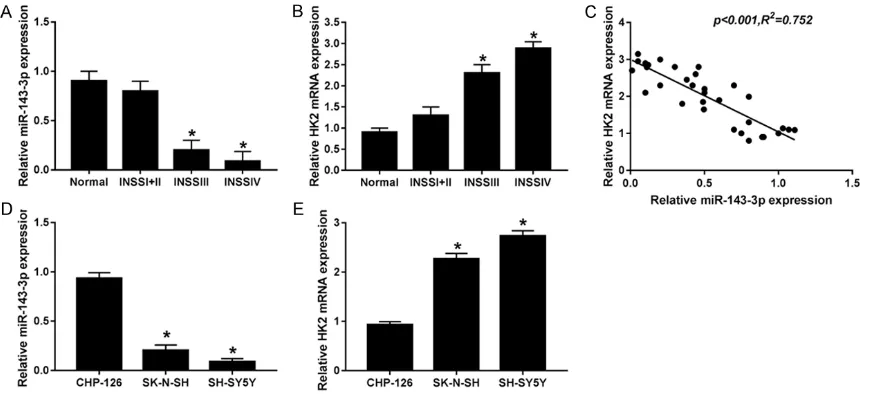
miR-143-3p was significantly downregulated while HK2 was upregulated in NB tissues and cells
[image:4.612.91.527.70.267.2]Present findings indicate that miR-143-3p and HK2 might be involved in the pathogenesis of NB.
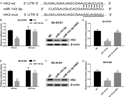
HK2 directly targeted by miR-143-3p
MiRNAs can function as an oncogene or anti-tumor factor in multiple cancers via targeting cancer-related molecules [21]. In view of the alteration of miR-143-3p and HK2 in NB tis-sues and cells, as well as the negative correla-tion between expression levels of the two genes, present researchers asked whether HK2 was mediated by miR-143-3p through complementary binding sites. Bioinformatics analysis predicted the putative binding sites of miR-143-3p within the 3’-UTR of HK2 using TargetScan online website (Figure 2A). To fur
[image:5.612.92.522.65.398.2]miR-143-3p inhibited glycolysis and prolifera-tion while inducing apoptosis in NB cells by targeting HK2
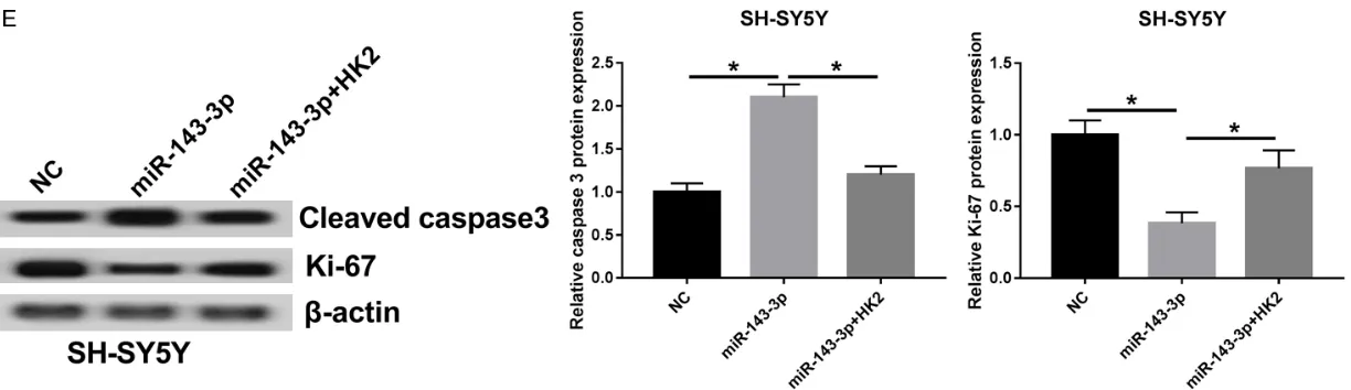
Since glycolysis, proliferation, and apoptosis are required for tumor progression, the current study probed the function and mechanisms of miR-143-3p in NB. Addition of miR-143-3p sup-pressed glucose consumption (Figure 3A) and cell proliferation (Figure 3B) in SH-SY5Y and SK-N-SH cells. This was overturned by HK2 res -toration (Figure 3A and 3B). In contrast, apop-tosis of SH-SY5Y and SK-N-SH cells was signifi -cantly induced in the presence of miR-143-3p, while HK2-overexpression abolished miR-143-3p-induced cell apoptosis (Figure 3C). Caspa- se-3 plays a key role in the regulation of cell apoptosis and Ki-67 has been considered a biomarker of cell proliferation. Protein levels of cleaved caspase-3 were strikingly upregulated, while Ki-67 was downregulated in miR-143-3p-transfected SH-SY5Y and SK-N-SH cells (Figure 3D and 3E). Introduction of HK2 abrogated stimulatory effects of miR-143-3p on cleaved caspase-3 expression, as well as inhibitory effects on Ki-67 expression (Figure 3D and
3E). Present findings indicate that miR-143-3p attenuated glycolysis and proliferation, while stimulating apoptosis in NB cells by directly tar-geting HK2.
miR-143-3p inhibited cell migration and inva-sion by directly interacting with HK2 in NB cells
Metastasis is a major threat to NB-related death. NB patients with distant-metastasis often develop malignant lesions. The current study verified the regulatory effects of miR-143-3p on cell migration and invasion in NB cells. Results revealed that migration and inva-sion abilities of SH-SY5Y and SK-N-SH cells were significantly suppressed following the transfection of miR-143-3p, compared with the NC group (Figure 4A and 4B). However, migra-tion and invasion abilities inhibited by miR-143-3p were induced with the restoration of HK2 in SH-SY5Y and SK-N-SH cells (Figure 4A and 4B). Results suggest that miR-143-3p inhibits cell migration and invasion via directly interacting with HK2.
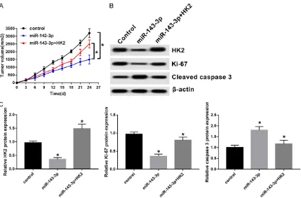
Introduction of miR-143-3p inhibited tumor growth via regulation of HK2 in vivo
In present study, effects of miR-143-3p on tumor growth in vivo were evaluated. SH-SY5Y
cells transfected with 3p or miR-143-3p+HK2 were subcutaneously injected into BALB/c nude mice. Volumes of NB tumors in different groups continually increased during the whole tumor growth period (Figure 5A). Presence of miR-143-3p lowered tumor vol-umes. They were inversed by the restoration of HK2 (Figure 5A). The abundance of HK2 and Ki-67 prominently decreased, while cleaved caspase-3 was increased in miR-143-3p-expressed NB tumors. HK2 introduction over-turned inhibitory effects of miR-143-3p on HK2 and Ki-67 expression, as well as stimulatory effects on cleaved caspase-3 expression (Fi- gure 5B and 5C). Present data suggests that miR-143-3p suppresses NB growth in vivo via negative modulation of HK2.
Discussion
Figure 3. miR-143-3p inhibited cell glycolysis and proliferation, while inducing apoptosis by targeting HK2. SH-SY5Y and SK-N-SH cells were transfected with NC, miR-143-3p, or miR-143-3p+HK2. (A) About 48 hours after transfection, glucose consumption was detected by colorimetry. Cell proliferation (B), apoptosis (C), and protein levels of cleaved caspase-3 and Ki-67 (D and E) in SH-SY5Y and SK-N-SH cells were measured by MTT, flow cytometry, or Western blot assay, respectively.
Figure 4. miR-143-3p attenuated cell migration and invasion by interacting with HK2. A and B. Migration and inva-sion abilities of SH-SY5Y and SK-N-SH cells were determined by Transwell assay after transfection of NC, miR-143-3p, or miR-143-3p+HK2. *P<0.05.
Figure 5. Presence of miR-143-3p repressed tumor growth in vivo via negatively regulating HK2. BABL/c nude mice were injected subcutaneously with NC, miR-143-3p, or miR-143-3p+HK2-transfected SH-SY5Y cells. A. Tumors were measured using Vernier calipers every three days after injection and volume was calculated as the formula of (length × width 2)/2. B and C. Abundances of HK2, Ki-67, and cleaved caspase-3 proteins were measured by
[image:9.612.93.522.373.656.2]Previous studies have indicated that the ag- gressive potential of cancer cells was inhibited after miR-143-3p overexpression. Moreover, overexpressed miR-143-3p inhibited prolifera-tion and motility, while inducing cell cycle arrest of triple-negative breast cancer (TNBC) cells via negative modulation of LIM domain kinase 1 (LIMK1) at mRNA and protein levels [17]. In addition, miR-143-3p was notably downregu-lated in esophageal squamous cell carcinoma (ESCC) and has been correlated with poor prog-nosis of patients. Functionally, upregulation of miR-143-3p recedes cell proliferation and mi- gration through directly binding to QKI-5 [19]. The addition of miR-143 significantly suppress -es the malignant progr-ession of epithelial ovar-ian carcinoma (EOC) by targeting connective tissue growth factor (CTGF). This was reflected by the decreased proliferation, migration, and invasion of EOC cells [25]. The current study provides the first evidence that miR-143-3p plays a prime role in the modulation of NB cell glycolysis. Moreover, miR-143-3p could retard the progression of NB, as demonstrated by decreased cell proliferation, migration, and invasion, as well as induced cell apoptosis. These findings support the view that miR-143-3p could function as a vital tumor-suppressor in NB. This view is in agreement with previous studies, suggesting that miR-143-3p suppress-es tumor progrsuppress-ession in various cancers. HK2, a major type of hexokinase family, is mo- stly overexpressed in several types of malig-nancies [26, 27]. Ectopic HK2 expression has been implicated with the occurrence and devel-opment of human cancers through catalyzing the first step of glycolysis. The current study confirmed HK2 as a functional target of miR-143-3p. Re-overexpression of HK2 abrogated miR-143-3p-suppressed cell glycolysis, prolif-eration, migration, and invasion, as well as miR-143-3p-induced apoptosis in NB cells. In vivo
experiments further illuminated the stimulatory effects of HK2 on miR-143-3p-inhibited tumor growth. In line with present findings, Patra et al. revealed HK2-induced tumor initiation and pro-gression in KRAS or ErbB2-driven mouse mod-els of lung cancer [28]. Zhu et al. suggested the important role of HK2 in miR-98-mediated sup-pression of glucose uptake, lactate production, and proliferation of colon cancer cells through acting as a molecule target of miR-98 [29]. Likewise, reduction of HK2 by miR-125a low-ered lactate production and glucose
consump-tion, as well as ATP and reactive oxygen species (ROS) expression, in HCC cells [30]. These find -ings, together with present results, verify the crucial roles of HK2 in miRNA-mediated cancer progression.
In summary, the current study indicates that miR-143-3p suppressed glycolysis, prolifera-tion, migraprolifera-tion, and invasion, while stimulating apoptosis in NB cells and weakening tumor growth in vivo. Mechanically, inhibitory effects of miR-143-3p on the malignant progression of NB cells were overturned by its target HK2. Present findings not only support the concept that aerobic glycolysis is required for tumor growth, but also provide a novel therapeutic avenue for NB patients, especially for those with advanced INSS stage.
Disclosure of conflict of interest
None.
Address correspondence to: Yuanhua Cen, Depart- ment of Pediatrics, Fenghua Hospital, 36 Gongyuan Road, Fenghua, Ningbo 315500, Zhejiang, China. Tel: +86-159-5823-8236; E-mail: cyh821207@sina. com
References
[1] Ward E, Desantis C, Robbins A, Kohler B, Jemal A. Childhood and adolescent cancer statistics, 2014. CA Cancer J Clin 2014; 64: 83-103. [2] Harrison J, Myers M, Rowen M, Vermund H.
Results of combination chemotherapy, sur-gery, and radiotherapy in children with neuro-blastoma. Cancer 2015; 34: 485-490. [3] Zhi F, Wang R, Wang Q, Xue L, Deng D, Wang S,
Yang Y. MicroRNAs in neuroblastoma: small-sized players with a large impact. Neurochem Res 2014; 39: 613-623.
[4] Weiss WA, Aldape K, Mohapatra G, Feuerstein BG, Bishop JM. Targeted expression of MYCN causes neuroblastoma in transgenic mice. Embo J 2014; 16: 2985-2995.
[5] Pajtler KW, Rebmann V, Lindemann M, Schulte JH, Schulte S, Stauder M, Leuschner I, Schmid KW, Köhl U, Schramm A. Expression of NTRK1/ TrkA affects immunogenicity of neuroblastoma cells. Int J Cancer 2013; 133: 908-919. [6] Vander Heiden MG, Cantley LC, Thompson CB.
Understanding the warburg effect: the meta-bolic requirements of cell proliferation. Science 2009; 324: 1029-1033.
[8] Tan VP, Miyamoto S. HK2/hexokinase-II inte -grates glycolysis and autophagy to confer cel-lular protection. Autophagy 2015; 11: 963-964.
[9] Kudryavtseva AV, Fedorova MS, Zhavoronkov A, Moskalev AA, Zasedatelev AS, Dmitriev AA, Sadritdinova AF, Karpova IY, Nyushko KM, Kalinin DV. Effect of lentivirus-mediated shRNA inactivation of HK1, HK2, and HK3 genes in colorectal cancer and melanoma cells. BMC Genetics 2016; 17: 117-125.
[10] Tao T, Ming C, Jiang R, Han G, Huang Y, Su H, Qiang H, Xu H, Xiao J. Involvement of EZH2 in aerobic glycolysis of prostate cancer through miR-181b/HK2 axis. Oncol Rep 2017; 17: 1430-1436.
[11] Botzer LE, Maman S, Sagiassif O, Meshel T, Nevo I, Yron I, Witz IP. Hexokinase 2 is a deter -minant of neuroblastoma metastasis. Br J Cancer 2016; 114: 759-766.
[12] Schulte JH, Horn S, Schlierf S, Schramm A, Heukamp LC, Christiansen H, Buettner R, Ber- wanger B, Eggert A. MicroRNAs in the patho-genesis of neuroblastoma. Cancer Lett 2009; 274: 10-15.
[13] Friedman RC, Farh KH, Burge CB, Bartel DP. Most mammalian mRNAs are conserved tar-gets of microRNAs. Genome Res 2008; 19: 92-105.
[14] Farazi TA, Hoell JI, Morozov P, Tuschl T. microR -NAs in human cancer. Adv Exp Med Bio 2013; 774: 1-20.
[15] Yoshino H, Enokida H, Itesako T, Kojima S, Kinoshita T, Tatarano S, Chiyomaru T, Naka-gawa M, Seki N. Tumor-suppressive microR -NA-143/145 cluster targets hexokinase-2 in renal cell carcinoma. Cancer Sci 2013; 104: 1567-1574.
[16] Fang R, Xiao T, Fang Z, Sun Y, Li F, Gao Y, Feng Y, Li L, Wang Y, Liu X. miR-143 regulates cancer glycolysis via targeting hexokinase 2. J Bio Chem 2012; 287: 23227-23235.
[17] Li D, Hu J, Song H, Hui X, Wu C, Zhao B, Dan X, Wu T, Zhao J, Lin F. miR-143-3p targeting LIM domain kinase 1 suppresses the progression of triple-negative breast cancer cells. Am J Transl Res 2017; 9: 2276-2285.
[18] Liu M, Jia J, Wang X, Liu Y, Wang C, Fan R. Long non-coding RNA HOTAIR promotes cervical cancer progression through regulating BCL2 via targeting miR-143-3p. Cancer Bio Ther 2018; 19: 391-399.
[19] He Z, Yi J, Liu X, Jing C, Han S, Li J, Chen L, Song H. MiR-143-3p functions as a tumor sup-pressor by regulating cell proliferation, inva-sion and epithelial-mesenchymal transition by targeting QKI-5 in esophageal squamous cell carcinoma. Mol Cancer 2016; 15: 51.
[20] Yang HJ, Ju F, Guo XX, Ma SP, Wang L, Cheng BF, Zhuang RJ, Zhang BB, Shi X, Feng ZW. RNA-binding protein RBM3 prevents NO-induced apoptosis in human neuroblastoma cells by modulating p38 signaling and miR-143. Sci Rep 2017; 7: 41738.
[21] Croce CM. Causes and consequences of mi-croRNA dysregulation in cancer. Eur J Cancer 2012; 48: S8-S9.
[22] Buechner J, Tømte E, Haug BH, Henriksen JR, Løkke C, Flægstad T, Einvik C. Tumour-sup-pressor microRNAs let-7 and mir-101 target the proto-oncogene MYCN and inhibit cell pro-liferation in MYCN-amplified neuroblastoma. Br J Cancer 2011; 105: 296-303.
[23] Lynch J, Fay J, Meehan M, Bryan K, Watters KM, Murphy DM, Stallings RL. Abstract 2290: MiR-335 suppresses neuroblastoma cell inva-siveness by direct targeting of multiple genes from the non-canonical TGF-beta signalling pathway. Carcinogenesis 2012; 72: 2290-2290.
[24] Althoff K, Beckers A, Odersky A, Mestdagh P, Köster J, Bray IM, Bryan K, Vandesompele J, Speleman F, Stallings RL. MiR-137 functions as a tumor suppressor in neuroblastoma by downregulating KDM1A. Int J Cancer 2013; 133: 1064-1073.
[25] Wang L, He J, Xu H, Xu L, Li N. MiR-143 targets CTGF and exerts tumor-suppressing functions in epithelial ovarian cancer. Am J Transl Res 2016; 8: 2716-2726.
[26] He HC, Bi XC, Zheng ZW, Dai QS, Han ZD, Liang YX, Ye YK, Zeng GH, Zhu G, Zhong WD. Real-time quantitative RT-PCR assessment of PIM-1 and hK2 mRNA expression in benign prostate hyperplasia and prostate cancer. Med Oncol 2009; 26: 303-308.
[27] Peschiaroli A, Giacobbe A, Formosa A, Markert EK, Bongiornoborbone L, Levine AJ, Candi E, D’Alessandro A, Zolla L, Finazzi AA. miR-143 regulates hexokinase 2 expression in cancer cells. Oncogene 2013; 32: 797-802.
[28] Patra K, Wang Q, Bhaskar P, Miller L, Wang Z, Wheaton W, Chandel N, Laakso M, Muller W, Allen E. Hexokinase 2 is required for tumor ini-tiation and maintenance and its systemic dele-tion is therapeutic in mouse models of cancer. Cancer Cell 2013; 24: 213-228.
[29] Zhu W, Huang Y, Qi P, Pei X, Xie N, Hao Y. MicroRNA-98 suppress warburg effect by tar-geting HK2 in colon cancer cells. Dig Dis Sci 2016; 62: 1-9.