Case Report
Available online www.ijsrr.org
ISSN: 2279–0543
International Journal of Scientific Research and Reviews
Soft Tissue Augmentation using modified Apically Repositioned Flap
(MARF) – A Case Report
Gupta Neetika
*, Chhina Shivjot and Mishra Shivesh
Department of Periodontics, I.T.S Dental College, Hospital and Research Centre, Greater Noida, Uttar Pradesh 201308
ABSTRACT
This case report describes a technique for increasing the width of the attached gingiva which is a modification of the apically repositioned flap technique. Patient reported with chief complaint of hypersensitivity in the lower anterior teeth. On examination, the case was diagnosed to be a case of class III miller’s classification, with an inadequate zone of AG .Treatment with MARF resulted in a significant increase (i.e 2 mm) in attached gingiva. There was no significant change in probing pocket depth. The advantages of this technique include: Minimal surgical trauma; it does not require a second surgical site; it is less time-consuming; and it results in a perfect gingival color match.
KEYWORDS
: gingival recession, Apically displaced flap, keratinized gingiva , attached gingiva , Root coverage.*Corresponding Author
Neetika Gupta
Final Year Postgraduate student
I.T.S. Dental College, Hospital and Research Centre,
Greater Noida, Uttar Pradesh 201308. India
INTRODUCTION
Orban1 first described the term attached gingiva as that part of the gingiva that is firmly attached to the underlying tooth and bone and is stippled on the surface. The width of the attached
gingiva (AG) is the distance between the mucogingival junction (MGJ) and the projection on the
external surface of the bottom of the gingival sulcus or the periodontal pocket2. The presence of an adequate zone of AG was considered critical for the maintenance of marginal tissue health.3. Lang and Loe in 1972 4 stated that the presence of adequate attached gingiva is necessary to maintain gingival health.
A multiplicity of operations have been devised, modified and remodified to correct problems
associated with lack of AG. One of the first surgical techniques designed to correct such problems was
an apically repositioned flap5 that allowed surgeons to increase or preserve the area of AG by moving the tissue apically and exposing a variable band of crestal bone.
But this technique has limitations6, apically repositioned flap technique leave 3–5 mm of denuded bone in the coronal portion which has a risk of bone resorption, marginal recession and regional accelerated
phenomenon. To overcome these disadvantages, Carnio and Miller 7in 1999 described the modified apically repositioned flap (MARF) technique for increasing the width of AG for single tooth and
perceived advantages includes minimal trauma, ease of execution, predictable color match, requires less
chair time. Carnio and Camargo in 2006 have proposed MARF technique for multiple teeth8.
This case report describes the cases in which augmentation of attached gingiva was done by MARF
technique.
CASE REPORT
A 32 years old female patient with an inadequate zone of AG reported to the outpatient
department of periodontics and implantology. Informed consent was obtained from the patient.
At baseline, the clinical parameters such as the probing depth (PD), the width of keratinized
tissue (KT), and the width of the AG were recorded ( Fig –b). Method employed for locating the
mucogingival junction is the visual method. The apicocoronal distance from MGJ to the gingival margin
is the width of KT. Pocket Depth was measured using the University of North Carolina‑15 periodontal
probe from the gingival margin to base of the sulcus. IOPA of this region (Fig –a) suggests, interdental
bone loss, hence the case was diagnosed to be a case of class III Miller’s classification. Prognosis was
planned. Etiotrophic phase included scaling, root planning, and oral hygiene instructions were given.
Patient was recalled after 2 weeks of Phase I therapy and then surgical phase ie. MARF procedure was
carried out.
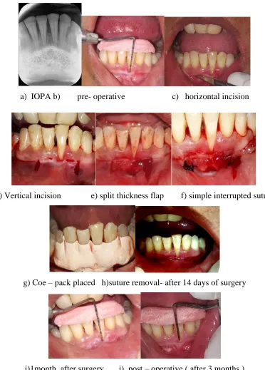
a) IOPA b) pre- operative c) horizontal incision
d) Vertical incision e) split thickness flap f) simple interrupted suture
g) Coe – pack placed h)suture removal- after 14 days of surgery
i)1month after surgery j) post – operative ( after 3 months )
Fig 1 - a) IOPA , b) pre- operative, c) horizontal incision d) Vertical incision e) split thickness flap ,f) simple interrupted
suture, g) Coe – pack placed ,h) suture removal- after 14 days of surgery , i) 1month after surgery, j) post – operative ( after
MODIFIED APICALLY DISPLACED FLAP
The surgical procedure was performed according to the protocol given by Carnio and Miller 7in 1999.Local anesthesia using 2% lignocaine hydrochloride with 1:2,00,000 epinephrine was
administered. A horizontal incision in the AG was made with no. 15 Bard-Parker blade, 0.5 mm coronal
to MGJ. (Fig – c) Horizontal incision was made parallel to MGJ, at an angle of 30 to 45 degrees, formed
by the blade and the portion of the gingival surface coronal to the blade. Therefore, the blade makes
contact with periosteum at a point slightly apical to the alveolar crest.
The gingiva present coronal to the initial incision remains intact around the teeth. The
mesiodistal extension of the initial horizontal incision should be extended by at least one half tooth
mesially and distally of the areas in which gingival augmentation is desired. Two vertical incisions were
placed on the mesial and distal ends connecting the horizontal incision. These incisions extended beyond
the mucogingival junction. (Fig – d) A split-thickness flap is elevated, and the dissection is extended in
the apical direction as far as deemed necessary (Fig – e). The flap is then secured to the periosteum with
simple interrupted sutures using 3-0 mersilk (Fig – f). For preventing dead space between the flap and
periosteal bed, a gentle finger pressure was applied and the periodontal pack was placed (Fig – g)
Postoperative care
Postoperative instructions and medications was given Amoxicillin (500 mg thrice daily for 5
days) and aceclofenac (100 mg thrice daily for 3 days) were given. Patient was instructed to avoid
brushing, flossing, manipulating the surgical site with tongue, lips, and fingers for 6 weeks and use
0.12% chlorhexidine mouth rinse twice daily for 3 weeks. Suture removal was done after 2 weeks (Fig –
h). All the clinical parameters were recorded at 3 months. (Fig – j)
OUTCOME
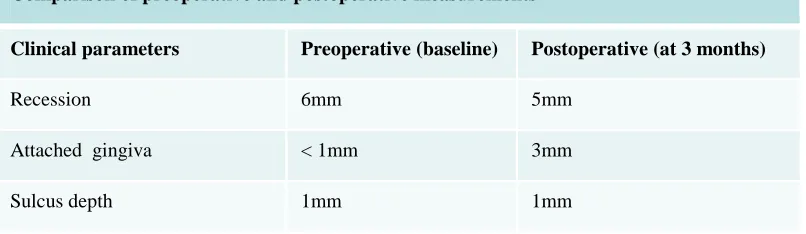
Pre- and post-operative (3 months) clinical measurements are compared. Preoperatively, there
was inadequate width of attached gingiva <1mm but after MARF procedure, there is gain of 2mm of
Table 1- Clinical parameters at baseline and at 3 months
DISCUSSION
AG is composed of keratinized epithelium and dense connective tissue and helps to stabilize the
gingival margin position. AG is bound to underlying periosteum and protects the periodontium.
Deficient AG with poor plaque control may lead to gingival recession.9Data suggests that 2 mm of the gingiva is an adequate width for maintaining gingival health10.Lang and Loe4 in 1972 reported that areas with 1 mm or <1 mm AG often presented with clinical signs of inflammation. According to Maynard11et al. in 1979, physiological dimensions of about 5 mm of KT with 3 mm of attached gingiva are needed
for maintaining gingival health when planning for subgingival restorations.
Friedman12 in 1962 said that an adequate amount of the gingiva is any dimension of the gingiva that is compatible with gingival health or that prevents gingival margin during movements of the
alveolar mucosa. According to Trombelli 13gingival augmentation should be taken into account whenever a change in mucogingival morphology will facilitate plaque control.Hall14 mentioned that areas with <2 mm of AG should be checked for active recession.
According to Karring et al.15the main factor determining the nature of the epithelial surface that will develop over the exposed periosteum is the origin of the epithelial cell that will migrate over the
wound and is eventually surrounded by the KT.As the epithelial cells migrating from margins of the
wound to cover the exposed connective tissue are keratinized in nature, which results in the formation
and maturation of KT. This will prevent the proliferation of non-keratinized cells originating from the
oral mucosa to the surgical area. Hence, it gives a predictable gingival color match with surrounding
tissue.
The results of this case report showed that MARF is an effective and efficient technique to
increase the keratinized and attached tissue width. A major limitation of the MARF technique is a need
Comparison of preoperative and postoperative measurements
Clinical parameters Preoperative (baseline) Postoperative (at 3 months)
Recession 6mm 5mm
Attached gingiva < 1mm 3mm
for ≥0.5 mm AG to be present pre-surgically. This is necessary to allow for the full perimeter of the
wound to be surrounded by KT and is important in origin of the granulation tissue during healing. The
presence of bone dehiscence is another factor contraindicated in MARF technique. If a distance of more
than 0.2 mm is present at the bottom of the pocket and bony crest, root dehiscence is likely to occur
when a flap is positioned apically, which enhances the probability of the gingival recession.
CONCLUSION
MARF is a reliable technique to increase the width of attached gingiva. MARF is a simple
surgical procedure when compared to other mucogingival procedures for gingival augmentation. It
offers considerable advantages such as good esthetic results and no requirement of a second surgical
site. MARF can be used as an alternative to other invasive procedures such as FGG with comparable and
reliable results and minimal patient discomfort. Definitely, more number of cases are needed to ensure
the predictability and success of this technique.
REFERENCES
1. Orban B. Clinical and histologic study of the surface characteristics of the gingiva. Oral Surg
Oral Med Oral Pathol 1948;1:827-41
2. Carranza F. The gingiva. Carranza’s Clinical Periodontology, 9th ed. Philadelphia: W.B.
Saunders; 2002;16.
3. Consensus report. Mucogingival therapy. Ann Periodontol 1996;1:702‑6.
4. Lang NP, Löe H. The relationship between the width of keratinized gingiva and gingival health.
J Periodontol 1972;43(10):623-7.
5. Friedman N. Mucogingival surgery: The apically repositioned flap. J Periodontol
1962;33:328‑40.
6. Matter J. Free gingival grafts for the treatment of gingival recession. A review of some
techniques. J Clin Periodontol 1982;9:103‑14.
7. Carnio J, Miller PD Jr. Increasing the amount of attached gingiva using a modified apically
repositioned flap. J Periodontol 1999;70:1110‑7.
8. Carnio J, Camargo PM. The modified apically repositioned flap to increase the dimensions of
attached gingiva: The single incision technique for multiple adjacent teeth. Int J Periodontics
9. Wennstrom JL, Zuccheli G, Prato P. Mucogingival therapy – Periodontal plastic therapy. In:
Lindhe J, Editor. Clinical Periodontology and Implant Dentistry 5th ed., Vol. 2. Oxford, UK:
Blackwell Munksgaard; 2008; 956
10.Wennström J, Lindhe J. Role of attached gingiva for maintenance of periodontal health. Healing
following excisional and grafting procedures in dogs. J Clin Periodontol 1983;10(2):206-21.
11.Ochsenbein C, Maynard JG. The problem of attached gingiva in children. ASDC J Dent Child
1974;41(4):263-72.
12.Friedman N. Mucogingival surgery: the apically repositioned flap. J Periodontol. 1962;33:328.
13.Trombelli L. Periodontal regeneration in gingival recession defects. Periodontol 2000
1999;19:138-50.
14.Hall WB. Establishing the adequacy of attached gingival. Critical Decisions in Periodontology,
Part.
15.Karring T, Lang NP, Löe H. The role of gingival connective tissue in determining epithelial
![N (3 Nitrophenyl) 1 oxo 2,6,7 trioxa 1 phosphabicyclo[2 2 2]octane 4 carboxamide dimethylformamide solvate](data:image/gif;base64,R0lGODlhAQABAIAAAP///wAAACH5BAEAAAAALAAAAAABAAEAAAICRAEAOw==)