R E S E A R C H
Open Access
High intravascular tissue factor
—
but not
extracellular microvesicles
—
in septic
patients is associated with a high SAPS II
score
Carolin Trepesch
1, Ramona Nitzsche
1, Aenne Glass
2, Bernd Kreikemeyer
1, Jochen K. Schubert
3and Sonja Oehmcke-Hecht
1*Abstract
Background:Sepsis is associated with coagulation abnormalities, and a high content of intravascular tissue factor (TF) may contribute to the development of multisystem organ failure. Circulating microvesicles (MVs) are increased during sepsis and characterized by their phosphatidylserine content. It is unclear whether MVs—as a part of the host response to the infection—are beneficial or rather contribute to systemic complications in sepsis. In the present prospective clinical pilot study, we investigated whether plasma TF and MVs are associated with the risk of multiple organ failure and mortality.
Methods:Thirty patients diagnosed with sepsis, severe sepsis, or septic shock were enrolled and classified as 19 survivors and 11 non-survivors. Blood samples were collected on the day of admission and then daily for up to 2 weeks. MVs and TF were quantified in plasma by ELISA.
Results:Non-survivors had significantly higher TF concentrations on day 3 compared to survivors. Logistic regression analysis revealed that patients with high amounts of TF had significantly increased risk for severity of disease, according to high Simplified Acute Physiology Score II (SAPS II) scores (odds ratio 18.7). In contrast, a higher content of phosphatidylserine-rich MVs were apparently associated with a lower risk for mortality and multiple organ failure, although this was only a trend and the odds ratios were not significant.
Conclusions:This study showed that a high amount of TF in septic patients is significantly associated with increased risk for disease severity, according to a high SAPS II score. Quantification of total MVs in plasma, independent from their cell origin, might be indicative for the outcome of patients in sepsis.
Keywords:Sepsis, Tissue factor, Phosphatidylserine, Extracellular vesicles
Background
Sepsis is a serious medical condition caused by an over-whelming immune response to an infection. It is often as-sociated with bacteremia and characterized by systemic signs and symptoms of inflammation [1].
Fifty to 70 % of patients with sepsis develop coagulation abnormalities that range from minor to hyper activation
of coagulation. Hyper activation of coagulation frequently causes excess fibrin deposition characterized by simultan-eous widespread microvascular thrombosis and profuse bleeding from various sites [2].
The principal initiator of coagulation is tissue factor. Tissue factor (TF) is a transmembrane glycoprotein that triggers blood coagulation via binding factor (F) VIIa and FX with subsequent thrombin generation and fibrin clot propagation. Following vessel injury, TF is exposed to blood and activates the clotting system, which prevents excess blood loss and seals the wound. Under normal con-ditions, TF is exclusively expressed in vascular and tissue * Correspondence:[email protected]
1
Institute of Medical Microbiology, Virology and Hygiene, Rostock University Medical Center, Schillingallee 70, 18057 Rostock, Germany
Full list of author information is available at the end of the article
cells that have no direct contact with blood. However, this concept is outdated as evidence supports the presence of intravascular TF, also described as“circulating”or“ blood-borne”TF [3]. The cellular source of TF in blood is still a matter of discussion, but it has been concluded that monocytes are the only blood cells that synthesize and express TF [4]. Several groups demonstrated higher levels of intravascular TF in septic patients [5–8].
It is unclear how TF circulates, although it has been described that TF is transported on membrane vesicles. Membrane vesicles are sphere-shaped structures, less than 1μm of diameter and limited by a lipid bilayer. They can be secreted from all human blood-born cells and are com-monly classified according to their formation mechanism and their size. Membrane vesicles with a size between 100 and 1000 nm are called microparticles or microvesicles and shed from the cell plasma membrane, while exosomes are 50–100-nm vesicles that are secreted by cells via exo-cytosis [9]. The outer surface of membrane vesicles and exosomes is enriched in negatively charged phosphatidyl-serine (PS), which provides a catalytic surface for many plasma proteins including contact and coagulation factors [10]. Beyond its function in coagulation, PS is also in-volved in clearance of apoptotic cells and modulation of the immune system [11]. In this paper, the term MVs will be used to designate all types of cell-derived microvesicles that are PS-rich and found in plasma, independent of their size. Depending on cell activation, MVs differ in their composition and function. Elevated levels of MVs have been related to pathological conditions such as bleeding and thrombotic disorders, cardiovascular diseases, cancer, and sepsis [12, 13].
Due to their small size, PS-rich MVs offer an add-itional phospholipid platform that has approximately 50-to 100-fold more pro-coagulant activity than activated platelets [14]. We recently reported that the number of total PS-rich MVs in plasma was significantly increased in patients suffering from sepsis [15]. Moreover, in another study, we found a significant increase of total MVs in a mouse streptococcal sepsis model, suggesting that their release is an immune response to the infection [16]. As described above, MVs are supposed to be the most important reservoir for intravascular TF; they may transfer and deliver TF to target cells, such as platelets and neutro-phils, thereby amplifying and disseminating the pro-coagulant response [10].
These findings raised the question whether MVs—as a part of the host response to the infection—are beneficial or rather contribute to systemic complications in sepsis. In the present prospective clinical pilot study, we investi-gated the concentration of plasma TF and total MVs in 30 septic patients and evaluated whether both parame-ters are associated with the risk of multiple organ failure and mortality.
Methods
Patients
Thirty patients with sepsis (n= 6), severe sepsis (n= 4), or septic shock (n= 20) were included in this pilot study. Patients were enrolled from the Intensive Care Medicine Unit at University Medical Center of Rostock. The proto-col had been approved by our Institutional Ethics commit-tee (A 201151), and informed consent was obtained from the patients or their caring relatives.
All patients were diagnosed according to the guide-lines of ACCP/SCCM-Consensus Conference (American College of Chest Physicians/Society of Critical Care Medicine), defined as those patients meeting two of the four systemic inflammatory response syndrome criteria together with an infectious process [17].
The following demographic data were collected at admission in the ICU: age, gender, Simplified Acute Physiology Score II (SAPS II) [10], and Sequential Organ Failure Assessment (SOFA) [12].
Sampling and methods
Blood samples were collected as patients were diagnosed with sepsis and then daily for up to 7 days, depending on the patient. After 1 week, sampling occurred every second day until day 13. A minimum of two samples and a max-imum of 10 samples were derived from a patient. The average study period was 8.6 days. Immediately after blood samples were taken, the blood was centrifuged at 1500×g, for 20 min at 21 °C. The resulting platelet-poor plasma obtained was centrifuged again, at 10,000×g, for 5 min and 21 °C. The platelet-free plasma obtained after the second centrifugation was stored at−80 °C until analysis.
Total MVs were quantified by a prothrombinase assay that detect PS exposed at the MV surface (ACTICHROME ®MP- Activity ELISA kit, American Diagnostica, USA), according to the instruction of the manufacturer. In this assay, PS-positive MVs bind to Annexin V and expose their PS-rich surface, allowing Factor Xa-Va in the presence of calcium to activate prothrombin into thrombin. The PS concentration is the limiting factor. There is a direct rela-tionship between the PS present on the MVs and the amount of thrombin generation, which is measured via its specific activity on the thrombin substrate. Results were expressed as nanomolar PS by reference to a standard curve established with liposomes of known concentration.
Western blot analyses were performed with sheep anti-bodies against human high molecular kininogen (Affinity Biologicals) and its degradation products as described pre-viously [18].
Statistics
All data were stored and analyzed using the SPSS statistical package 20.0 (SPSS Inc. Chicago, Illinois, USA). Descriptive statistics were computed for continuous and categorical variables. The computed statistics included mean and standard deviations or median and interquartile range of continuous variables, frequencies, and relative frequencies of categorical factors. Testing for the differences of continu-ous variables between the study groups was accomplished by the two-sample t test for independent samples or the Mann-WhitneyU test, as appropriate. Test selection was based on evaluating the variables for normal distribution employing the Kolmogorov-Smirnov test.
The logistic regression model was used to assess whether TF or MVs can predict risk of mortality, high SAPS II, and SOFA score. According to their average con-tent of PS and TF, patients were divided into three groups and odds ratios as well as 95 % confidence intervals (95 % CI) were calculated for the outcome of survival, high SAPS II, and SOFA score while one of the groups was used as a reference group. All values resulted from two-sided statistical tests, and p≤0.05 was considered to be significant.
Availability of data and materials
The dataset supporting the conclusions of this article is included within the article. Raw data are available by the corresponding author.
Results
Characteristics of patients
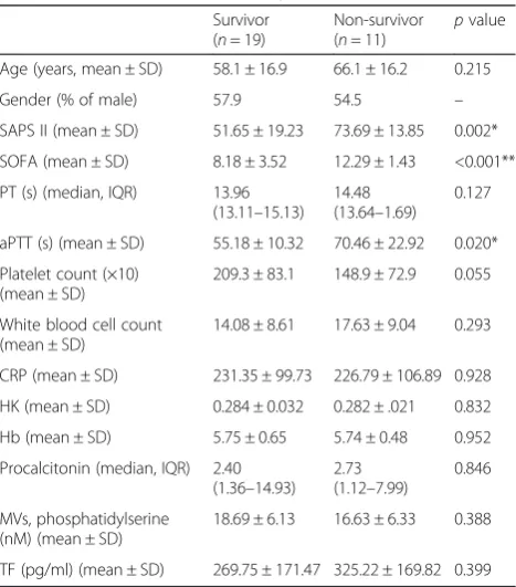
The clinical and laboratory data of our patient population are summarized in Table 1. Thirty patients were investi-gated, 13 (43.3 %) were female and 17 (56.7 %) male. The age ranged between 21 and 84 years; the mean age was 61.03 years. Survivors were younger than non-survivors but not significantly (Table 1). From 30 patients, 19 (63.3 %) survived, whereas 11 (36.7 %) died on ICU. The SAPS II and SOFA scores of non-survivors were significantly higher compared to those of survivors (Table 1). Further on, the activated partial thromboplastin time (aPTT) as a single parameter was significantly prolonged in non-survivors. The aPTT is a marker for the intrinsic coagulation path-way, and the data suggest a consumption of contact factors in non-survivors. However, the prolonged aPTT was not associated with degradation of high molecular weight kinin-ogen in these patients (data not shown).
Other parameters such as platelet count, white blood cell count, and procalcitonin were not significantly differ-ent between the two groups (Table 1).
In most of the patients, multiple infectious sources and bacterial isolates were identified (Table 2).
Tissue factor
In septic patients, a systemic activation of the extrinsic coagulation pathway by TF may contribute to the devel-opment of multisystem organ failure and subsequent mortality [19]. TF circulates in the blood in two different forms: (i) associated with the cellular membrane of white blood cells, platelets, and cell-derived microvesicles and (ii) as a soluble protein lacking the transmembrane domain generated by an alternatively spliced messenger RNA (mRNA) [20]. The ELISA used in the present study detects both forms of TF in plasma [21].
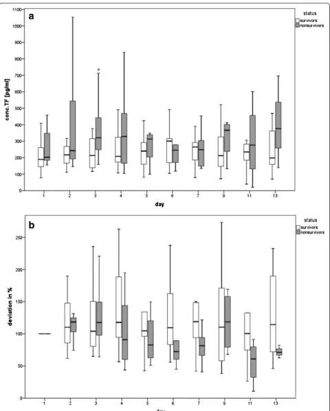
Figure 1a depicts the TF concentrations of survivors and non-survivors on several days. Hereby, a significant difference between survivors and non-survivors was de-tected on day 3 (p= 0.031), whereby non-survivors had higher concentrations (Fig. 1a). The baseline calculation showed no significant differences (Fig. 1b).
A logistic regression analysis was done. First, the risk of mortality based on TF quantity was calculated, and patients were divided into three groups, (i) patients with high TF concentrations (>300 pg/ml), (ii) patients with middle TF concentrations (220–300 pg/ml), and (iii)
Table 1Baseline characteristics of patients
Survivor (n= 19)
Non-survivor (n= 11)
pvalue
Age (years, mean ± SD) 58.1 ± 16.9 66.1 ± 16.2 0.215
Gender (% of male) 57.9 54.5 –
SAPS II (mean ± SD) 51.65 ± 19.23 73.69 ± 13.85 0.002*
SOFA (mean ± SD) 8.18 ± 3.52 12.29 ± 1.43 <0.001**
PT (s) (median, IQR) 13.96 (13.11–15.13)
14.48 (13.64–1.69)
0.127
aPTT (s) (mean ± SD) 55.18 ± 10.32 70.46 ± 22.92 0.020*
Platelet count (×10) (mean ± SD)
209.3 ± 83.1 148.9 ± 72.9 0.055
White blood cell count (mean ± SD)
14.08 ± 8.61 17.63 ± 9.04 0.293
CRP (mean ± SD) 231.35 ± 99.73 226.79 ± 106.89 0.928
HK (mean ± SD) 0.284 ± 0.032 0.282 ± .021 0.832
Hb (mean ± SD) 5.75 ± 0.65 5.74 ± 0.48 0.952
Procalcitonin (median, IQR) 2.40 (1.36–14.93)
2.73 (1.12–7.99)
0.846
MVs, phosphatidylserine (nM) (mean ± SD)
18.69 ± 6.13 16.63 ± 6.33 0.388
TF (pg/ml) (mean ± SD) 269.75 ± 171.47 325.22 ± 169.82 0.399
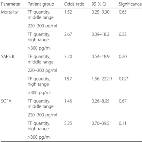
patients with low TF concentrations (110–220 pg/ml), who served as the reference group. Patients with middle and high TF concentrations had a 1.5-fold or 2.6-fold higher risk to die, compared to patients with a low TF content (Table 3). Both odds ratios were not significant, but there is a trend towards increased mortality in pa-tients with high TF concentrations.
The patients were divided by their SAPS II score, whereby high SAPS II scores (>60) are accompanied with a higher risk of mortality. The logistic regression analysis was done, and patients with low TF quantity (110–220 pg/ml) were conduced as reference group. Patients with middle TF quantities (220–300 pg/ml) had a 3.2-fold higher risk for a high SAPS II score (Table 3), compared to patients with low TF (110–220 pg/ml). Patients with high amounts of TF (>300 pg/ml) had a statistically significant 18.7-fold increased risk for a SAPS II score above 60 (Table 3), compared to patients with low TF (110–220 pg/ml).
Analogous to the SAPS II score, the patients were divided by their SOFA scores and a value between 11 and 20 was the negative event. The logistic regression analysis showed a 1.46-fold higher risk for patients with middle TF quantity (220–300 pg/ml) for a high SOFA score (Table 3). In patients with high TF (>300 pg/ml), the risk for a SOFA score above 10 (Table 4) was 5.25-fold higher, compared to the reference group (patients with low TF 110–220 pg/ml). Although these odds ratios are not significant, the trend supports the following hypothesis that a high content of intravascular TF seems to be associated with a high risk for mortality.
Table 2Sources and microorganism isolates of patients
Survivors Non-survivors
Respiratory tract Candida albicans 5 Candida albicans 2
Candida galbrata 2
Candida tropicalis 1 Candida tropicalis 1
Aspergillus fumagatus 2 Strepotoccus anginosus 1
Viridans streptococci 1 Staphylococcus aureus 2
Staphylococcus coagulase negative 2
Staphylococcus coagulase negative 1
Stenothrophomonas maltophila 1
MRSA 1
Enterocabter cloacae 1
Escherichia coli 3
Enterococci 1
Proteus mirabilis 1
Serratia marcescens 1
Intra-abdominal/ pelvis
Candida albicans 3 Candida albicans 3
Candida krusei Candida galbrata 1
Candida tripolis 1 Candida tropicalis 1
Staphylococcus capitis 1 MRSA 1
Staphylococcus epidermidis 3
Staphylococcus epidermidis 2
Staphylococcus warneri 1 Staphylococcus haemolyticus 1
Streptococcus anginosus 1
Enterococci ssp.
Enterococcus faecalis 4 Enterococcus faecalis 2
Enterococcus faecium 4 Enterococcus faecium 5
Enterobacter cloacae 1
Escherichia coli 3 Escherichia coli 1
Bacteroides fragilis 3
Proteus mirabilis 1
Pseudomonas aeruginosa 1
Pseudomonas aeruginosa 1
Anaerob mixed flora 2 Anaerob mixed flora 1
Urinary tract Escherichia coli 1 Enterococcus faecium 1
Proteus vulgaris 1
Klebsiella oxytoca 1 Klebsiella pneumoniae ssp. 1
Trichosporon asahii 1
Heart and vascular system
Enterococcus faecium 1
Proprionibacterium acnes 1
Streptococcus gallolyticus 1
Table 2Sources and microorganism isolates of patients
(Continued)
Staphylococcus epidermidis 1
Streptococcus alactolyticus 1
Blood/catheter Proprionibacterium acnes 1
Proprionibacterium acnes 1
Staphylococcus epidermidis 1
Staphylococcus epidermidis 2
Staphylococcus coagulase negative 2
Staphylococcus capitis 1
Staphylococcus hominis 1
Wound/soft tissue Candida albicans 1 Escherichia coli 1
Staphylococcus epidermidis 1
Staphylococcus aureus 1
Streptococcus pyogenes 1
Main sources and organisms detected in septic patients are represented
PS-positive MVs in plasma
We measured the concentration of PS-positive MVs in plasma of the septic patients on several days but found no significant differences in MV concentration between survivors and non-survivors (Fig. 2a). The baseline calculation showed no significant differences, although the mean MV quantity in the non-survivor group was lower at all days (Fig. 2b).
To calculate the risk of mortality based on MV quantity, again, a logistic regression analysis was done. Patients were divided into three groups, (i) patients with high MV concentrations (>18 nM) as the reference group, (ii) patients with MV concentrations in the middle range (15– 18 nM), and (iii) patients with MV concentrations in the low range (0–15 nM, Table 4). Taken all together, odds ratios implicate that patients with MV concentrations in the middle and low range had a higher risk for mortality or a high SAPS II and SOFA score, when compared to the reference group (Table 4). Although these odds ratios were not significant; the trend supports the hypothesis that a higher content of PS-rich MVs could be associated with a lower risk for mortality.
Discussion
It is now accepted that TF circulates in the blood: (i) as-sociated with white cells and platelets, (ii) asas-sociated with cell-derived MVs, and (iii) as a soluble protein lack-ing the transmembrane domain generated by an alterna-tively spliced mRNA [20]. Only the last two forms are
detectable in plasma. We found significantly elevated TF concentrations on day 3 in non-survivors of septic pa-tients in comparison with the survivors. This corre-sponds to the findings in earlier studies comparing survivors with non-survivors. Green et al. found a sig-nificant increased TF concentration even on day 3 in children with sepsis [6]. Also Gando et al. showed a sig-nificantly higher TF content on several days in non-survivors [21]. Assuming that the SAPS II score reflects severity of sepsis, patients with TF concentrations above 300 pg/ml in our study had a significantly higher risk for a serious course of sepsis. This was supported by a ten-dency for higher risk of mortality and SOFA score of such patients, though these odds ratios were not signifi-cant. Although several studies reported significant higher TF concentrations in plasma of septic patients [5–8], only one study showed a significant correlation between high TF concentrations and increased mortality [6].
An increase in intravascular TF is an indication of tis-sue factor production from activated monocytes. MVs derived from monocytes express and deliver intravascu-lar TF to sites of pathogen exposure, which initiates immunothrombosis inside a blood vessel [22]. The re-lease of MV-associated TF by monocytes may be there-fore regarded as a physiological element of intravascular immunity, to detect and protect against pathogens in the vasculature [23]. However, as we did not found any cor-relation between TF concentration and total MVs (data not shown), we assume that only a minor part of the cir-culating MVs contains TF. Another possible reason may be that the ELISA used in the present study detects both MV-associated TF as well as the soluble form of TF.
The pro-coagulant activity of MVs has mainly been attributed to TF but MVs also elicit phospholipid-dependent pro-coagulant activity due to the exposure of PS on their membrane [24]. In an earlier study, we found increased concentrations of PS-rich MVs in patients suffering from sepsis compared to healthy persons [15]. As pro-coagulant MVs seem to play a key role in multi-organ dysfunction and septic shock, it was, therefore, tempting to speculate that a high amount of total PS-rich MVs in septic patients is associated with a higher risk for mortality. Surprisingly, the data of our present study sug-gest the opposite; a higher content of total MVs was asso-ciated with a trend to lower risk for mortality in septic patients, which is in contrast to TF. One could speculate that the MVs are trapped in the organs; therefore, their amount in the circulation is lower. But it should be con-sidered that—behind pathological functions—MVs may be also beneficial at the early stage of sepsis, as they can com-pensate for some of the host’s systemic reactions [25]. It has been reported that elevated levels of platelet, endothe-lium-, and leukocyte-derived MVs predict a more favor-able outcome in severe sepsis with regard to mortality and Table 3TF antigen concentration and the risk of mortality, high
SAPS II, or SOFA score calculated by logistic regression analysis
Parameter Patient group Odds ratio 95 % CI Significance
Mortality TF quantity, middle range
1.52 0.25–9.30 0.65
220–300 pg/ml
TF quantity, high range
2.67 0.39–18.2 0.32
>300 pg/ml
SAPS II TF quantity, middle range
3.20 0.54–18.9 0.20
220–300 pg/ml
TF quantity, high range
18.7 1.56–222.9 0.02*
>300 pg/ml
SOFA TF quantity,
middle range
1.46 0.26–8.05 0.67
220–300 pg/ml
TF quantity, high range
5.25 0.70–39.5 0.11
>300 pg/ml
organ dysfunction. The authors support the hypothesis that an early increased inflammatory response is associated with improved survival rates [26]. Our recent and actual findings promote this thesis, showing that formation and release of PS-rich MVs are a part of the immune response to bacterial infections [15, 16]. Thus, a high amount of MVs may be a marker of cell activation and inflammation, reflecting a potent inflammatory response, necessary to fight against the infection. A beneficial function for MVs in sepsis was also supported by a study of Mostefai et al., who showed that the total number of circulating and platelet MVs in patients with septic shock was increased. These MVs exert a protective effect against vascular hyporeactivity by main-taining a tonic pressure in septic shock [27]. Also, Guervilly et al. found that high levels of circulating leukocyte MVs are associated with better outcome in acute respiratory distress syndrome [28]. Although the mechanism of
protection is not clear, one could hypothesize that the antibacterial effect of MVs, released from human neutro-philic granulocytes, restricts bacterial growth and dissem-ination during sepsis [29]. In a previous study, we have shown that pro-coagulant MVs can bind toStreptococcus
pyogenes bacteria and promote clotting, entrapment, and killing of the bacteria in a fibrin network. Thus, an inter-action of MVs with bacteria may protect the host [16], which supports the hypothesis above.
A limitation of our pilot study is the relatively small sample size, which reflects typical cases of a Northeast German center for intensive care. This means that the power to detect and label MVs as statistically significant risk factors for mortality was limited. Thus, our data must be validated in an independent larger cohorts of sepsis patients because of the heterogeneity of patients with sepsis and the fact that disease outcome is related to several
a
b
Fig. 2Course of MVs in plasma of survivors and non-survivors, depicted asaphosphatidylserine-concentration (nM) andbbaseline
baseline characteristics [30, 31]. In the future, elucidation of protective mechanisms of MVs is an emerging challenge to design new therapeutic strategies.
Conclusions
This study showed that high amounts of intravascular TF in septic patients significantly increased the risk of disease severity, according to SAPS II scores above 60. In contrast, a high amount of PS-rich MVs is not associated with disease severity or mortality.
Abbreviations
MVs, microvesicles; PS, phosphatidylserine; SAPS II, Simplified Acute Physiology Score II; SOFA, Sequential Organ Failure Assessment; TF, tissue factor
Acknowledgements
This work was supported by a grant from the Deutsche
Forschungsgemeinschaft (project OE 547/2-1, awarded to SOH). The funding agency had no role in the design of the study and collection, analysis, and interpretation of data or in writing the manuscript. We appreciate the Critical Care Team for their cooperation and support.
Authors’contributions
CT collected the samples and analyzed the data. RN performed experiments regarding contact activation. AG performed the statistical analysis. BK participated in the design of the study. JKS participated in the design of the study and interpreted the data. SOH conceived the study and wrote the manuscript. All authors read and approved the final manuscript.
Competing interests
The authors declare that they have no competing interests.
Author details
1
Institute of Medical Microbiology, Virology and Hygiene, Rostock University Medical Center, Schillingallee 70, 18057 Rostock, Germany.2Institute for
Biostatistics and Informatics in Medicine and Ageing Research, Rostock University Medical Center, Rostock, Germany.3Department of Anaesthesia
and Intensive Care Medicine, Rostock University Medical Center, Rostock, Germany.
Received: 10 February 2016 Accepted: 16 May 2016
References
1. Angus DC, van der Poll T. Severe sepsis and septic shock. N Engl J Med. 2013;369:2063.
2. Levi M, Poll TVD. Coagulation in patients with severe sepsis. Semin Thromb Hemost. 2015;41:9–15.
3. Mackman N. The many faces of tissue factor. J Thromb Haemost. 2009;7 Suppl 1:136–9.
4. Osterud B. Tissue factor expression in blood cells. Thromb Res. 2010;125 Suppl 1:S31–4.
5. Gando S, Nanzaki S, Sasaki S, et al. Significant correlations between tissue factor and thrombin markers in trauma and septic patients with disseminated intravascular coagulation. Thromb Haemost. 1998;79:1111–5. 6. Green J, Doughty L, Kaplan SS, et al. The tissue factor and plasminogen
activator inhibitor type-1 response in pediatric sepsis-induced multiple organ failure. Thromb Haemost. 2002;87:218–23.
7. Stief TW, Ijagha O, Weiste B, et al. Analysis of hemostasis alterations in sepsis. Blood Coagul Fibrinolysis. 2007;18:179–86.
8. Gando S, Nanzaki S, Sasaki S, et al. Activation of the extrinsic coagulation pathway in patients with severe sepsis and septic shock. Crit Care Med. 1998;26:2005–9.
9. Théry C, Ostrowski M, Segura E. Membrane vesicles as conveyors of immune responses. Nat Rev Immunol. 2009.
10. van der Poll T, Herwald H. The coagulation system and its function in early immune defense. Thromb Haemost. 2014;112:640–8.
11. Frey B, Gaipl US. The immune functions of phosphatidylserine in
membranes of dying cells and microvesicles. Semin Immunopathol. 2011;33: 497–516.
12. György B, Szabó TG, Pásztói M, et al. Membrane vesicles, current state-of-the-art: emerging role of extracellular vesicles. Cell Mol Life Sci. 2011;68: 2667–88.
13. Nomura S, Shimizu M. Clinical significance of procoagulant microparticles. J Intensive Care. 2015;3:2.
14. Sinauridze EI, Kireev DA, Popenko NY, et al. Platelet microparticle membranes have 50- to 100-fold higher specific procoagulant activity than activated platelets. Thromb Haemost. 2007;97:425–34.
15. Oehmcke S, Mörgelin M, Malmström J, et al. Stimulation of blood mononuclear cells with bacterial virulence factors leads to the release of pro-coagulant and pro-inflammatory microparticles. Cell Microbiol. 2012;14:107–19.
16. Oehmcke S, Westman J, Malmström J, et al. A novel role for pro-coagulant microvesicles in the early host defense against streptococcus pyogenes. PLoS Pathog. 2013;9:e1003529.
17. Reinhart K, Brunkhorst F, Bone H. Prävention, Diagnose, Therapie und Nachsorge der Sepsis. Intensivmed. 2010;47:185–207. DOI:10.1007/s00390-010-0169-2.
18. Nitzsche R, Rosenheinrich M, Kreikemeyer B, et al. Streptococcus pyogenes triggers activation of the human contact system by streptokinase. Infect Immun. 2015;83:3035–42.
19. Hack CE. Tissue factor pathway of coagulation in sepsis. Crit Care Med. 2000;28:S25–30.
20. Cimmino G, Golino P, Badimon JJ. Pathophysiological role of blood-borne tissue factor: should the old paradigm be revisited? Intern Emerg Med. 2011;6:29–34.
21. Gando S, Kameue T, Morimoto Y, et al. Tissue factor production not balanced by tissue factor pathway inhibitor in sepsis promotes poor prognosis. Crit Care Med. 2002;30:1729–34.
22. Engelmann B, Massberg S. Thrombosis as an intravascular effector of innate immunity. Nat Rev Immunol. 2013;13:34–45.
23. Hickey MJ, Kubes P. Intravascular immunity: the host-pathogen encounter in blood vessels. Nat Rev Immunol. 2009;9:364–75.
24. Spronk HMH, ten Cate H, van der Meijden PEJ. Differential roles of tissue factor and phosphatidylserine in activation of coagulation. Thromb Res. 2014;133 Suppl 1:S54–6.
25. Reid VL, Webster NR. Role of microparticles in sepsis. Br J Anaesth. 2012;109:503–13.
Table 4MV quantity and the risk of mortality, high SAPS II, or SOFA score calculated by logistic regression analysis
Parameter Patient group Odds ratio 95 % CI Significance
Mortality MV quantity, low range
3.6 0.62–21.03 0.16
0–15 nM
MV quantity, middle range
1.2 0.15–9.77 0.87
15–18 nM
SAPS II MV quantity, low range
1.7 0.32–8.76 0.54
0–15 nM
MV quantity, middle range
3.5 0.47–25.9 0.22
15–18 nM
SOFA MV quantity,
low range
3.5 0.63–19.5 0.153
0–15 nM
MV quantity, middle range
2.7 0.39–18.2 0.32
26. Soriano AO, Jy W, Chirinos JA, et al. Levels of endothelial and platelet microparticles and their interactions with leukocytes negatively correlate with organ dysfunction and predict mortality in severe sepsis. Crit Care Med. 2005;33:2540–6.
27. Mostefai HA, Meziani F, Mastronardi ML, et al. Circulating microparticles from patients with septic shock exert protective role in vascular function. Am J Respir Crit Care Med. 2008;178:1148–55.
28. Guervilly C, Lacroix R, Forel J-M, et al. High levels of circulating leukocyte microparticles are associated with better outcome in acute respiratory distress syndrome. Crit Care. 2011;15:R31.
29. Timár CI, Lorincz AM, Csépányi-Kömi R, et al. Antibacterial effect of microvesicles released from human neutrophilic granulocytes. Blood. 2013;121:510–8.
30. Mansur A, Mulwande E, Steinau M, et al. Chronic kidney disease is associated with a higher 90-day mortality than other chronic medical conditions in patients with sepsis. Sci Rep. 2015;5:10539.
31. Mansur A, Steinau M, Popov AF, et al. Impact of statin therapy on mortality in patients with sepsis-associated acute respiratory distress syndrome (ARDS) depends on ARDS severity: a prospective observational cohort study. BMC Med. 2015;13:128.
• We accept pre-submission inquiries
• Our selector tool helps you to find the most relevant journal
• We provide round the clock customer support
• Convenient online submission
• Thorough peer review
• Inclusion in PubMed and all major indexing services
• Maximum visibility for your research
Submit your manuscript at www.biomedcentral.com/submit