Bioremediation: Copper Nanoparticles
from Electronic-waste
D. R. MAJUMDER*
Head of the Department of Microbiology, Abeda Inamdar Senior College, 2390-B, K.B. Hidayatullah Road, Azam Campus, Pune-411001, India
Tel. No.: 9822309381
E-mail id: [email protected]
Abstract
A single-step eco-friendly approach has been employed to synthesize copper nanoparticles. The superfast advancement in the field of electronics has given rise to a new type of waste called electronic waste. Since the physical and chemical recycling procedures have proved to be hazardous, the present work aims at the bioremediation of e-waste in order to recycle valuable metals. Microorganisms such as Fusarium oxysporum
and Pseudomonas sp. were able to leach copper (84-130 nm) from integrated circuits present on electronic boards under ambient conditions. Lantana camara, a weed commonly found in Maharashtra was also screened for leaching copper. The characteristics of the copper nanoparticles obtained were studied using X-ray diffraction analysis, energy-dispersive spectroscopy, scanning electron microscopy, Fourier Tranform Infrared analysis, Transmission electron microscopy, Thermogravimetric analysis and Cyclic Voltammetry. Copper nanoparticles were found to be effective against hospital strain Escherichia coli 2065.
Keywords: Bioremediation, E-waste, copper nanoparticles, Pseudomonas sp., Fusariumoxysporum, Lantana camara.
Abbreviations: X-ray diffraction analysis (XRD), energy-dispersive spectroscopy (EDX), scanning electron microscopy (SEM), Fourier Tranform Infrared (FT-IR) analysis, Transmission electron microscopy (TEM), Thermogravimetric analysis (TGA) and Cyclic Voltammetry
1. INTRODUCTION
Electronic waste popularly known as ‘e-waste’ consists of integrated circuits from obsolete and discarded electronic goods such as mobile phones, computers, printers, iPods and batteries etc. Around 90-92% of the components of an integrated circuit can be recycled and reused. Heavy metals such as silicon, arsenic, iron, copper, aluminum, lead, zinc, chromium, cadmium, mercury and barium are viable options for recycling. Physical incineration and chemical processes using strong acids are hazardous as well as expensive for treatment of e-waste causing major concern for environmentalists today1. Unprecedented growth in the area of nanoscience has revolutionized nanotechnology especially in the field of medicine2-5. Biological approaches using microorganisms and plant extracts for synthesis of metal nanoparticles have been suggested as valuable alternatives to traditional methods6-11.
In the present study, leaf extracts of the weed Lantana camara and microorganisms such as Fusarium and
Pseudomonas were screened for extracting copper from integrated circuits and obtaining it in nano form. Hence, this work gives a solution to bioremediation as well as the recovery of valuable metal nanoparticles from e-waste.
2. Materials and Methods
2.1 Synthesis of copper nanoparticles using microorganisms 2.1.1 Screening and isolation of microorganisms
Organisms were isolated from garden soil as well as from a dumpsite of e-waste in Chennai, India. These were plated on Nutrient agar and Potato Dextrose agar. The integrated circuits were taken from the circuit boards, ripped apart, cut into small pieces and sprinkled over the plates. The plates were incubated at room temperature for 48 hours and checked for growth and visible color change to brown.
2.1.2 Shake flask studies
2.1.3 Identification of fungi
The bacterial and fungal cultures were identified from Agharkar Research Institute, Pune.
2.1.4 Particle size analysis
The size of copper obtained after bioleaching was determined using a Particle Size Analyzer, Centre for Materials for Electronics Technology (C-MET), Pashan, Pune. The incubated suspension (copper, distilled water and organism) was centrifuged at 10,000 rpm and the supernatant was further subjected to ultrasonication. This was then used for particle size analysis.
2.2 Synthesis of copper nanoparticles using weed Lantana camara 2.2.1 Plant material and preparation of the extract
To prepare aqueous extract of Lantana camara, 25 g of leaves was taken and washed thoroughly in distilled water, dried, cut into fine pieces, crushed and added to 100 ml sterile double distilled water. It was then heated at 60oC for 10 minutes and filtered through Whatman filter paper No.1.
2.2.2 Synthesis of Copper Nanoparticles
5 ml of extract was added to 20 ml of 0.025 M copper sulphate solution. Bioreduction of cupric ions was monitored by periodic sampling.
2.2.3 Ultra Violet-Visible absorbance spectroscopy study 12
The optical absorbance was recorded on Ultra Violet-Visible spectrophotometer (Systronics 118 double beam model) over a wavelength range of 200–800 nm. The solution containing the signatory color of copper nanoparticles (dark green) was then poured out into petri dishes and left in the oven for drying at 40◦C for 24 hours.
2.2.4 Scanning Electron Microscopy and Energy-Dispersive Spectroscopy study 13
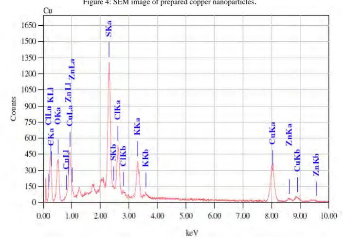
Lantana camara extract pre and post reduction was coated with a thin layer of graphite and examined in a scanning electron microscope (SEM) (JEOL-JSM-6360A) with an energy-dispersive elemental analyzer. The chemical composition of the products was determined by energy-dispersive X-ray spectroscopy using a JEOL-JSM-6360A instrument.
2.2.5 Transmission electron microscopy
Samples of copper nanoparticles were prepared by placing drops of the product solution onto carbon-coated copper grids and allowing the solvent to evaporate. Transmission electron microscopy (TEM) measurements were performed on a Technia G2 ZOU Twin instrument operated at an accelerating voltage of 200 kV.
2.2.6 X- Ray Diffraction Analysis
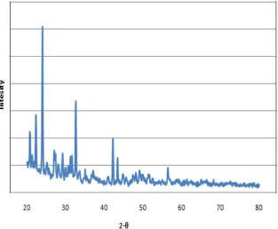
The formation and quality of nanoparticles were checked by X-ray Diffraction (XRD) technique. XRD measurements of drop-coated films of synthesized nanoparticles on glass substrate were recorded in a wide range of Bragg angles 2θ at a scanning rate of 2omin-1, carried out on a Brucker –AXS D8 advanced instrument that was operated at a voltage of 20 kV and a current of 30 mA with copper Kα radiation.
2.2.7 Fourier transform infrared (FT-IR) analysis
Synthesized copper nanoparticles were tested for antimicrobial activity against pathogenic bacterium E. coli 2065 by agar disc-diffusion method. A 0.1 ml aliquot of test organism was spread on Muller Hinton agar. Paper discs loaded with copper nanoparticles and reference drug (Chloramphenicol, 100μg/ml) were placed on the surface of agar plates and incubated at 37oC for 24 hrs after which diameters of inhibition zones were measured.
3. RESULT AND DISCUSSION
3.1 Synthesis of nanoparticles by microorganisms
3.1.1 Screening, Identification and shake flask studies for bioleaching of copper from e-waste using microorganisms
A bacterial colony which produced a diffusible blue-green pigment on nutrient agar, showed a visible color change within 48 hours in presence of pieces of integrated circuits. In case of fungi, mycelial growth was observed on and surrounding the integrated circuits on plate after approximately 20 days. The bacterial and fungal cultures were identified from Agharkar Reseach Institute, Pune. The bacterial colony was identified as Pseudomonas sp. while the fungal isolate was identified as Fusarium oxysporum.
In shake flask studies, Fusarium oxysporum and Pseudomonas sp. showed bioremediation of copper with a distinct colour change.
3.1.2 Particle size analysis of metals
Bioremediation of copper from integrated circuits of e-waste was carried out using one bacterial and a fungal isolate. The bioleached copper was obtained in the nano size, which was determined by the particle size analyzer.
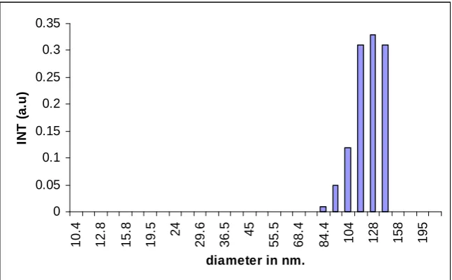
Particle size studies showed that Pseudomonas sp. leached out copper nanoparticles of size 84-130 nm (Fig. 1). While F. oxysporum leached out copper in the range of 93-115 nm (Fig. 2).
0 0.05 0.1 0.15 0.2 0.25 0.3 0.35 10. 4 12. 8 15. 8 19. 5 24 29. 6 36. 5 45 55. 5 68. 4 84. 4
104 128 158 195
diameter in nm.
IN T ( a .u )
0
0.01
0.02
0.03
0.04
0.05
0.06
0.07
0.08
0.09
10
.4
14.
24
19.
49
26.
68
36.
53
50
68.
44
93.
69
12
8.
25
17
5.
56
diameter in nm
IN
T(
a
.u)
Figure 2: Size analysis of copper nanoparticles synthesized by F. oxysporum
3.2 Synthesis of nanoparticles by Lantana camara
3.2.1 Ultra Violet-Visible spectra of copper nanoparticles
Reduction of copper ions to obtain copper nanos on exposure to plant leaf extracts was observed by color change and detected by Ultra Visible spectroscopy. It is generally recognized that Ultra Violet-Visible spectroscopy could be used to examine size and shape-controlled nanoparticles in aqueous suspensions. UV–Visible spectra were recorded at different time intervals for monitoring the reaction. Fig. 3 shows the change in absorbance spectrum at different wavelengths. The absorption peaks arise from the localized surface plasmon resonance (SPR), which can be predicted by the well-known resonance condition14. The Surface Plasmon band in the copper nanoparticles solution remains close to 300 nm throughout the reaction period, suggesting that the nanoparticles were dispersed in aqueous solution with no evidence of aggregation in Ultra Violet-Visible absorption spectrum.
Figure 4: SEM image of prepared copper nanoparticles.
Figure 5: EDX image of prepared copper nanoparticles
3.2.3 Transmission Electron Micrograph
Figure 6: TEM image of a prepared sample of copper nanoparticles
3.2.4 X-Ray Diffraction analysis
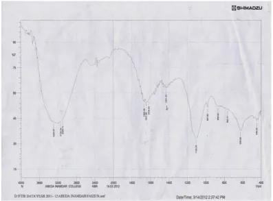
copper nanoparticles. The peaks near 3290.67 cm-1, 3138.29 cm-1, 1668.48 cm-1 correspond to N-H stretching bond, C-H stretching bond and -C=C stretch bond respectively (Fig. 8). The weaker band at 1668.48cm-1 corresponds to amide arising due to carbonyl stretch in proteins. The peak at 1105.25 cm -1
corresponds to C-O stretch stretching vibration of the carboxy group13,15, 16, 17.
Figure 8: FT-IR spectra of copper nanoparticles synthesized by plant extract
3.2.6 Cyclic voltammogram
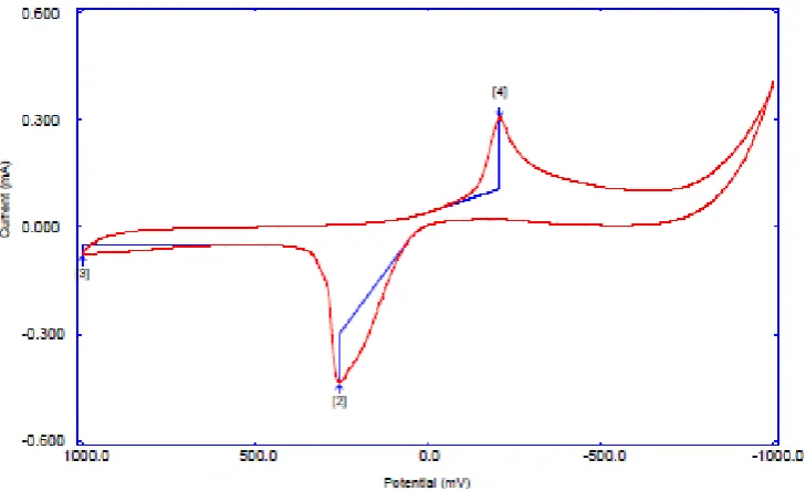
Figure 9: Cyclic voltammogram of extracted nanoparticles
3.2.7 Thermal study
nanoparticles18. Heavy metal ions have diverse effects on bacterial cell function. For copper ions, the mechanism may involve oxidative stress12. The exact mechanism behind bactericidal effect of copper nanoparticles is not clear. The results summarized in Fig. 11 indicate the inhibition of E. coli 2065 by copper nanoparticles with an inhibition zone of diameter 0.5cm. Comparatively the zone of inhibition obtained by the reference drug (Chloramphenicol) was found to be 2cm.
Figure 11: Zones of inhibition around discs impregnated with copper nanoparticles and reference drug (Chloramphenicol)
4. CONCLUSION
Biosynthesis of nanometals was attempted for the first time from integrated circuits with successful results. Here we have reported for the first time, the synthesis of copper nanoparticles of varying sizes using the leaf extract of a notorious weed Lantana camara and microorganisms Fusarium oxysporum and
Pseudomonas sp. at ambient temperature. This process employs environmentally benign natural resources as an alternative to chemical synthesis protocols and proves to be a low cost candidate for synthesizing copper nanoparticles. The process of reduction is extracellular and rapid which leads to the development of feasible biosynthesis of nanoparticles. This green chemistry approach towards the synthesis of metal nanoparticles has many advantages such as feasibility in scale-up, economic viability etc. Applications of such eco-friendly nanoparticles in antimicrobials, wound healing and other medical and electronic applications make this method potentially exciting for the large-scale synthesis of other inorganic nanoparticles holding true to the phrase ‘Waste to Health’.
ACKNOWLEDGEMENT
I would like to thank Dr E.M Khan, Principal, Abeda Inamdar Senior College for providing the necessary infrastructure conducive for research.
REFERENCES
[1] ENDS Environment Daily, Adopted By The Trans-Atlantic Network For Clean Production, 1999.
[2] W. H. De Jong, J. A. B. Paul, Borm, Drug delivery and nanoparticles: applications and hazards, Int. J. Nanomedicine, 2011, 3(2), pp.
133-149.
[3] M. Patil., D. S. Mehta and S. Guvya, Future impact of nanotechnology on medicine and dentistry, J. Indian Soc. Periodontol, 2008,
12(2), pp. 34-40.
[4] J. Y. Song.; B. S. Kim, Rapid biological synthesis of silver nanoparticles using plant leaf extracts, Bioprocess Biosyst Eng., 2009,
32(1), pp. 79-84.
[5] H. Bar.; D. K. Bhui, G. P. Sahoo, P. Sarkar, S. P. De, A. Misra, Green synthesis of silver nanoparticles using latex of Jatrophacurcas,
Colloids Surf A Physicochem Eng Asp., 2009, 339, pp. 134-139.
[6] L. Rassaei, M. Sillanpaa, R. W. French, R. G. Compton, F. Markenv, Arsenite determination in the presence of phosphate at electro
[7] E. Bae; H. J. Park, J. Lee, Y. Kim, J. Yoon, K. Park, Bacterial cytotoxicity of the silver nanoparticle related to physicochemical metrics and agglomeration properties, Envion Toxico Chem., 2010, 29(10), pp. 2154-2160.
[8] D. Mishra and Y. H. Rhee, Current Research Trends of Microbiological Leaching for Metal Recovery from Industrial Wastes, Current
Research, Technology and Educational topics in Applied Microbiology and Microbial Biotechnology, A. Mendez- Vilas (Ed.). 2010, pp. 1289-1296.
[9] H. Shimazono, The Biochemistry of Wood Rotting Fungi, J. Jap. For. Soc., 1951, 33(2), pp. 393-397.
[10] S. Gurunathan, K. Kalishwaralal, R. Vaidyanathan, D. Venkataraman, S R. Pandian, J. Muniyandi, Biosynthesis, purification and
characterization of silver nanoparticles using Escherichia coli, Colloids Surf B Biointerfaces, 2009, 74(1), pp. 328-335.
[11] N. Krumov, I. P. Nochta, S. Oder, V. Gotcheva, A. Angelov, C. Posten, Production of Inorganic Nanoparticles by Microorganisms,
Chem. Eng. Technol., 2009, 32(7), pp. 1026–1035.
[12] D. Bhattacharya, R K. Gupta, Nanotechnology and potential of microorganisms, Crit Rev Biotechnol., 2005, 25, pp. 199-204.
[13] N. Cioffi, L. Torsi, N. Ditaranto, G. Tantillo, L. Ghibelli, L. Sabbatini, T. B. Zacheo, M. D'Alessio, P. G. Zambonin, E. Traversa,
Copper nanoparticle/polymer composites with antifungal and bacteriostatic properties, Chem. Mater,. 2005, 17 (21), pp. 5255–5262.
[14] S. K. Das and E. Marsili, Bioinspired Metal Nanoparticle: Synthesis, Properties and Application, Nanomaterials 2011, pp. 253-278.
[15] A.Thirumurugan, N. A. Tomy, H. P. Kumar, P. Prakash, Biological synthesis of silver nanoparticles by Lantana camara leaf extracts,
International Journal of Nanomaterials and Biostructures, 2011, 1 (2), pp. 22-24.
[16] M. M. Priya, B. Karunai, J. A. Selvia, J. Paul, Green synthesis of silver nanoparticles from the leaf extracts of Euphorbia hirta and
Nerium indicum, Digest Journal of Nanomaterials and Biostructures, 2011, 6 (2), pp. 869 – 877.
[17] P.C. Nagajyothi and K. D. Lee, Synthesis of Plant-Mediated Silver Nanoparticles Using Dioscorea batatas Rhizome Extract and
Evaluation of Their Antimicrobial Activities, Journal of Nanomaterials, 2011, pp. 1-7.
[18] K. Yoon, J. H. Byeon, J. Park, J. Hwang, Susceptibility constants of Escherichia coli and Bacillus subtilis to silver and copper