Proximity-Dependent Biotinylation
Brooke Morriswood,aKatharina Havlicek,aLars Demmel,aSevil Yavuz,aMarco Sealey-Cardona,aKeni Vidilaseris,aDorothea Anrather,b Julius Kostan,cKristina Djinovic´-Carugo,c,dKyle J. Roux,eGraham Warrena
Max F. Perutz Laboratories, University of Vienna and Medical University of Vienna, Vienna, Austriaa
; Department for Biochemistry and Cell Biology, Max F. Perutz Laboratories, University of Vienna, Vienna, Austriab
; Department for Structural and Computational Biology, Max F. Perutz Laboratories, University of Vienna, Vienna, Austriac
; Department of Biochemistry, Faculty of Chemistry and Chemical Technology, University of Ljubljana, Ljubljana, Sloveniad
; Children’s Health Research Center, Sanford Research/USD, Sioux Falls, South Dakota, USAe
The trypanosomes are a family of parasitic protists of which the African trypanosome,Trypanosoma brucei, is the best charac-terized. The complex and highly ordered cytoskeleton ofT. bruceihas been shown to play vital roles in its biology but remains difficult to study, in large part owing to the intractability of its constituent proteins. Existing methods of protein identification, such as bioinformatic analysis, generation of monoclonal antibody panels, proteomics, affinity purification, and yeast two-hy-brid screens, all have drawbacks. Such deficiencies—troublesome proteins and technical limitations—are common not only to T. bruceibut also to many other protists, many of which are even less well studied. Proximity-dependent biotin identification (BioID) is a recently developed technique that allows forward screens for interaction partners and near neighbors in a native environment with no requirement for solubility in nonionic detergent. As such, it is extremely well suited to the exploration of the cytoskeleton. In this project, BioID was adapted for use inT. brucei. The trypanosome bilobe, a discrete cytoskeletal struc-ture with few known protein components, represented an excellent test subject. Use of the bilobe protein TbMORN1 as a probe resulted in the identification of seven new bilobe constituents and two new flagellum attachment zone proteins. This constitutes the first usage of BioID on a largely uncharacterized structure, and demonstrates its utility in identifying new components of such a structure. This remarkable success validates BioID as a new tool for the study of unicellular eukaryotes in particular and the eukaryotic cytoskeleton in general.
T
he trypanosomes are a family of uniflagellated kinetoplastids. All species are parasitic, and a number are human pathogens. The African trypanosomeTrypanosoma bruceiis one such species and is responsible for considerable morbidity and mortality in both people and livestock. It is also the best-studied member of the Trypanosomatida order.T. bruceiexhibits a highly organized cytoskeletal architecture (1). The cell body is a tapered cylinder, maintained by a subpel-licular corset of microtubules aligned with their plus ends at the cell’s posterior pole (2). In the trypomastigote form, the stage in whichT. bruceispends the majority of its life cycle, the single flagellum emerges near the posterior end of the cell and is attached lengthwise as far as the anterior cell tip (3). The base of the flagel-lum is present in a bulb-shaped invagination of the plasma mem-brane termed the flagellar pocket, which is the site of all exo- and endocytic traffic (4). The basal body that nucleates the flagellar axoneme abuts the flagellar pocket, with the paired probasal body lying orthogonally to it (5). At the top of the flagellar pocket is an electron-dense cytoskeletal barrier element termed the flagellar pocket collar (6). Between the flagellar pocket collar and the site of flagellum exit from the cell body is a vase-shaped region named the flagellar pocket neck (7). The flagellar pocket neck, flagellar pocket, and flagellar membrane are all contiguous with the plasma membrane and represent distinct subdomains of it (3). Originat-ing between the basal body and probasal body is an array of four specialized microtubules that follow a helical path around the fla-gellar pocket, pass through the flafla-gellar pocket collar and flafla-gellar pocket neck, and parallel the path of the flagellum to the cell’s anterior pole (8). A proteinaceous filament runs alongside this microtubule quartet from the flagellar pocket neck region
on-wards (9). The protein filament and the microtubule quartet to-gether comprise the flagellum attachment zone (FAZ). The FAZ is responsible for adhering the flagellum to the cell body.
Numerous studies have established that the cytoskeleton is es-sential for normal cell growth ofT. brucei in vitro. Loss of cyto-skeletal proteins cause a variety of morphological, motility, or replicative phenotypes, ranging from aberrant cell shapes and de-tached flagella to impaired or entirely abrogated cell locomotion and compromised organelle biogenesis and cytokinesis (10–13). Despite this wealth of evidence supporting the role of the cytoskel-eton in trypanosome biology, it remains poorly understood due to the intractability of many of its protein components. A good ex-ample is the trypanosome bilobe, an enigmatic cytoskeletal struc-ture of unclear function (14). The roughly 2-m-long bilobe is present in the flagellar pocket neck and partially overlaps with the posterior end of the FAZ (15). There are only two proteins known to exclusively localize to it—TbMORN1 (Tb927.6.4670) and TbL-RRP1 (Tb11.01.0680) (15,16). TbCentrin4 and TbCentrin2 have also been localized to the bilobe but are additionally present on the
Received21 November 2012Accepted17 December 2012 Published ahead of print21 December 2012
Address correspondence to [email protected]. M.S.-C. and K.V. contributed equally to the manuscript.
Supplemental material for this article may be found athttp://dx.doi.org/10.1128 /EC.00326-12.
Copyright © 2013, American Society for Microbiology. All Rights Reserved.
doi:10.1128/EC.00326-12
on September 8, 2020 by guest
http://ec.asm.org/
basal and probasal bodies at the base of the flagellum (17,18). Recent light microscopy and electron microscopy work has shown that TbMORN1 and TbLRRP1 colocalize on a fishhook-shaped structure whose hook encircles the flagellar pocket above the flagellar pocket collar and whose stem projects toward the cell anterior. TbCentrin4 is present on a bar that lies alongside the anterior stem of the fishhook-shaped structure, completing a hair-pin arrangement (19). It is not clear if the bilobe ensemble of TbMORN1, TbLRRP1, and TbCentrin4 represents two separate structures (fishhook plus bar) or a single entity with discrete sub-domains. Despite extensive effort, no new bilobe proteins have been discovered to date (our unpublished data).
The recently developed proximity-dependent biotin identifi-cation (BioID) technique shows considerable promise for redress-ing this methodological shortfall (20). It enables forward bio-chemical screens for binding partners and near neighbors in a native environment without any requirement for detergent solu-bility. BioID involves tagging a protein of interest with a modified form of the 35-kDa bacterial biotin ligase BirA, which has been previously adapted for expression in eukaryotic cells (20). The normal reaction mechanism of BirA consists of conjugating nu-cleotide (in the form of ATP) to biotin in order to generate a highly reactive biotinoyl-5=-AMP intermediate (21). This reactive intermediate is then retained in the active site until the enzyme locates a specific lysine residue in the target sequence on its protein substrate. The sequestered intermediate then reacts with the pre-sented lysine residue, resulting in biotinylation of the target pro-tein. In BioID, a mutant form of the BirA enzyme is used which exhibits an extremely low affinity for the reactive biotinoyl-5= -AMP intermediate (22). Consequently this modified enzyme, des-ignated BirA*, effectively acts as a biotinoyl-5=-AMP synthase and releases molecule after molecule of the intermediate into the sur-rounding milieu. The principle of BioID is therefore to tag the protein of interest with BirA* and to incubate the cells in excess biotin. The BirA* module proceeds to spit out highly reactive mol-ecules of biotinoyl-5=-AMP, leading to the indiscriminate biotiny-lation of all proteins in the immediate vicinity of the tagged bait in a proximity-dependent fashion (20). These putative binding part-ners and near neighbors of the protein of interest can then be affinity purified using streptavidin-coated beads. Owing to the remarkable affinity of the biotin-streptavidin bond (Kd,⬃10⫺14 M) (Kd, dissociation constant), far harsher lysis conditions can be used than in a conventional affinity purification. As such, cyto-skeletal proteins, which normally partition into the detergent-in-soluble fraction when weak nonionic detergents such as NP-40 are used, are efficiently solubilized and can be captured. These puri-fied proteins can then be identipuri-fied by mass spectrometry. A key strength of BioID is therefore the ability to preserve information about interactions while employing very strong detergents for cell lysis. By carrying out Western blots of eluted proteins using horse-radish peroxidase (HRP)-conjugated streptavidin, the molecular weights of the most prevalent binding partners and near neighbors can be inferred in advance and compared with the mass spectrom-etry data (see Fig. S1 in the supplemental material).
The work described here concerns the adaptation of BioID for T. brucei. As a discrete and stable cytoskeletal structure with a largely uncharacterized protein composition, the bilobe—via its marker protein TbMORN1—represented an excellent test case for the BioID system. The goal therefore was to identify novel struc-tural components of this cytoskeletal entity. Using BioID, a large
number of TbMORN1 candidate binding partners and near neighbors were identified by mass spectrometry. Localization of a selection of the highest-scoring candidates relative to controls re-sulted in the assignment of seven new bilobe proteins and two new FAZ proteins. Integrating these localizations into our existing ul-trastructural knowledge of the bilobe has produced a new model of the structure and supports the notion that it is composed of discrete subdomains.
MATERIALS AND METHODS
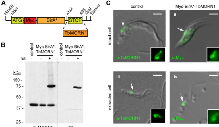
Antibodies and reagents.The rabbit polyclonal anti-Myc antibody and mouse monoclonal anti-Myc antibody were purchased from Abcam and Invitrogen, respectively. The anti-TbBILBO1 antibodies have been de-scribed previously (19). The anti-TbMORN1 rabbit polyclonal antibodies used in this paper, which were raised against recombinant full-length TbMORN1, are new. In immunoblots of whole-cell lysates fromT. brucei, the antibodies recognized a single protein of approximately 40 kDa (Fig. 1B). In indirect immunofluorescence assays the antibodies produced a labeling pattern indistinguishable from that observed using the older antibodies (Fig. 1C). These specific signals in immunoblotting and immu-nofluorescence were both lost when assaying samples from cells depleted of TbMORN1. As such, their performance in immunoblotting and indi-rect immunofluorescence was identical to or better than that of the older original anti-TbMORN1 antibodies, and no specificity issues are evident (reference15and data not shown). Biotin, M280 streptavidin-conjugated Dynabeads, HRP-conjugated streptavidin, AlexaFluor488-conjugated streptavidin, and Dyna-Mag magnet were purchased from Invitrogen. The rabbit anti-LdCentrin4 polyclonal antibody, mouse monoclonal an-ti-Ty1 (BB2) antibody, and 1B41 anti-FAZ antibody were gifts kindly received from Hira Nakhasi (FDA), Cynthia He (National University of Singapore), and Linda Kohl (Museum National d’Histoire Naturelle).
Generation of the pLEW100_Myc_BirA* plasmid.The Myc_BirA* module was amplified from the pcDNA3.1(⫺) cloning vector with incor-poration of additional restriction enzyme sites by PCR. The module was ligated into the pLEW100 expression vector between the HindIII and BamHI sites to generate the new pLEW100_Myc_BirA* plasmid (Fig. 1A). The complete TbMORN1 open reading frame was subcloned into the vector between the XhoI and AflII sites.
Cell lines, culture, and generation.The procyclic 29-13 strain ofT. brucei bruceiwas used for the generation of cells inducibly expressing Myc-BirA*-TbMORN1 (23). The pLEW100_Myc_BirA*-TbMORN1 construct was linearized by digestion with NotI and introduced into 29-13 cells by electroporation. Stable transformants were selected by growth in medium containing 5g/ml phleomycin and cloned by limiting dilution. Putative clones were screened as described in Results. The procyclic 427 Lister strain ofT. brucei bruceiwas used for candidate protein localization. Cells were cultured in SDM-79 medium supplemented with 7.5g/ml hemin and 20% heat-inactivated fetal calf serum (Sigma-Aldrich) at 27°C. 29-13 cells additionally required 15g/ml neomycin and 50g/ml hy-gromycin to maintain T7 polymerase and tetracycline repressor trans-genes.
Immunofluorescence microscopy.Cells were attached to coverslips by centrifugation (1,800⫻g, 5 s). Detergent-extracted cells, when re-quired, were prepared by incubating cells in PEME buffer {2 mM EGTA, 1 mM MgSO4, 0.1 mM EDTA, 0.1 M PIPES [piperazine-N,N=- bis(2-eth-anesulfonic acid)]-NaOH, pH 6.9} supplemented with 0.5% NP-40 (vol/ vol) for 5 min at room temperature (RT) and then washed with phos-phate-buffered saline (PBS). Cells were fixed in 4% paraformaldehyde in PBS (20 min, RT). After fixation, cells were washed with PBS, permeabil-ized with 0.25% Triton X-100 in PBS (vol/vol) (5 min, RT), washed again with PBS, and blocked in 3% bovine serum albumin (BSA) in PBS (wt/ vol) (30 min, RT). Antibody binding steps, coverslip mounting, image acquisition, and image processing have been described previously (19).
Purification of biotinylated proteins.Biotin stock solution (1 mM) in SDM-79 tissue culture medium was made fresh for each experiment.
on September 8, 2020 by guest
http://ec.asm.org/
allel cultures of cells (experimental and control) were grown in either the presence or the absence of 10 ng/ml tetracycline overnight to induce Myc-BirA*-TbMORN1 expression. This drug concentration produces mild overexpression of the ectopic copy of the protein. The cultures were then scaled up to final volumes, supplemented with biotin stock solution to a final concentration of 50M, and incubated for a further 24 h. The cells were harvested by centrifugation (1,800⫻g, 5 min, 4°C), washed three times with PBS, and transferred to Eppendorf tubes. The cells were ex-tracted in PEME buffer supplemented with 0.5% NP-40 (vol/vol) (15 min, RT, gentle mixing), and a 5% sample (E1) was taken. Detergent-soluble and -insoluble fractions were then separated by centrifugation (3,400⫻g, 2 min, RT); a 5% sample was taken of the supernatant (S1). The detergent-insoluble pellet was extracted in lysis buffer (0.4% SDS, 500 mM NaCl, 5 mM EDTA, 1 mM dithiothreitol [DTT], 50 mM Tris-HCl, pH 7.4) (30 min, RT, gentle mixing) and a 5% sample taken (P1). The solubilized material was collected by centrifugation (16,000⫻g, 10 min, RT) and a 5% sample taken of the supernatant (S2). Streptavidin-coated Dynabeads (approximately 100l per 50 ml of cell culture) were separately added to fractions S1 and S2 and incubated with gentle mixing (4 h, 4°C). The beads were separated from the unbound fraction by magnetic separation using a Dyna-Mag magnet. A 5% sample was taken of the two free unbound fractions (F1, F2). The Dynabeads were washed extensively with either PBS alone or the full wash procedure as described by Roux and coauthors (20). The Dynabeads were pelleted by centrifugation (6,000⫻g, 2 min, RT). The beads (representing 100% of fractions B1 and B2) were resus-pended in SDS-PAGE loading buffer and used directly for blotting. Equal fractions from each sample were separated by SDS-PAGE, transferred to nitrocellulose, and blotted with HRP-conjugated streptavidin. In bulk preparations intended for mass spectrometry analysis, the Dynabeads were added only to fraction S2. After the final centrifugation step, a 10% sample of the beads (fraction B2) was taken and separated electrophoreti-cally by SDS-PAGE, and the protein concentration was estimated by silver staining. The remaining 90% was analyzed by mass spectrometry. Of the
three bulk preparations, the first was conducted using the full wash pro-cedure as detailed by Roux et al., the latter two using PBS washes only (20). Small-scale preparations were typically 50-ml cell cultures (approxi-mately 5⫻108cells); bulk preparations were 500-ml cell culture (approx-imately 5⫻109cells).
Mass spectrometry (nano-liquid chromatography-tandem mass spectrometry [LC/MS-MS]) analysis.Beads were washed 5 times with 50 mM ammonium bicarbonate and then suspended in 100l ammonium bicarbonate. Disulfide bonds were reduced with 10% DTT (by weight of the estimated amount of protein) (30 min, 56°C) and subsequently alky-lated with 50% iodoacetamide (by weight of the estimated amount of protein) (20 min, RT, in the dark). Then, 5% DTT (wt/wt of the estimated amount of protein) was added to consume excess iodoacetamide and proteins were digested with 5% trypsin (by weight of the estimated amount of protein) (recombinant, proteomics grade; Roche) (37°C, over-night). Digests were stopped by the addition of trifluoroacetic acid to approximately pH 2. Digests (5 to 10% of total) were analyzed on an UltiMate 3000 high-performance liquid chromatography (HPLC) system (Dionex; Thermo Fisher Scientific) coupled to an LTQ-Orbitrap Velos mass spectrometer (Thermo Fisher Scientific) via a nanoelectrospray ion-ization source (Proxeon; Thermo Fisher Scientific). Peptides were loaded on a trapping column (PepMap C18, 5-m particle size, 300m inner diameter, 5 mm length; Thermo Fisher Scientific) with 0.1% trifluoro-acetic acid and separated on an analytical column (PepMap C18, 3-m particle size, 75m inner diameter, 150 mm length; Thermo Fisher Sci-entific) with a flow rate of 300 nl/min applying a 90-min linear gradient from 2.5% up to 40% acetonitrile. The voltage of the ion source was set to 1,500 V. The mass spectrometer was operated in the data-dependent mode: 1 full scan in the orbitrap (m/z, 400 to 1,800; resolution, 60,000) with lock mass (m/z, 445.120025) enabled was followed by a maximum of 20 MS-MS scans. Monoisotopic precursors were selected; singly charged signals were excluded from fragmentation. The collision energy was set at
FIG 1Characterization of Myc-BirA*-TbMORN1-expressing cells. (A) Schematic drawing showing cloning of theTbMORN1open reading frame into the modified pLew100_Myc_BirA* plasmid for tetracycline-inducible expression. (B) Myc-BirA*-TbMORN1 is tightly and inducibly expressed. 29-13 cells stably transfected with the pLew100_Myc_BirA*-TbMORN1 plasmid were incubated in the presence or absence of 10 ng/ml tetracycline. Whole-cell lysates were analyzed by immunoblots with either anti-TbMORN1 or anti-Myc antibodies. In untransfected (control) cells and uninduced cells, only the endogenous TbMORN1 (40 kDa) is detected. In the presence of tetracycline, an additional approximately 75 kDa polypeptide was detected by both antibodies. (C) Myc-BirA*-TbMORN1 recapitulates the localization and morphology of the endogenous protein. Untransfected (control) cells or Myc-BirA*-TbMORN1-expressing cells were analyzed by immunofluorescence assay. Intact (panels i and ii) and detergent-extracted (panels iii and iv) cells were labeled with anti-TbMORN1 or anti-Myc antibodies. The bilobe structure is indicated (white arrows, area shown enlarged in insets). Scale bars, 2m.
on September 8, 2020 by guest
http://ec.asm.org/
35%, the Q value at 0.25, and the activation time at 10 ms. Fragmented ions were excluded from further selection for 30 s.
Mass spectrometry data interpretation.Raw spectra were interpreted by Mascot 2.2.04 (Matrix Science) using Mascot Daemon 2.2.2. Spectra were searched against the TriTryp database for T.brucei427 and T.brucei927 (18,355 entries, 23 April 2012;http://tritrypdb.org/) (24). The database was manually supplemented with the sequences of strepta-vidin and a set of frequent contaminants such as keratins and proteases. Search parameters were as follows: peptide tolerance, 2 ppm; fragment tolerance, 0.8 Da; trypsin specificity with two missed cleavages; carbam-idomethyl cysteine as static; oxidation of methionine as variable modifi-cation. MASCOT results were loaded into Scaffold (version 3.00.02; Pro-teome Software). Peptide identifications were accepted when a minimum of two peptides were identified with a probability greater than 95% as assigned by the Protein Prophet algorithm; this resulted in a false discov-ery rate of 0%.
Candidate screening.Candidate genes were cloned into the pXS2 ex-pression vector with a Ty1 epitope tag. The plasmids were introduced into cells by electroporation, and localizations were determined by immuno-fluorescence assay after an overnight incubation. Endogenous replace-ment of candidate genes to generate stably transfected cell lines was ac-complished using previously published methods (19). The endogenous replacements encoded a triple Ty1 epitope tag at the 5=end of the target allele and were screened by 10-g/ml blasticidin selection. Integration of the targeting construct at the endogenous allele locus by double homolo-gous recombination was confirmed by PCR amplification of genomic DNA, using primers that annealed within the epitope tag and outside the targeting fragment (see Fig. S2A in the supplemental material).
RESULTS
In order to generate trypanosome cells that inducibly expressed transgenes for BioID, the Myc-BirA* module was subcloned into the pLEW100 expression vector (23). This vector permits trans-genes to be stably integrated into a ribosomal DNA (rDNA) locus and provides tetracycline-inducible control over expression of the encoded protein. The new expression vector was termed pLEW100_Myc_BirA*. The TbMORN1 open reading frame was then cloned into pLEW100_Myc_BirA* (Fig. 1A). Trypanosome cells from the 29-13 cell line were transfected with the construct, and stably transfected clones were obtained. The presence of the pLEW100_Myc_BirA*-TbMORN1 construct in the genomic DNA of the clones was confirmed by PCR (data not shown). Ad-dition of tetracycline to the culture medium caused expression of an approximately 75-kDa polypeptide that was recognized by both anti-TbMORN1 and anti-Myc antibodies. No expression was seen in untransfected 29-13 control cells or uninduced cul-tures (Fig. 1B). It was concluded that Myc-BirA*-TbMORN1 was faithfully and inducibly expressed. Indirect immunofluorescence analysis confirmed the localization and morphology of Myc-BirA*-TbMORN1. Endogenous TbMORN1 has a fishhook-shaped expression pattern and localizes close to the point of fla-gellum entry into the cell (Fig. 1C, panel i). In intact cells there is also a small cytoplasmic fraction of TbMORN1. In detergent-ex-tracted cells, TbMORN1 is present only at the bilobe (Fig. 1C, panel iii). Myc-BirA*-TbMORN1 was found to recapitulate the localization and morphology of endogenous TbMORN1 in both intact cells (Fig. 1C, panel ii) and detergent-extracted cells (Fig. 1C, panel iv). In the absence of tetracycline, no labeling was detected with the anti-Myc antibodies (data not shown). It was concluded that addition of the Myc-BirA* module did not inter-fere with correct localization. The induction of expression and correct localization of Myc-BirA*-TbMORN1, respectively,
shown by immunoblotting and immunofluorescence assay, were demonstrated in two other independent clonal cell lines (data not shown). Culturing Myc-BirA*-TbMORN1-expressing cells at tet-racycline concentrations between 0 and 10 ng/ml showed no effect on growth (data not shown). The Myc-BirA* module therefore appears to be nontoxic to trypanosome cells.
To determine whether the expressed Myc-BirA*-TbMORN1 caused biotinylation of candidate binding partners and near neighbors, the cells were cultured for 24 h in the presence of excess biotin and the presence or absence of tetracycline. Intact and de-tergent-extracted cells were labeled with anti-Myc antibodies and fluorescently conjugated streptavidin. In the presence of tetracy-cline, intact cells showed good labeling in both channels and strong overlap at the bilobe was clearly visible (Fig. 2A, arrows and insets). Both the anti-Myc and streptavidin labeling patterns closely resembled that seen for endogenous TbMORN1. Consis-tent with the data presented inFig. 1C, in detergent-extracted cells the labeling was confined to the bilobe area. The anti-Myc and streptavidin signals strongly overlapped (Fig. 2C, arrows and in-sets). In the absence of tetracycline, intact cells showed no anti-Myc labeling and only a background signal from streptavidin (Fig. 2B). Detergent-extracted cells showed no significant signal with either label in the absence of tetracycline (Fig. 2D). Whole-cell lysates from both Whole-cell populations were analyzed by Western blotting. An HRP-conjugated streptavidin probe confirmed a sig-nificant increase in the number and quantity of biotinylated pro-teins in cells expressing Myc-BirA*-TbMORN1 (Fig. 2E, upper panel). Equal loading of lysates was confirmed by immunoblot-ting with antibodies specific for the flagellar pocket collar protein TbBILBO1 antibodies (Fig. 2E, lower panel). It was concluded that expression of Myc-BirA*-TbMORN1 in the presence of ex-cess biotin caused biotinylation of candidate binding partners and near neighborsin vivo. These results were verified in two other independent clonal cell lines (data not shown).
It was decided to separately purify candidates from the deter-gent-soluble (cytoplasmic) and detergent-insoluble (cytoskeletal) fractions, as the primary goal of the project was to identify struc-tural components of the bilobe. Such components were predicted to be tightly associated with the cytoskeleton, as is the case for TbMORN1 (15). A biochemical fractionation protocol was de-signed to accomplish this two-part purification (Fig. 3A). Comparison of equal fractions from cells expressing Myc-BirA*-TbMORN1 suggested that the protocol functioned according to design (Fig. 3B). The biotinylated proteins in the whole-cell input (E1) were efficiently partitioned into detergent-soluble (S1) and detergent-insoluble (P1) fractions. The detergent-insoluble frac-tion was effectively solubilized by SDS (S2). The unbound fracfrac-tion of the cytoplasmic, detergent-soluble population (F1) contained a number of proteins indicating that in this preparation the strepta-vidin beads’ binding capacity was probably saturated (compare F1 and S1). The unbound fraction of the cytoskeletal, detergent-in-soluble population (F2) was largely depleted of biotinylated pro-teins, indicating good capture (compare F2 and S2). Despite the relatively good capture rates, both elutions (B1 and B2) displayed considerably less material relative to the inputs (S1 and S2). This was likely to be due to inefficient elution of the biotinylated pro-teins from the streptavidin beads. In both eluates, an approxi-mately 75-kDa protein was clearly visible—immunoblots with an-ti-Myc antibodies showed that this was Myc-BirA*-TbMORN1 (data not shown). A large percentage of Myc-BirA*-TbMORN1
on September 8, 2020 by guest
http://ec.asm.org/
partitions into the detergent-soluble fraction, due to the mild lev-els of overexpression. This is consistent with the amount of TbMORN1 present at the bilobe being a finite and saturable quan-tity (our unpublished observations). Comparison of purifications under experimental (⫹Tet) and control (⫺Tet) conditions re-vealed an extremely good signal-to-noise ratio (Fig. 3C). Eluates from control (⫺Tet) conditions showed virtually no biotinylated proteins whatsoever (Fig. 3C, left panel, fractions B1 and B2). Loading of larger fractions showed the presence of a number of candidates of defined molecular weights in the experimental con-dition elutions (Fig. 3C, right panel). Of particular interest, puri-fications from the detergent-insoluble, cytoskeletal fraction (B2) revealed three good candidates of approximately 79-, 90-, and 130-kDa molecular sizes in addition to Myc-BirA*-TbMORN1 (Fig. 3C, right panel). It was concluded that candidate TbMORN1 binding partners and near neighbors could be efficiently purified from Myc-BirA*-TbMORN1 cells and that uninduced (⫺Tet) cells represent an excellent control for nonspecific binding and background levels.
To identify candidate TbMORN1 binding partners and near neighbors from the detergent-insoluble, cytoskeletal fraction, bulk purifications were carried out in the presence of biotin and in the presence or absence of tetracycline. The eluates (B2) from both control and experimental samples were analyzed by mass spec-trometry. To avoid the problem of inefficient elution of biotinyl-ated proteins from the streptavidin-conjugbiotinyl-ated beads, the cap-tured proteins were directly trypsinized on the beads. Three independent bulk purifications were carried out. Comparison of fractions from each of these purifications confirmed a high level of reproducibility (see Fig. S2B in the supplemental material). Prom-ising candidate proteins were identified on the basis of having high scores (in terms of number of assigned spectra and number of unique peptides) in the experimental samples and low-to-zero scores in the control samples. The top-scoring protein in all three
data sets was TbMORN1 itself. Significantly, TbLRRP1, which has been shown to colocalize with TbMORN1, scored as a strong hit (Table 1) (19). The flagellar pocket collar protein TbBILBO1, which only partially overlaps with TbMORN1, was not present. A total of 10 proteins were selected for screening, based on the fol-lowing criteria: (i) high scores in terms of assigned spectra and unique peptides in all three experimental samples; (ii) low-to-zero scores in all three control samples; (iii) uncharacterized localiza-tion (Table 1). Many of these candidates had molecular weights corresponding to those of the proteins observed in the B2 elution fraction (Fig. 3C, right panel). A complete list of all high-scoring proteins across all three experiments is also provided (see Table S1 in the supplemental material). Included in the table are RNA in-terference (RNAi) phenotypes, as determined in the recent high-throughput screen ofT. brucei, and information on whether the proteins are also represented in theT. bruceiflagellum proteome (10,25).
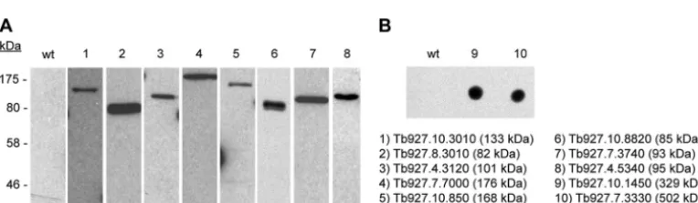
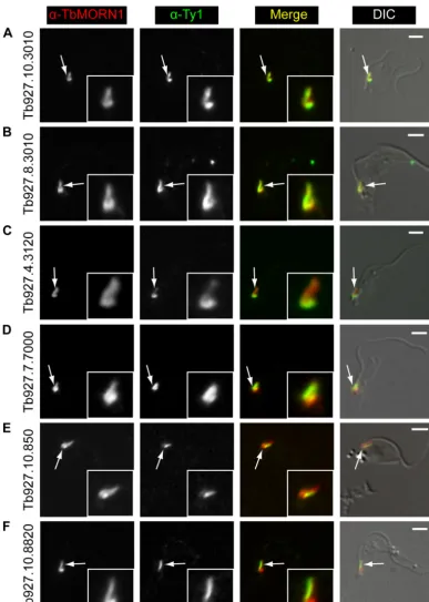
The localization of all 10 proteins in detergent-extracted cells was determined. This was accomplished either by transient ex-pression of a Ty1 epitope-tagged version of the candidate or, in the case of larger or poorly transfecting candidates, by generation of endogenous replacement cell lines expressing the candidate with a triple Ty1 epitope tag. Expression of the tagged proteins was con-firmed by immunoblots of whole-cell lysates with Ty1 anti-bodies (Fig. 4A). For the largest two proteins (Tb927.10.1450 and Tb927.7.3330; 329 and 502 kDa, respectively), expression was confirmed by dot blot (Fig. 4B). All but one of the candidates exhibited a localization consistent with its designation as a TbMORN1 binding partner or near neighbor. Six proteins showed good overlap with TbMORN1 (Fig. 5). Two proteins (Tb927.10.3010 and Tb927.08.3010) strongly overlapped with TbMORN1 throughout its whole fishhook expression pattern (Fig. 5AandB). One protein was present throughout the whole fishhook shape but gave the strongest signal from the hooked pos-FIG 2Biotinylation by Myc-BirA*-TbMORN1in vivo. Myc-BirA*-TbMORN1 cells were incubated with excess biotin in the presence or absence of 10 ng/ml tetracycline (Tet). (A and B) Immunofluorescence assay of intact cells labeled with anti-Myc antibodies and fluorescently conjugated streptavidin. In the presence of tetracycline (A), the two labels strongly overlapped. The bilobe (arrows, area enlarged in insets) was clearly visible. In the absence of tetracycline (B), only endogenously biotinylated proteins were labeled as a background signal. (C and D) Immunofluorescence assay of detergent-extracted cells labeled as in panels A and B. In the presence of tetracycline (C), colocalization between the anti-Myc and streptavidin labels was seen at the bilobe (arrows, area enlarged in insets). In the absence of tetracycline (D), no labeling was observed in either channel. Images are maximum intensity z-projections; overlap was confirmed in single 0.1-m sections. Identical exposure times and channel level settings were used in image acquisition and processing. Scale bars, 2m. (E) Immunoblot of whole-cell lysates using HRP-conjugated streptavidin. In the presence of tetracycline, many additional polypeptides are strongly biotinylated. Equal loading was confirmed by blotting the samples with anti-TbBILBO1 (lower panel).
on September 8, 2020 by guest
http://ec.asm.org/
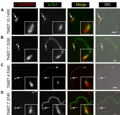
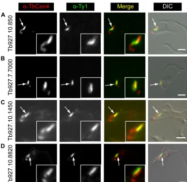
terior part (Tb927.4.3120) (Fig. 5C). Two proteins (Tb927.7.7000 and Tb927.10.850) partially overlapped with TbMORN1 but showed an additional localization alongside it (Fig. 5DandE). Another protein (Tb927.10.8820) was localized solely to the stem anterior part of TbMORN1 (Fig. 5F). Three of the remaining pro-teins showed a localization that could be readily interpreted as indicating a binding partner or near neighbor of TbMORN1 (Fig. 6). One (Tb927.10.1450) was positioned alongside the stem anterior part of TbMORN1, much like TbCentrin4 (Fig. 6A). Two (Tb927.7.3330 and Tb927.4.5340) were localized to a punctate filamentous structure whose posterior end partially overlapped with the anterior stem of TbMORN1 (Fig. 6BandC). Only one candidate (Tb927.7.3740) appeared to be a false positive, which was defined as a protein whose localization disfavored its being a TbMORN1 binding partner or near neighbor. This candidate (Tb927.7.3740) was localized to the flagellum (Fig. 6D). The fla-gellum passes through the hook posterior part of TbMORN1 (producing an apparent partial overlap by immunofluorescence), but the flagellar proteins and bilobe proteins are insulated from one another by the flagellum membrane and flagellar pocket neck membrane. The localization of Tb927.7.3330 and Tb927.4.5340, with a partial overlap with the TbMORN1 bilobe, a punctate fila-mentous morphology, and a trajectory from the bilobe to the an-terior end of the cell but not the tip of the flagellum, suggested FAZ localization. This was confirmed by colabeling with the FAZ-spe-cific 1B41 monoclonal antibody (see Fig. S2C and D in the sup-plemental material).
Three of the candidates (Tb927.7.7000, Tb927.10.850, Tb927.10.1450) had localizations that suggested they might also partially or strongly overlap TbCentrin4. TbCentrin4 is primarily present on a bar that lies alongside the TbMORN1 fishhook, with a small amount extending toward the basal and probasal bodies where it is also localized. In addition, the protein (Tb927.10.8820) overlapping solely with the stem anterior part of TbMORN1 had an expression pattern strongly reminiscent of TbCentrin4.
Cola-beling experiments using anti-Ty1 and anti-TbCentrin4
antibodies confirmed a partial overlap with TbCentrin4 for Tb927.10.850 and Tb927.7.7000 (Fig. 7AandB). As expected, Tb927.10.1450 strongly overlapped with the TbCentrin4 bar, con-sistent with its localization alongside the stem anterior part of TbMORN1 (Fig. 7C). Tb927.10.8820 was found to mirror the TbCentrin4 bar (Fig. 7D).
DISCUSSION
BioID is a new and very powerful technique for identifying bind-ing partners and near neighbors and was first used in mammalian tissue culture cells (20). The work described here demonstrates the successful adaptation of this technique to the parasitic protist T. brucei, using TbMORN1 to discover novel structural compo-nents of the trypanosome bilobe. The Myc-BirA* biotinylation module was found to be nontoxic and permissive for normal lo-FIG 3Purification of candidate TbMORN1 binding partners and near
neigh-bors. (A) Schematic of purification protocol. Following incubation in excess biotin and the presence or absence of tetracycline (⫹/⫺Tet), the cells are incubated with 0.5% NP-40 (E1) and separated by centrifugation into detergent-soluble (S1) and detergent-insoluble (P1) fractions. The detergent-insoluble fraction is then further extracted with 0.4% SDS. Fractions S1 and S2 are incubated with streptavidin-conjugated paramagnetic beads, and the free unbound fractions (F1 and F2, re-spectively) are separated magnetically. Fractions B1 and B2 are, respectively, the bound cytoplasmic and cytoskeletal eluates and will contain candidate TbMORN1 binding partners and near neighbors. (B) Representative purification. Myc-BirA*-TbMORN1-expressing cells were processed as described for panel A. Samples were immunoblotted using HRP-conjugated streptavidin. Equal fractions were loaded in each lane. (C) Representative purification from Myc-BirA*-TbMORN1 cells
incubated with excess biotin in either the presence or absence of tetracycline. Samples were immunoblotted using HRP-conjugated streptavidin; equal frac-tions in each lane. Comparison of⫹/⫺tetracycline conditions shows clear enrichment of biotinylated proteins in fractions E1, S1, and P1 and purifica-tion with minimal background in elupurifica-tions B1 and B2 (left panel). Loading larger fractions of elutions B1 and B2 shows efficient capture of candidate TbMORN1 binding partners and near neighbors. Background signal from⫺ Tet cells remains minimal (right panel).
on September 8, 2020 by guest
http://ec.asm.org/
calization of tagged protein (Fig. 1). At approximately 35 kDa it is slightly larger than GFP, but it seems reasonable to assume that any protein whose localization is unaffected by GFP tagging could be utilized without handicap.
Incubation of Myc-BirA*-TbMORN1-expressing cells in ex-cess biotin leads to the reproducible biotinylation of candidate proteins in the vicinity of the tagged bait (Fig. 2). Biotinylated proteins from both cytoplasmic and cytoskeletal fractions were efficiently purified and the latter subjected to analysis by mass spectrometry (Fig. 3). Comparison of the mass spectrometry data obtained from purifications in experimental/induced (⫹Tet) and control/uninduced (⫺Tet) conditions allowed subtraction of background binding and consequently the rapid selection of promising candidates. Screening a shortlist of candidates con-firmed partial or strong overlap with TbMORN1 in the over-whelming majority of cases.
Most work on the trypanosome cytoskeleton has relied on a combination of light microscopy and electron microscopy cou-pled with phenotypic analysis following protein depletion by RNAi. Such work is, however, innately hampered by the difficulty of identifying candidate proteins to study and the inability to read-ily infer direct physical relationships between them. Both of these drawbacks relate to the fact that a large fraction of cytoskeletal and
cytoskeleton-associated proteins are detergent insoluble and thus refractory to conventional affinity purification techniques. Tan-dem affinity purification (TAP) tagging techniques have become the benchmark method for identifying protein complexes but have an unavoidable requirement for detergent solubility of the complexes and sufficient strength and persistence of intracomplex interactions to withstand washing. The amount of material re-quired may also be a limiting factor due to limited preservation of interacting partners. In the trypanosome cytoskeleton field, there-fore, most protein identification efforts have focused on three ap-proaches: (i) bioinformatic screens to identify orthologues of pre-viously characterized proteins (26), (ii) generation of panels of monoclonal antibodies against entire elements of the cytoskeleton (27), and (iii) proteomic analysis of entire substructures of the cytoskeleton (10,16,28). The first method has the obvious draw-back of being limited to identifying candidates with sequence/ structural homology to other proteins, and this difficulty can also be compounded by the considerable evolutionary time separating trypanosomes (and other protists) from the better-studied mem-bers of the Eukaryota taxon. By definition, therefore, it cannot identify trypanosome-specific proteins. The second method is rel-atively unfocused owing to the large number of antigens present in even a substructure of the cytoskeleton, and identifying the target TABLE 1Candidates shortlisted for evaluation of localizationa
Accession no.
Molecular size (kDa)
No. of assigned spectra (no. of unique peptides)
% SC Statusb Featuresc
Expt 1 Expt 2 Expt 3
⫹Tet ⫺Tet ⫹Tet ⫺Tet ⫹Tet ⫺Tet
Tb927.6.4670 41 235 (37) 0 (0) 136 (25) 0 (0) 324 (28) 2 (2) 71 TbMORN1 15⫻MORN
Tb927.10.3010 133 108 (63) 0 (0) 187 (59) 1 (1) 132 (54) 0 (0) 60 Unc 2⫻CC
Tb927.7.3330 502 63 (43) 0 (0) 21 (16) 2 (1) 34 (20) 2 (1) 7 Unc 13⫻CC
Tb11.01.0680 79 43 (26) 0 (0) 52 (25) 0 (0) 52 (24) 0 (0) 41 TbLRRP1 7⫻LRR, 1⫻CC
Tb927.7.3740 93 32 (27) 0 (0) 23 (19) 0 (0) 25 (18) 0 (0) 35 Unc 5⫻CC
Tb927.7.7000 176 31 (26) 0 (0) 41 (34) 0 (0) 29 (22) 0 (0) 20 Unc
Tb927.10.1450 329 30 (23) 0 (0) 30 (19) 2 (1) 26 (22) 2 (2) 9 Unc 4⫻CC
Tb927.10.8820 85 24 (17) 0 (0) 60 (21) 1 (1) 26 (16) 1 (1) 27 Unc
Tb927.10.850 168 20 (18) 0 (0) 27 (19) 0 (0) 26 (18) 0 (0) 15 Unc 5⫻WD40, 7⫻CC
Tb927.4.3120 101 18 (15) 0 (0) 29 (21) 0 (0) 23 (21) 0 (0) 27 Unc
Tb927.4.5340 95 10 (10) 0 (0) 7 (7) 0 (0) 7 (7) 0 (0) 14 Unc 5⫻CC
Tb927.8.3010 82 3 (3) 0 (0) 9 (7) 1 (1) 11 (8) 1 (1) 10 Unc 3⫻CC
aHits were ranked according to the number of assigned spectra in experiment 1; % sequence coverage (% SC) is shown, along with scores for assigned spectra and unique peptides
under⫹/⫺Tet conditions.
bUnc, uncharacterized. c
MORN, membrane occupation and recognition nexus repeats; CC, predicted coiled-coil regions; LRR, predicted leucine-rich repeats; WD40, predicted WD40 repeat.
FIG 4Confirmation of expression of tagged candidate proteins. Whole-cell lysates from cells expressing candidate TbMORN1 binding partners and near neighbors were analyzed by immunoblotting with anti-Ty1 antibodies. (A) Western blot of candidates. Expected molecular size is shown next to the figure. Addition of the Ty1 epitope tag causes a distinct decrease in electrophoretic mobility. (B) Dot blot of the two larger candidates.
on September 8, 2020 by guest
http://ec.asm.org/
antigen of a given monoclonal antibody remains far from trivial (29). The third method is comprehensive in that it can faithfully provide an entire inventory of constituent proteins but provides no information on the relationships between these proteins and
no easy way to evaluate possible contaminants. Given that there are hundreds of different proteins in the flagellum alone, estab-lishing such relationships can become a formidable task. Another method of screening for direct interactions of detergent-insoluble FIG 5Localization of candidate proteins that overlap TbMORN1. Candidates were expressed with the Ty1 epitope tag in 427 cells either by transient transfection (first five rows) or endogenous replacement (last row). Detergent-extracted cells were labeled with anti-TbMORN1 and anti-Ty1 antibodies and analyzed by fluorescence microscopy. Single 0.1-m z-slices are shown. Arrows indicate bilobe structure; area enlarged in insets. Fluorescence images were superimposed on differential interference contrast (DIC) images. The candidates localized either to the whole TbMORN1 bilobe (Tb927.10.3010, Tb927.8.3010) (A and B), primarily to the hook posterior part of TbMORN1 (Tb927.4.3120) (C), to both the bilobe and the adjacent TbCentrin4 arm (Tb927.7.7000, Tb927.10.850) (D and E), and to the posterior stem of TbMORN1 (Tb927.10.8820) (F). Scale bars, 2m.
on September 8, 2020 by guest
http://ec.asm.org/
proteins, the yeast two-hybrid technique, requires nonnative con-ditions and frequently generates false positives.
These results constitute an emphatic validation of BioID as a method for forward biochemical screens of binding partners and near neighbors. TbMORN1 has provided an exemplary test case for the technique. It is tightly associated with the trypanosome cytoskeleton, and it localizes to the bilobe—a discrete (approxi-mately 2-m long) structure of unclear function and almost com-pletely unknown composition. Attempts to identify other bilobe proteins on the basis of homology have proved fruitless to date (our unpublished data). Here, using BioID has permitted the identification of at least seven new bilobe-resident proteins. In addition, two novel FAZ proteins were identified. All of the pro-teins here identified were previously experimentally uncharacter-ized, and none are homologous to TbMORN1 and TbLRRP1. All are specific to either the trypanosomes or the TriTryps, and they exhibit no common structural features other than a preponder-ance of coiled-coil and repeat regions. As such, it is highly unlikely that they would ever have been detected usingin silicomethods. All are detergent insoluble and consequently are highly unlikely to
have ever been detected using conventional affinity purification techniques.
The presence of at least two FAZ proteins in this data set rein-forces the idea that there is a close physical association of the bilobe and the FAZ. An overlap has previously been noted be-tween the stem anterior part of TbMORN1 and the posterior end of the FAZ at both immunofluorescence and electron microscopy levels (15,19). Similarly, the varied and overlapping distribution of the new bilobe proteins lends weight to the hypothesis that the structure is composed of subdomains rather than being a single homogeneous entity. It is curious that TbCentrin4 was not pres-ent in the list of candidates, especially given that Tb927.10.1450 overlapped with it. This could be due to a combination of the high number (i.e., 18) and small size of its predicted tryptic peptides within its 149-amino-acid primary sequence, disfavoring it for mass spectrometry identification. However, it also could not be detected in Western blots of the eluates, so it seems likely that a more complex explanation is involved (data not shown). On a related note, the absence of the flagellar pocket collar protein Tb-BILBO1 indirectly supports the notion that the flagellar pocket FIG 6Localization of candidate proteins to the bilobe, FAZ, and flagellum. Candidates were expressed with the Ty1 epitope tag in 427 cells either by endogenous replacement (first two rows) or transient transfection (last two rows). Detergent-extracted cells were labeled with anti-TbMORN1 and anti-Ty1 antibodies and analyzed by fluorescence microscopy. Tb927.10.1450 was localized adjacent to the anterior stem part of TbMORN1 (A). Tb927.7.3330 and Tb927.4.5340 were present on the FAZ and showed partial overlap with TbMORN1 (B and C). Tb927.7.3740 was present on the flagellum (D). Single 0.1-m z-slices are shown. Arrows indicate bilobe structure; area enlarged in insets. Fluorescence images were superimposed on DIC images. Scale bars, 2m.
on September 8, 2020 by guest
http://ec.asm.org/
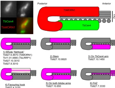
collar and the bilobe are distinct structures (19). The various lo-calizations at the bilobe of the screened candidates have been sum-marized schematically (Fig. 8). It seems likely that the bilobe is composed of at least four subdomains—the whole fishhook-shaped structure (Fig. 8, panel 1), its anterior stem (Fig. 8, panel 2), its posterior hook (Fig. 8, panel 4), and the TbCentrin4 bar (Fig. 8, panel 3). In addition, a number of proteins are present at least partially on both the TbMORN1 fishhook and the TbCen-trin4 bar, covering the entire bilobe (Fig. 8, panel 5). The anterior parts overlap the posterior end of the FAZ (Fig. 8, panel 6). These new findings extend our previous morphological work (19). Ex-ploring these subdomains and teasing out the functional roles of the individual proteins within them represents an exciting future program of work. The priority in this study was to identify core structural components of the bilobe rather than more labile ones, but it will be interesting also to determine the composition of the detergent-soluble biotinylated fractions obtained in the experi-ments.
A final curiosity is why Tb927.7.3740 scored so highly when it
is not localized to the bilobe. The protein registered as a good hit in all three independent purifications, better even than a number of the confirmed hits. A possible explanation is that the protein is in fact a binding partner of TbMORN1 but only a transient one. During early stages of the cell cycle, TbMORN1 and TbLRRP1 have been localized to tendrillar protein fibers that connect the bilobe and the basal body and probasal body (19). It is possible that there is a transient association between TbMORN1 and Tb927.7.3740 in this interval. Again, further work is required to clarify the question.
In conclusion, the results here validate the use of the BioID methodology inT.brucei. Although promiscuous biotinylation by BirA* was first demonstrated inEscherichia coli, the data here con-stitute—to our knowledge—the first demonstration of proximi-ty-dependent BioID in nonhuman eukaryotic cells (30). Unlike the proof-of-principle experiments conducted on lamin-A in the comparatively well-characterized mammalian nuclear envelope, they are also the first application of BioID to an extranuclear struc-ture about which very little is known. The success of BioID in FIG 7Localization of candidate TbMORN1 binding partners relative to TbCentrin4. Candidates were expressed with the Ty1 epitope tag in 427 cells either by transient transfection (first two rows) or endogenous replacement (last two rows). Detergent-extracted cells were labeled with anti-TbCentrin4 and anti-Ty1 antibodies and analyzed by fluorescence microscopy. Single 0.1-m z-slices are shown. Arrows indicate bilobe structure; area enlarged in insets. Fluorescence images were superimposed on DIC images. As expected, Tb927.10.850 and Tb927.7.7000 showed a partial overlap with TbCentrin4 (A and B). Tb927.10.1450 (C) and Tb927.10.8820 (D) showed good and no overlap, respectively. Scale bars, 2m.
on September 8, 2020 by guest
http://ec.asm.org/
identifying new components of that structure is very encouraging. Given that the only requirement for the technique is the capability for transgene expression, it is likely to be particularly suited to the study of protists (parasitic and otherwise) and other simpler eu-karyotes where the level of genome annotation is low and the amount of evolutionary time separating them from standard model organisms such as mice and fruit flies inhibits easy bioin-formatic identification of orthologues. Starting with only a single probe protein, BioID could in principle be used to bootstrap through the members of a protein complex or interaction net-work. The cytoskeletal field, frequently burdened with large and insoluble proteins, should benefit in particular.
ACKNOWLEDGMENTS
Funding was from the FWF Austrian Science Fund (FWF grant P22276-B12).
We thank Gustav Ammerer for assistance with mass spectrometry and Rainer Gith and other members of the mass spectrometry facility. We thank also Brian Burke (Institute of Medical Biology, Singapore) and the other members of the Warren lab for critical discussion of the manuscript.
REFERENCES
1.Gull K.1999. The cytoskeleton of trypanosomatid parasites. Annu. Rev. Microbiol.53:629 – 655. doi:10.1146/annurev.micro.53.1.629.
2.Robinson DR, Sherwin T, Ploubidou A, Byard EH, Gull K. 1995. Microtubule polarity and dynamics in the control of organelle position-ing, segregation, and cytokinesis in the trypanosome cell cycle. J. Cell Biol.
128:1163–1172.
3.Lacomble S, Vaughan S, Gadelha C, Morphew MK, Shaw MK, McIn-tosh JR, Gull K.2009. Three-dimensional cellular architecture of the
flagellar pocket and associated cytoskeleton in trypanosomes revealed by electron microscope tomography. J. Cell Sci.122:1081–1090. doi:10.1242 /jcs.045740.
4.Engstler M, Thilo L, Weise F, Grunfelder CG, Schwarz H, Boshart M, Overath P. 2004. Kinetics of endocytosis and recycling of the GPI-anchored variant surface glycoprotein in Trypanosoma brucei. J. Cell Sci.
117:1105–1115. doi:10.1242/jcs.00938.
5.Lacomble S, Vaughan S, Gadelha C, Morphew MK, Shaw MK, McIn-tosh JR, Gull K.2010. Basal body movements orchestrate membrane organelle division and cell morphogenesis in Trypanosoma brucei. J. Cell Sci.123:2884 –2891. doi:10.1242/jcs.074161.
6.Gull K.2003. Host-parasite interactions and trypanosome morphogene-sis: a flagellar pocketful of goodies. Curr. Opin. Microbiol.6:365–370. 7.Henley GL, Lee CM, Takeuchi A.1978. Electron microscopy
observa-tions on Trypanosoma brucei: freeze-cleaving and thin-sectioning study of the apical part of the flagellar pocket. Z Parasitenkd.55:181–187. 8.Sherwin T, Gull K.1989. The cell division cycle of Trypanosoma brucei
brucei: timing of event markers and cytoskeletal modulations. Philos. Trans. R. Soc. Lond. B Biol. Sci.323:573–588.
9.Vickerman K.1969. On the surface coat and flagellar adhesion in trypano-somes. J. Cell Sci.5:163–193.
10. Broadhead R, Dawe HR, Farr H, Griffiths S, Hart SR, Portman N, Shaw MK, Ginger ML, Gaskell SJ, McKean PG, Gull K.2006. Flagellar motility is required for the viability of the bloodstream trypanosome. Nature440: 224 –227. doi:10.1038/nature04541.
11. Ralston KS, Kisalu NK, Hill KL.2011. Structure-function analysis of dynein light chain 1 identifies viable motility mutants in bloodstream-form Trypanosoma brucei. Eukaryot. Cell10:884 – 894. doi:10.1128/EC .00298-10.
12. Zhou Q, Liu B, Sun Y, He CY.2011. A coiled-coil- and C2-domain-containing protein is required for FAZ assembly and cell morphology in Trypanosoma brucei. J. Cell Sci.124:3848 –3858. doi:10.1242/jcs.087676. 13. May SF, Peacock L, Almeida Costa CI, Gibson WC, Tetley L, Robinson DR, Hammarton TC.2012. The Trypanosoma brucei AIR9-like protein is
FIG 8Schematic summarizing localizations of candidate TbMORN1 binding partners and near neighbors. The image at top left shows a close-up of the bilobe area from a detergent-extracted cell labeled with anti-TbMORN1 and anti-TbCentrin4. Dimensions of the schematic are taken from immunofluorescence images. TbCentrin4 localizations to the basal body and probasal body and the extra density extending from the bar toward the basal body are excluded for clarity.
on September 8, 2020 by guest
http://ec.asm.org/
cytoskeleton-associated and is required for nucleus positioning and accu-rate cleavage furrow placement. Mol. Microbiol.84:77–92. doi:10.1111/j .1365-2958.2012.08008.x.
14. He CY, Pypaert M, Warren G.2005. Golgi duplication in Trypanosoma brucei requires Centrin2. Science 310:1196 –1198. doi:10.1126/science .1119969.
15. Morriswood B, He CY, Sealey-Cardona M, Yelinek J, Pypaert M, Warren G.2009. The bilobe structure of Trypanosoma brucei contains a MORN-repeat protein. Mol. Biochem. Parasitol.167:95–103. doi:10.1016 /j.molbiopara.2009.05.001.
16. Zhou Q, Gheiratmand L, Chen Y, Lim TK, Zhang J, Li S, Xia N, Liu B, Lin Q, He CY.2010. A comparative proteomic analysis reveals a new bi-lobe protein required for bi-lobe duplication and cell division in Trypanosoma brucei. PLoS One 5:e9660. doi:10.1371/journal.pone .0009660.
17. Shi J, Franklin JB, Yelinek JT, Ebersberger I, Warren G, He CY.2008. Centrin4 coordinates cell and nuclear division in T. brucei. J. Cell Sci.
121:3062–3070. doi:10.1242/jcs.030643.
18. Wang M, Gheiratmand L, He CY.2012. An interplay between Centrin2 and Centrin4 on the bi-lobed structure in Trypanosoma brucei. Mol. Mi-crobiol.83:1153–1161. doi:10.1111/j.1365-2958.2012.07998.x.
19. Esson HJ, Morriswood B, Yavuz S, Vidilaseris K, Dong G, Warren G.
2012. Morphology of the trypanosome bilobe, a novel cytoskeletal struc-ture. Eukaryot. Cell11:761–772. doi:10.1128/EC.05287-11.
20. Roux KJ, Kim DI, Raida M, Burke B.2012. A promiscuous biotin ligase fusion protein identifies proximal and interacting proteins in mammalian cells. J. Cell Biol.196:801– 810. doi:10.1083/jcb.201112098.
21. Lane MD, Young DL, Lynen F.1964. The enzymatic synthesis of holo-transcarboxylase from apoholo-transcarboxylase and (⫹)-biotin. I. Purifica-tion of the apoenzyme and synthetase; characteristics of the reacPurifica-tion. J. Biol. Chem.239:2858 –2864.
22. Kwon K, Beckett D.2000. Function of a conserved sequence motif in biotin holoenzyme synthetases. Protein Sci.9:1530 –1539. doi:10.1110/ps .9.8.1530.
23. Wirtz E, Leal S, Ochatt C, Cross GA.1999. A tightly regulated inducible
expression system for conditional gene knock-outs and dominant-negative genetics in Trypanosoma brucei. Mol. Biochem. Parasitol.99: 89 –101.
24. Aslett M, Aurrecoechea C, Berriman M, Brestelli J, Brunk BP, Car-rington M, Depledge DP, Fischer S, Gajria B, Gao X, Gardner MJ, Gingle A, Grant G, Harb OS, Heiges M, Hertz-Fowler C, Houston R, Innamorato F, Iodice J, Kissinger JC, Kraemer E, Li W, Logan FJ, Miller JA, Mitra S, Myler PJ, Nayak V, Pennington C, Phan I, Pinney DF, Ramasamy G, Rogers MB, Roos DS, Ross C, Sivam D, Smith DF, Srinivasamoorthy G, Stoeckert CJ, Subramanian S, Thibodeau R, Tivey A, Treatman C, Velarde G, Wang H.2010. TriTrypDB: a functional genomic resource for the Trypanosomatidae. Nucleic Acids Res.38: D457–D462. doi:10.1093/nar/gkp851.
25. Alsford S, Turner DJ, Obado SO, Sanchez-Flores A, Glover L, Berriman M, Hertz-Fowler C, Horn D.2011. High-throughput phenotyping using parallel sequencing of RNA interference targets in the African trypano-some. Genome Res.21:915–924. doi:10.1101/gr.115089.110.
26. Absalon S, Blisnick T, Kohl L, Toutirais G, Dore G, Julkowska D, Tavenet A, Bastin P.2008. Intraflagellar transport and functional analysis of genes required for flagellum formation in trypanosomes. Mol. Biol. Cell
19:929 –944. doi:10.1091/mbc.E07-08-0749.
27. Bonhivers M, Nowacki S, Landrein N, Robinson DR.2008. Biogenesis of the trypanosome endo-exocytotic organelle is cytoskeleton mediated. PLoS Biol.6:e105. doi:10.1371/journal.pbio.0060105.
28. Portman N, Lacomble S, Thomas B, McKean PG, Gull K.2009. Com-bining RNA interference mutants and comparative proteomics to identify protein components and dependences in a eukaryotic flagellum. J. Biol. Chem.284:5610 –5619. doi:10.1074/jbc.M808859200.
29. Vaughan S, Kohl L, Ngai I, Wheeler RJ, Gull K. 2008. A repetitive protein essential for the flagellum attachment zone filament structure and function in Trypanosoma brucei. Protist 159:127–136. doi:10.1016/j .protis.2007.08.005.
30. Cronan JE.2005. Targeted and proximity-dependent promiscuous pro-tein biotinylation by a mutant Escherichia coli biotin propro-tein ligase. J. Nutr. Biochem.16:416 – 418. doi:10.1016/j.jnutbio.2005.03.017.