Available online on 15.11.2018 at http://jddtonline.info
Journal of Drug Delivery and Therapeutics
Open Access to Pharmaceutical and Medical Research
© 2011-18, publisher and licensee JDDT, This is an Open Access article which permits unrestricted non-commercial use, provided the original work is properly cited
Open Access
Research Article
In-vitro
cytotoxicity of java tea mediated selenium nanoballs against L6
cell lines
Chitra Sivakumar 1*, Karthikeyan Jeganathan2
1 Research Scholar, Research and Development Centre, Bharathiar University, Coimbatore, India
2 Associate Professor and Head, Department of Biochemistry, (PG and Research) Kongunadu Arts and Science College, (Autonomous),
Coimbatore, India
ABSTRACT
The emergence of Nanotechnology has provided a catholic research in recent years by intersecting with assorted branches of science and forming collision on all forms of life. At present there has been a prodigious excitement in the field of Nano pharmacology to seek the role of Nano selenium in individual healthcare and explains how selenium is a double edged sword in the pathologies of chronic diseases like Diabetes mellitus because of their inferior toxicity and ability to gradually release the bioactive principle and free selenium after ingestion. Thus the present study is an embryonic attempt and was aimed at the adroitful synthesis of selenium nanoballs from the aqueous extract of well known herbal tea leaves namely Java tea (Orthosiphon stamineus) using selenious acid solution. The synthesized Selenium nanoparticles were then subjected to various characterization techniques such as UV, FTIR, FESEM, EDAX and Zeta potential respectively. Finally the green synthesized SeNps were tested for their cytotoxic effect against L6 rat skeletal muscle cell lines. The pre clinical studies are underway to prove the insulin mimic activity of the selenium nanoparticles.
Keywords: Selenium Nanoparticles, Java tea, L6 cell lines.
Article Info:Received 03 Oct, 2018; Review Completed 06 Nov 2018; Accepted 09 Nov 2018; Available online 15 Nov 2018
Cite this article as:
Chitra Sivakumar, Karthikeyan Jeganathan, In-vitro cytotoxicity of java tea mediated selenium nanoballs against L6 cell
lines, Journal of Drug Delivery and Therapeutics. 2018; 8(6):195-200 DOI: http://dx.doi.org/10.22270/jddt.v8i6.2046
*Address for Correspondence:
Chitra Sivakumar, Research Scholar, Research and Development Centre, Bharathiar University, Coimbatore, India. Email: [email protected]
1. INTRODUCTION
The field of Nanotechnology devours great enthusiasm in recent years because of its anticipated impact on science, industry, economy and our everyday life. Today nanoparticles of both metallic and non metallic origin are under research and development in various fields of biology and therapeutics 1. Due to the inimitable characteristics exhibited by nanoparticles, they are employed in nanomedicine and nanotherapeutics which are the supreme aspects of nanotechnology to be implemented in human health.
Metal nanoparticles are vital part of future nano-therapeutics which has wide-ranging applications in diverse areas such as chemistry, physics, and biomedical and material sciences2. They have tremendous applications in the area of catalysis, optoelectronics, diagnostic biological probes and display devices 3. Production of nanoparticles can be achieved through conventional chemical methods and physical methods4,5. While chemical approaches are the most popular methods for the
Biogenic synthesis of Se nanoparticles is frequently achieved by reduction of selenate/selenite in presence of bacterial proteins or plant extracts containing phenols, flavonoids amines, alcohols, proteins and aldehydes. Very few studies have been reported the synthesis of selenium nanoparticles using higher plants. The biomaterials of higher plants used for the synthesis of selenium nanoparticles were Capsicum annum10, dried raisin extract11, leaves of lemon12, Terminaliaarjuna13 and seed extract of fenugreek14, flower broth of Catharanthu
sroseus15, Flower of Bougainvilleaspectabilis16 in the
synthesis of selenium nanoparticles. In the present study
Orthosiphon stamineus Benth which is a popular traditional
folk medicine is selected for synthesising selenium nanoparticles for the first time as an maiden attempt. It is used in Indonesia for rheumatism, diabetes, hypertension, tonsillitis, epilepsy, menstrual disorders, gonorrhea, syphilis, renal calculus and gallstones; in Vietnam for urinary lithiasis, edema, eruptive fever, influenza, hepatitis, jaundice and biliary lithiasis; and in Myanmar to alleviate diabetes and urinary tract and renal diseases17-21. Phytochemical studies 22,23and Pharmacological studies 24 of this plant have been conducted since many years. study Thus the present investigation is aimed to evaluate the ability of bioreductive potential of the aqueous leaf extract of Orthosiphon stamineus in the synthesis of selenium nanoparticles and also evaluated the cytotoxicity of selenium nanoparticles against L6 rat skeletal muscle cell lines.
2. MATERIALS AND METHODS
2.1. Collection of Plant Material
The fresh plant of (Orthosiphon stamineus) was collected and was authenticated by Dr. S. Sahaya Sathish, Associate Professor, Department of Botany, St. Joseph’s College, Trichy. Dried Java tea leaves were purchased from Organic farm, Salem and was used as the sample for the biosynthesis of the Selenium nanoparticles.
2.2. Preparation of Extract
The dried leaves were blended using a blender and the powder was stored in a clean glassware container for further analysis.50 gms of powdered leaves were mixed in 300 ml of distilled water in a clean beaker and was stored in a glass container .The contents were mixed by stirring for 15 minutes. It was kept undisturbed for overnight after 24 hours the mixture was then filtered using filter paper (whatman no1).The filtrates were then used for further assay. The aqueous extract of the leaves were screened qualitatively for the presence of secondary metabolites using the routine phytochemical analysis.
2.3. Synthesis of Selenium Nanoparticles
For the green synthesis of selenium nanoparticles, 1 ml of plant extract was mixed with 10 ml of 30 mM selenious acid solution along with 200 ul of 40mM ascorbic acid which was used as an initiator of reduction reaction. Standard positive control was maintained using 1 ml of 0.2% sodium alginate + 10 ml of 30 mM selenious acid and 200 ul of 40mM ascorbic acid for the synthesis of selenium nanoparticles. 1% Orthosiphon leaf extract +200 µl of 40mM ascorbic acid was used as negative control16.The preparations were incubated at room temperature. After 24 hrs of incubation the preparation was centrifuged at 10000 rpm for 30 minutes. The pellet was washed with double distilled water and then with absolute alcohol three times. This washed ethanol pellet was dried overnight. The red selenium nanoparticles were suspended in PBS
(pH-7.4) by ultra sonication and then centrifuged. The powder form of the extract was used for further analysis.
2.4. Characterization of Nanoparticles
The formation of selenium nanoparticles was confirmed by the visual observation. The synthesized SeNps were analyzed for the rate of absorption by using UV-Visible spectrometer. The UV- Visible spectral study was done in the range of 200 to 400 nm. FT-IR measurements was carried out for the synthesized SeNps to identify the possible bioactive molecules responsible for the reduction of the selenium and the capping ability of the bio reduced selenium nanoparticles by the aqueous leaf extract of O.
Stamineus using KBr pellets and the spectra was recorded
in the wavelength interval 4000 to 400cm-.The FESEM and EDAX analysis revealed average shape, size and elemental composition of the synthesized SeNps. The charge distribution of selenium nanoparticles was confirmed with zeta potential analysis.
2.5. In-vitro Cytotoxicity Analysis MTT assay
The monolayer cell culture of L6 rat skeletal muscle cell lines was trypsinized and the cell count was adjusted to 1.0 x 10⁵ cells/ml using respective media containing 10% FBS. To each well of the 96 well micro titer plates, 100 µl of the diluted cell suspension (50,000cells/well) was added. After 24 h, when a partial monolayer was formed, the supernatant was flicked off, washed the monolayer once with medium and 100 µl of different test concentrations (10,20,40,80,160,320 µg/ml) of selenium nanoparticles were added on to the partial monolayer in micro titer plates. The plates were then incubated at 37oC for 24hrs in 5% CO2 atmosphere. After incubation the test solutions in the wells were discarded and 100 µl of MTT (5 mg/10 ml of MTT in PBS) was added to each well. The plates were incubated for 4 h at 37o C in 5% CO2 atmosphere. The supernatant was removed and 100 µl of DMSO was added and the plates were gently shaken to solubilize the formed formazan. The absorbance was measured using a micro plate reader at a wavelength of 590 nm. The percentage of cell viability was calculated using the following formula. % Cell Viability= 100–(OD of sample/OD of Control) x 100. 3. RESUTS AND DISCUSSION
3.1. Bioactive profiling of Aqueous extracts (O. Stamineus)
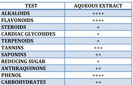
Table 1: Phytochemical screening of Aqueous extract of O.stamineus
TEST AQUEOUS EXTRACT
ALKALOIDS ++++
FLAVONOIDS ++++
STEROIDS +
CARDIAC GLYCOSIDES +
TERPENOIDS +
TANNINS +++
SAPONINS ++
REDUCING SUGAR +
ANTHRAQUINONE ++
PHENOL ++++
CARBOHYDRATES ++
Bioactive profiling was performed in aqueous extract of
O.Stamineus leaves. The phytochemical screening showed
protein, carbohydrate, cardiac glycosides and saponins in aqueous extract of Java tea leaves (Table-1). It is rich in flavanoids, terpenoids and other secondary metabolites having reducing functional groups which might have played a role in reducing selenious acids to SeNPs. O.
stamineus has wide traditional and pharmacological uses in
various pathophysiological conditions. It has been shown that plant extract containing phenol and flavonol derivatives act as reducing agents and nanoparticle stabilizer. FTIR spectrum of java tea extract shows the presence of various functional groups (Figure-3)
3.2. Visual Observation
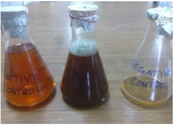
Figure 1: Visual Observation of Formation of SeNps
The bio reduction of the selenious acid using aqueous leaf extract of O. stamineus was monitored and the appearance of brownish orange colour indicates the formation selenium nanoparticles. Initially the plant extract was light brown in colour. But after the addition of colourless
selenious acid solution the colour gradually changes from light brown to dark brownish orange as shown in the (Figure-1).The appearance of brownish orange colour indicates the reduction of selenious acid and formation of SeNps similar to the results reported earlier16.
3.3. UV-Visible spectroscopy
Figure 2: UV–Vis Spectra of Se Nanoparticles Synthesized by Using Java tea leaf Extract
The above (Figure-2) shows the UV-Vis Spectra of the as- formed SeNps exhibiting the maximum absorption peak at about 209 nm. This is due to the surface plasmon resonance of selenium nanoparticles. The red color of the
3.4. Fourier Transform Infrared Spectroscopy (FTIR) analysis
Figure 3: FTIR Spectrum of the aqueous leaf extract and SeNps
FTIR measurements of both the aqueous java tea leaves extract and the synthesized dried SeNps were carried out to identify the possible biomolecules responsible for the reduction, capping and efficient stabilization of the bio reduced SeNps. The FTIR spectra of both the leaf extract as well as SeNps are shown in (Figure-3). The aqueous Java tea leaf extract displays a number of absorption peaks reflecting its complex nature. The spectrum was recorded in the wavelength region between 400 cm-1to 4000 cm-1. The spectrum of aqueous Java tea leaves extract shows the peaks at wave numbers 3388 cm-1,2928 cm-1,2856 cm-1,1637 cm-1,1385 cm-1,1321 cm-1,1265 cm-1,1158 cm-1,1069 cm-1 respectively. The broad peak at 3388 was due to the presence of alcohol (O-H) stretching of Phenolic compound. The peak at 2928 cm-1 and 2856 cm-1 showed a sharp peak and strong alkane C-H stretching. The peak at 1636 cm-1 and 1384 cm-1showed a variable alkene C=C and alkane (C-H)
stretch. The other peaks at 1265 cm-1, 1158 cm-1, 1069 cm-1were due to amine (C-N), ether, alkene, C-Cl sretching. After the reduction process the absorption peaks at 3381 cm-1,2960 cm-1,1718 cm-1,1629 cm-1,1516 cm-1 were observed as shown in (Figure-3).Corresponds to alcohol(O-H), alkene (C-H), carbonyl (C=O), alkene C=C and aromatic C=C stretching of Phenolic compound. The peaks at 2928 cm-1 and 2856 cm-1 were disappeared and shifted to 2960 cm-1.The peak corresponds to alkene C=C was disappeared and shifted to 1718 cm-1 . A sharp peak at 1384 cm-1was disappeared and was shifted to 1441 cm-1corresponds to aromatic C=C stretch. Based on the above observations it is inferred that the biosynthesized SeNps might be surrounded by any one of these organic molecules such as polyphenols, alkaloids, terpenoids which are in accordance with the facts already reported in the earlier reports16.
3.5. FESEM and EDAX analysis
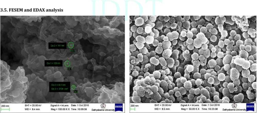
Figure 4: FESEM Analysis of Selenium nanoparticles
The surface morphology and size of the nanoparticles were obtained by Frontier Scanning Electron Microscopy analysis. The above (Figure-4) shows the SeNPs
of selenium nanoparticles using plant extract. The image clearly indicates the spherical nature of the selenium nanoparticles as shown in the above figure and they merely look like balls. The relatively uniform shape of the SeNps was confirmed in the range of 88 nm -141nm.The obtained results were also supported in the earlier studies.
Figure 5: Edax Spectrum of SeNps
An elemental composition analysis employing FESEM– EDAX showed the presence of a strong signal from Se atoms (12.62%) (Figure-5). This analysis indicated that the nanostructures were composed of selenium. Other EDAX peaks such as C, K, Na, Mg, O, Si were also found, suggesting that they were mixed precipitates of the selenium salt.
3.6. Zeta potential value of Selenium Nanoparticles:
Figure 6: Zeta Potential of SeNps
The zeta potential measurements indicate negative charge (-34.9 mV) on the selenium nanoparticles as shown in the above (Figure-6). If all the particles in suspension have a negative or positive zeta potential, then they will tend to repel each other and there is little tendency for the particles come together. The slightly negative charge on Se particles is probably resulting in the high stability of the selenium nanoparticles without forming aggregates and these particles do not transform to black amorphous form when kept for prolonged period of time of more than a month.
3.7. MTT Assay
Table 2: MTT Assay
L6 cell lines
Compound name Conc. µg/ml OD at 590nm % Cell viability
Control 0 0.596 0
SeNp 10 0.574 3.76
20 0.543 8.95
40 0.502 15.83
80 0.456 23.54
160 0.422 29.24
320 0.387 35.11
Figure 7: Invitro Cytotoxicity of Selenium nanoparticles
Thus the successfully synthesized SeNp was screened for the cytotoxicity against L6 Cell lines by MTT Assay. From the above Table-2, it was understood that the optical
nontoxic effect and the % of cell viability was notably increased. These results clearly demonstrate that the phytochemicals within these herbs provide nontoxic coating on SeNPs and corroborate the results of the internalization studies discussed above. The lack of any noticeable toxicity of Java tea mediated selenium nanoparticles provides new opportunities for the safe application in molecular imaging and therapy.
In modern science, biomedical technology is well developed and growing in recent times. Biological synthesis of metal nanoparticles is an important role in the field of modern nanotechnology. Green synthesis of nanomedicine plays key role in bio Medical science. In this study, biological synthesis of selenium using java tea leaves and their cytotoxic study is taken into consideration. The nanoparticles obtained are characterized by UV-spectral analysis, FESEM, EDAX, Zeta potential. The functional groups of the selenium nanoparticles in Java tea leaves extract were characterized by Fourier Transform Infrared Spectroscopy. The in vitrocytotoxicity analysis of selenium nanoparticles showed good cell viability against L6 rat skeletal muscle cell line. From this study, it is found that the biomedical properties are present in Selenium nanoparticles.
4. CONCLUSION
In the present study, selenium nanoballs were successfully synthesized using the plant extract of Orthosiphon
stamineus. The synthesized nanoparticles were found to
increase the cell viability in L6 cell lines. The preclinical studies for treating diabetes mellitus and to prove the insulin mimic activity of selenium nanoparticles are underway. Thus the green synthesized nanoparticles were found to be cost-effective, simpler, and environmentally safe. As the nanotechnology is an emerging field in medicine, the biological synthesis of nanoparticles helps in the other way. In Future, it is a promising contender for various clinical applications in a safe and ecofriendly manner.
ACKNOWLEDGEMENT
The authors would like to thank Dr. R. Rajamurugan (late) for his valuable guidance, and also grateful to Dr. S. Sahaya Sathish, Associate Prof., Dept. of Botany, St. Joseph college, Trichy, Dr. C. Narendhar, Assistant Prof., Dept. of Nano Science and technology, Sri Ramakrishna Engineering College, Coimbatore, Shri. G. Arihara Sivakumar, Associate Prof., Dept. of Pharmacology, KMCH College of Pharmacy, Coimbatore, Shri. D. Ramachandran, Scientist –C, Centre for Nano science and technology, Sathyabama University, Chennai for their technical assistance and support.
REFERENCES
1. Sengupta J, Ghosh S, Datta P, Gomes A. Gomes A. Physiologically Important Metal Nanoparticles and Their Toxicity. J. Nanosc. Nanotechnol, 2014; 14(1):990-1006. 2. Prasad KS, Pathak D, Patel A, Dalwadi P, Prasad R, Patel P,
Selvaraj K.P. Biogenic synthesis of silver nanoparticles using Nicotiana tobaccum leaf extract and study of their antibacterial effect. Afr.J. Biotechnol, 2011; 10: 8122. 3. Sathishkumar G, Gobinath C, Karpagam K, Hemamalini V,
Premkumar K, Sivaramakrishnan S, Phyto-synthesis of silver nanoscale particles using Morinda citrifolia L. and its inhibitory activity against human pathogens.Colloids Surf, 2012; B 95:235.
4. Murray CB, Kangan CR, Bawendi MG.Synthesis and characterization of monodisperse nanocrystals and
close-packed nanocrystal assemblies. Annu. Rev. Mater. Sci. 2000; 30:545.
5. Ayyub P, Chandran R, Taneja RP, Sharma A, Pinto R. Synthesis of nanocrystalline material by sputtering and laser ablation at low temperature. Appl. Phys. A, 2001; 73:67.
6. Ren F, He X, Wang K, Yin JJ. Biosynthesis of gold nanoparticles using Catclaw Buttercup (Radix Ranunculi Ternati) and evaluation of its colloidal stability, Biomed.Nanotechnol, 2012; 8:586.
7. Zhang Y, Zhang J, Wang HY, Chen HY. Synthesis of selenium nanoparticles in the presence of polysaccharides. Lett, 2004; 58: 2590.
8. Yost DA, Russel JC, Yang H, Evidence on the size-dependent absorption spectral evolution of selenium nanoparticles U.S. Patent 4954452 (1990).
9. Sasidharan S, Balakrishnaraja R. Comparison studies on the synthesis of selenium nanoparticles by various microorganisms.Int. J. Pure. Appl. Biosci, 2014; 2:112-117. 10. Li, Shen SK, Xie YH, Yu AJ, Zhang XR, Yang XZ, Li LB, Rapid,
room-temperature synthesis of amorphous selenium/protein compositesusing Capsicum annuumL. extract.Nanotechnol, 2007; 18:405101–405109.
11. Sharma G, Sharma AR, Bhavesh R, Park J, Ganbold B, Nam J, Lee S. Biomolecule-mediated synthesis of selenium nanoparticles using dried Vitisvinifera (raisin) extract. Molecules.2014; 19:2761-2770.
12. Prasad KS, Patel H, Patel T, Patel K, Selvaraj K. Biosynthesis of Se nanoparticles and its effect on UV-induced DNA damage.Colloids Surf. B. Biointerfaces, 2013; 103:261-266. 13. Prasad KS, Selvaraj K, Biogenic synthesis of selenium
nanoparticles and their effect on As(III)-induced toxicity on human lymphocytes. Biol Trace Elem Res, 2014, 157:275-283.
14. Ramamurthy CH, Sampath KS, Arunkumar P, Kumar M.S, Sujatha V, Premkumar K, Thirunavukkarasu C. Green synthesis and characterization of selenium nanoparticles and its augmented cytotoxicity with doxorubicin on cancer cells. Bioprocess Biosyst Eng, 2013; 36:1131-1139.
15. Ganesan V, et al., Bioinspiredsynthesis of selenium nanoparticles using flowers of Catharanthus roseus(L.) G.Don.and Peltophorum pterocarpum(DC.)Backer ex Heyne – a comparison. Int.J. ChemTech Res. 2015; 7(2):725-733 16. Ganesan V. Biogenic synthesis and characterization of
selenium nanoparticles using the flower of Bougainvillea spectabilis Willd. Int J Sci Res, 2015; 4:690–695.
17. Chin JH, Abas HH, Sabariah I. Effect of Orthosiphon stamineus leaf extracts on hepatic cytochrome P450, UGT and GST activity in STZ-induced diabetic rats. J Adv Sci Arts 2009; 1:1-8.
18. Suresh A, Yasuhiro T, Arjun H, Banskota S, Shimoji KT, Shigetoshi K. Siphonols A-E: Novel Nitric Oxide Inhibitors from Orthosiphon stamineus of Indonesia. Bioorg Med Chem Lett 2003; 13:31-35.
19. Bwin DM, Gwan US. Ministry of Health, Health and Myanmar traditional medicine. In: Burmese Indigenous Medicinal Plant: 1. Plants with reputed Hypoglycemic Action; Burma Medical Research Institute: Yangon, Burma, 1967; 126-28. 20. Eisai PT. Indonesia Medicinal Herb Index in Indonesia, 2nd
ed. University Press: Godjah Mada, 1995: 239-263.
21. Tran K. WHO Regional Office for the Western Pacific Manila and Institute of Material Medica Hanoi, in Medicinal Plants in Vietnam; Science and Technology Publishing House: Hanoi, Vietnam 1970.
22. Dhanjal S, Cameotra SS. Aerobic biogenesis of selenium nanospheres by Bacillus cereus isolated from coalmine soil. Microbial Cell Factories, 2010; 9:52-61.
23. Oremland RS, Herbal MJ, Blum JS, Langely S, Beveridge TJ, Ajayan PM, Sutto T, Ellis AV. Structural and Spectral Features of Selenium Nanospheres Produced by Se-Respiring Bacteria,Appl. Environ. Microbiol, 2004; 70:52.