Positional Preference: Prevalence in Infants and Follow-Up After Two
Years
Magda M. Boere-Boonekamp, MD, PhD*, and Lida T. van der Linden-Kuiper, MD‡
ABSTRACT. Objectives. 1) To determine the preva-lence of positional preference in the general population of infants up to the age of 6 months; 2) to gather infor-mation on possible risk factors; 3) to determine the per-centage of children with positional preference undergo-ing diagnostic evaluation and/or treatment; and 4) to assess the overall outcome of positional preference in infants and toddlers, with currently used diagnostic and treatment practices.
Setting. Infant health care centers in The Nether-lands.
Methods. Seven thousand six hundred nine infants below the age of 6 months were screened for positional preference (cases:nⴝ623). Anamnestic data and physical signs of asymmetry of the range of motion and the shape of the head were recorded. These data were also regis-tered of an immediate next child visiting the infant health care center with the same sex and about the same age but without positional preference (controls:nⴝ554). In a first follow-up study, 6 to 8 months after the original study, 468 of the 623 children with positional preference were reexamined for asymmetry of the range of motion and the shape of the head. In a second follow-up study, 24 to 32 months after the original study, 129 of 259 chil-dren who still had shown signs of asymmetry in the first follow-up study were again reexamined.
Results. The prevalence of positional preference was 8.2% and was highest in children below 16 weeks of age. The boy:girl ratio was 3:2. Firstborns, premature children, and children with breech position at the time of delivery proved to have a higher risk for positional preference. The supine sleeping position of the child and a strong preference in offering the feeding always from the right or the left side were positively correlated with positional preference. In the first follow-up study, 12% still showed restricted active range of motion, 8% restricted passive range of motion, 47% asymmetric flattening of the occi-put, and 23% of the forehead. Thirty-two percent of the children with positional preference had been referred for diagnostical/therapeutical intervention. In the second follow-up study, active range of motion was restricted in 6%, passive rotation in 2%, 45% had an asymmetric flat-tening of the occiput, and 21% of the forehead.
Conclusion. Positional preference is frequently ob-served (8.2%) in The Netherlands. It leads to referral, additional diagnostics and, if necessary, treatment of
al-most 1 of every 3 affected children. Extrapolated to the original population in 1995, 2.4% of all children would still have a restricted range of motion and/or flattening of the skull at the age of 2 to 3 years. The high prevalence of positional preference in infancy, the persistency of ac-companying signs, the large number of children referred for further diagnostic and/or treatment, and the resulting high medical expenses strongly call for a primary pre-ventive approach.Pediatrics2001;107:339 –343;positional preference, deformational plagiocephaly, asymmetry, in-fants, population-based study.
ABBREVIATION. IHC, infant health care.
P
ositional preference in infants has become more prevalent in The Netherlands since the early 1990s. In children with this condition, the head is turned toward one side most of the time. Active movements of the head to the opposite side are scarce and passive mobility is often restricted.1Inaddition to this, deformational plagiocephaly with asymmetrical flattening of the occipital skull, pro-truding of the forehead, and a so-called bat ear on the ipsilateral side is often observed.2Less frequent
ac-companying signs in infants with positional prefer-ence are scoliosis, limited abduction of the contralat-eral hip, and foot abnormalities.
Depending on the perspective of the author, the combination of signs is described under several and different headings: torticollis,3 scoliosis of infancy,4
molded baby syndrome,5habitual unilateral supine
position,6squint baby syndrome,7turned head,
ad-ducted hip, truncal curvature syndrome,8and
Kopf-gelenk Induzierte Symmetrie Sto¨rungen syndrome.9
Prevalence figures of positional preference in in-fants have never been published. However, several authors did report an increased frequency of refer-rals of children with deformational plagiocephaly to tertiary craniofacial centers in the past decade. As a consequence of this, the therapeutic management of these children was extensively discussed.10 –16In the
United States, the rising number of referrals has been attributed to the increased frequency of supine sleep-ing in response to the “Back to Sleep” campaign, triggered by the American Academy of Pediatrics in 1992.11,15,17 In The Netherlands, a comparable
cam-paign in which prone and side sleeping positions for infants are strongly discouraged to prevent the oc-currence of sudden infant death syndrome was started in 1989.18
The infant health care (IHC) program in The
Neth-From the *Organization for Home Care, Hengelo, The Netherlands and Centre for Health Care Research, University of Twente, Enschede, The Netherlands; and ‡Organization for Home Care, Maastricht, The Nether-lands.
Received for publication Dec 13, 1999; accepted May 8, 2000.
erlands is set apart from curative health care. The physicians on the program are mainly involved in monitoring and surveillance of normal development and have a counseling role. They are not permitted to initiate diagnostic procedures or to treat children under their care. If specialist involvement of any sort is deemed necessary, the child must be referred for this. It has become common practice in the IHC program to communicate handling suggestions to parents of children with positional preference. Chil-dren with serious motion deficits and those who do not improve on handling suggestions are referred for additional diagnostic evaluation to exclude underly-ing pathology (cervical anomalies, neurological de-fects, ocular torticollis, developmental dysplasia of the hip). If no pathology can be identified, usually an expectative attitude is adopted or physical therapy to stimulate symmetric development is started.
In 1995, the setting of the nonclinical surveillance environment of the regular Dutch IHC program was chosen to perform an epidemiologic survey with the following objectives: 1) to determine the prevalence of positional preference in the general population of infants up to the age of 6 months; 2) to gather infor-mation on possible risk factors; 3) to determine the percentage of children with positional preference un-dergoing diagnostic evaluation and/or treatment; and 4) to assess the overall outcome of positional preference in infants and toddlers, with currently used diagnostic and treatment practices.1,17
METHODS Original Survey
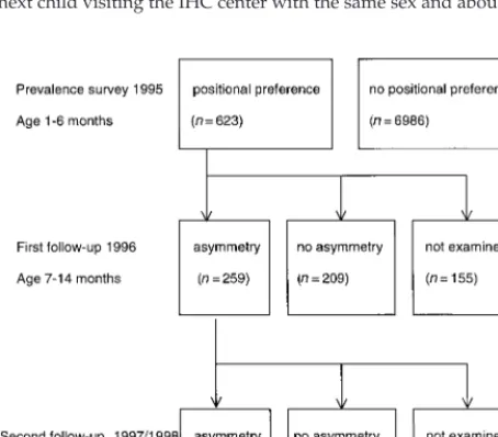
On their regular visit to the IHC center, a total of 7609 infants below the age of 6 months were screened for the presence of positional preference in the month of September 1995 (Fig 1). A large number of 167 IHC physicians across The Netherlands took part in this effort. Of every infant with positional preference (cases: n ⫽ 623), anamnestic data and physical signs were re-corded. The same data were also to be registered of an immediate next child visiting the IHC center with the same sex and about the
same age but without positional preference (controls:n⫽554). Within the time fixed, it was not feasible on these conditions to find a matching control child for every case child.
First Follow-Up Study
In March and April 1996, ie, 6 to 8 months after the original survey, it proved to be possible to reexamine approximately three quarters of the 623 children (n⫽468) with positional preference (Fig 1). The IHC physicians were requested to look for persistent signs of a restricted range of motion of the head as well as for signs of deformational plagiocephaly.
Second Follow-Up Study
Between September 1997 and April 1998, ie, 24 to 32 months after the original survey, a second follow-up examination was undertaken (Fig 1). Through this, follow-up data became available for approximately one half of the group of the 259 children (n⫽
129) who still had shown a restricted range of motion and/or signs of deformational plagiocephaly in the first follow-up study.
Definition of Positional Preference
Positional preference was defined as the condition in which the infant, in supine position, shows head rotation to either the right or the left side for approximately three quarters of the time of observation. Active rotation of the head over a range of 180 degrees cannot be accomplished; this is tested by provoking the child to follow a person or an object with its eyes and head. Passive rotation of the head to the nonpreferent side is usually possible but the range of motion is restricted.
Data Registration
In the original survey the following data were collected: 1) baseline characteristics: gender, birth rank (including stillbirths); 2) general characteristics on pregnancy and delivery: single/mul-tiple birth, duration of pregnancy, amount of amniotic fluid (oli-gohydramnion, normal, hydramnion, unknown), delivery mech-anism (vaginal, cesarean section), presentation at delivery (breech, other), use of ventouse or forceps on delivery, complications (frac-ture of the clavicle, shoulder dystocia); and 3) specific character-istics on nursing habits: sleeping position in the first week of life, after the first week, and in the days preceding the examination (supine, prone, or side); the position of the principal fosterer when taking care of the child (always from same side, alternate posi-tions, infant’s feet pointing toward the fosterer), the method of feeding (breast or bottle), right-/left-handedness of the principal fosterer when offering the bottlefeeding.
By means of physical examination the following data were collected by the IHC physician: 1) spontaneous movements, the active range of motion when following a person or object with the eyes and the head, passive range of motion of the head; 2) defor-mational plagiocephaly: visual assessment of the symmetry of the occipital skull and forehead and of the position of the ears; and 3) presence of a muscular torticollis (a shortened sternomastoid mus-cle, palpable and visible as a tight rope), scoliosis, limited abduc-tion of the hips or abnormalities of the feet (club feet, pes adduc-tus).
It was also recorded whether any handling suggestions were communicated to the parents with regard to the side and/or position in which the infant should be nursed. Any additional diagnostic evaluation and/or treatment that had taken place was registered as well. In the first and second follow-up study, data were collected on the spontaneous movements of the infant, the active and passive range of motion, and the symmetry of the occipital skull and forehead.
Analysis
The observed frequency distributions were compared using the Pearson2analysis. Odds ratios were calculated for the possible risk factors.
RESULTS Prevalence
The prevalence of positional preference in infants below the age of 6 months was 8.2% (n⫽ 623). The
prevalence in children younger than 8 weeks was 10%, between 8 and 16 weeks was 11%, and between 16 and 26 weeks was 3%. The boy:girl ratio was 3:2. In 68% of the 623 children, the preference was to the right side, in 27% to the left side, in 5% the direction was not reported. In 30% of the 623 children, the positional preference was accompanied by a restric-tion of the passive range of morestric-tion of the head.
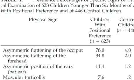
The prevalence of specific physical signs of the condition in children with defined positional prefer-ence compared with control children is presented in Table 1. A muscular torticollis could be held respon-sible for the positional preference in 7.6% of the 623 children. Based on the percentages in cases and con-trols presented in Table 1, the prevalence of flatten-ing of the occiput in the population of 7609 infants can be calculated at 9.9%.
({76%ⴱ623}⫹{4%ⴱ(7609⫺623)}/7609).
Baseline, General, and Specific Characteristics
The supine sleeping position of the child (after the first week of life and in the days before the exami-nation) and a strong preference in offering the feed-ing always from the right or the left side were posi-tively correlated with positional preference (Table 2). Firstborns, premature children, and children with a breech presentation at delivery proved to have a higher risk for positional preference.
Multiple/single pregnancy, amount of amniotic fluid, delivery mechanism, use of ventouse or for-ceps, complications in vaginal delivery, sleeping po-sition of the child in the first week of life, and posi-tion of the fosterer when taking care of the child were not related to the prevalence of positional preference.
Follow-Up
The first follow-up study of 468 children at the age 7 to 14 months who had shown positional preference in the first half year of life demonstrated substantial improvement of the condition. However, 55% of the children (n ⫽ 259) still had signs of asymmetry of the shape and/or the movements of the head: 12% still showed a restricted active range of motion, 8% a restricted passive range of motion, and 47%
asym-metric flattening of the occiput and 23% of the fore-head.
In the second follow-up study at the age of 2 to 3 years of 129 children of the group of 259 children who had shown signs of asymmetry of the move-ments and/or shape of the head in the first follow-up study, 53% (n ⫽ 68) still had signs of asymmetry: active range of motion was restricted in 6%, passive range of motion in 2%, and 45% had an asymmetric flattening of the occiput and 21% of the forehead.
Despite the fact that a considerable number of children were not followed up, there was no proof of any obvious positive or negative selection of cases in the first and second follow-up study. Therefore, the prevalence data of the second follow-up study were used to extrapolate the expected prevalence figures of the 623 cases at the age of 2 to 3 years. In the last column of Table 3 the thus extrapolated prevalences are presented to allow comparison with the preva-lence figures in the original survey and in the first follow-up study. Of all the 623 cases, 3% would still have a restricted active range of motion, 1% a re-stricted passive range of motion, 25% an asymmetric flattening of the occiput, and 12% of the forehead at the age of 2 to 3 years. In the original aselect popu-lation, 2.4% of the 7609 children would still have one or more symptoms of a restricted range of motion of the head and/or deformational plagiocephaly at the age of 2 to 3 years.
({0.53ⴱ259/468}ⴱ{623/7609}⫽2.4%).
Additional Diagnostics and Treatment
As was expected, handling suggestions with re-gard to the position in bed and the side and/or position in which the infant should be taken care of were communicated to 96% of the parents of children with positional preference. In the original survey, it was recorded that 107 of the 623 children (17%) with positional preference underwent some form of addi-tional diagnostic evaluation and/or treatment. Be-tween the original survey and the first follow-up study, another 15% of the children had been referred. For a number of 149 children, who underwent addi-tional diagnostics, data on the nature of a therapeutic intervention became available: 66 children received physical therapy, 3 children were treated with a hel-met, and at least 13 children proved to have devel-opmental dysplasia of the hip and were treated accordingly. As could be expected, the signs of asym-metry were more serious and persistent among those children who received treatment for the condition.
DISCUSSION
The 8.2% prevalence of positional preference in this population-based study of children younger than 6 months of age in The Netherlands proved to be very high. In a previously published report the prevalence of deformational plagiocephaly was esti-mated at .3%.20 The prevalence of flattening of the
occiput (nearly 10%), the most common sign of de-formational plagiocephaly, was far higher than ex-pected. Of all children with signs of plagiocephaly, none proved to have the nondeformational type
(cal-TABLE 1. Prevalence Percentages of Specific Signs on Physi-cal Examination of 623 Children Younger Than Six Months of Age With Positional Preference and of 446 Control Children
Physical Sign Children
With Positional Preference (n⫽623)
Control Children (n⫽446*)
Asymmetric flattening of the occiput 76.0 4.0 Asymmetric flattening of the
forehead
34.8 2.0
Asymmetric position of the ears (bat ear)
11.4 1.2
Muscular torticollis 7.6 .2
Scoliosis 18.4 1.1
Limited abduction of the hip(s) 8.0 1.4
Foot abnormality 2.4 .2
culated prevalence in literature: .003%16). These
find-ings strongly support the opinion that the prevalence of positional preference and deformational plagio-cephaly has risen dramatically in the past decade. The observation that boys are more frequently af-fected than girls and the more common preferential direction to the right side are in line with other published data.4,6,10,21
The higher prevalence of positional preference in firstborn children and after breech delivery is attrib-uted to a higher compression of the fetus toward the end of pregnancy.2 Positional preference was
ob-served more frequently in children who are always bottle-fed with either the right or the left hand; of-fering the feeding (bottle or breast) alternately from the right or left side proved to be a protective factor. In premature children, the lower tonus, a limited possibility to move, and an asymmetrical nursing care are plausible explanations for a more frequent occurrence of positional preference.22,23 The prone
and side sleeping position seemed to have a protec-tive effect for positional preference. In an unselected population in Sweden, 2.4% of the children with a prone sleeping position and 19% of children with a supine sleeping position showed positional prefer-ence.6Supine sleeping was described as a
predispos-ing factor for positional preference by several other authors.4,10,12,15,16,23,24
Although not proven by this observational study, a causal relationship between supine sleeping posi-tion and posiposi-tional preference seems likely. Whether the relationship between preferential side of feeding and positional preference is also of a causal nature is not clear. One may argue that a child with positional preference will typically nurse better on one side and the mother will continue to nurse the infant on this side. A preferential feeding pattern may thus be ei-ther the result of a positional preference or a cause for it.
In this study, the evaluation of the long-term
ef-TABLE 2. Data on Pregnancy, Delivery, Sleeping Position, and Method of Feeding of 623 Chil-dren Younger Than Six Months of Age With Positional Preference and of 554 Control ChilChil-dren, With Odd Ratios
Characteristic Percentage of Children Odds Ratio
(95% CI) With
Positional Preference (n⫽623)
Control Children (n⫽554)
First child 48.1 39.6 1.4 (1.1–1.8)
Second and next child 51.9 60.4
Prematurity 10.7 6.3 1.8 (1.2–2.8)
At term 89.3 93.7
Breech position 8.6 5.2 1.7 (1.1–2.7)
Other position 91.4 94.8
Supine sleeping position after first week 99.8 96.6 20.9 (2.8–157)
Other position .2 3.4
Supine sleeping position in the days before the examination
97.1 93.7 2.3 (1.2–4.1)
Other position 2.9 6.3
Preferential side of feeding 64.2 54.0 1.5 (1.2–2.9)
No preference (including breastfeeding) 35.8 46.0
CI indicates confidence interval.
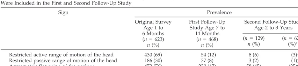
TABLE 3. Prevalence of Asymmetry of the Head in 623 Infants With Positional Preference: at the Age of 1 to 6 Months,17 to 14 Months,1and 2 to 3 Years19; Only the Children With Asymmetry of the Range of Motion of the Head and/or Deformational Plagiocephaly Were Included in the First and Second Follow-Up Study
Sign Prevalence
Original Survey Age 1 to 6 Months (n⫽623)
n(%)
First Follow-Up Study Age 7 to
14 Months (n⫽468)
n(%)
Second Follow-Up Study Age 2 to 3 Years
(n⫽129)
n(%)
(n⫽623) (%)*
Restricted active range of motion of the head 430 (69) 54 (12) 8 (6) (3)†
Restricted passive range of motion of the head 186 (30) 37 (8) 3 (2) (1)‡
Asymmetric flattening of the occiput 473 (76) 220 (47) 58 (45) (25)§
Asymmetric flattening of the forehead 217 (35) 108 (23) 27 (21) (12)㛳
* The prevalences for the whole group of 623 children at the age of 2 to 3 years were calculated through extrapolation to compensate for the children that were normal in the first follow-up study or not examined in both follow-up studies: the percentages of the second follow-up study were first converted into absolute numbers for the total of 259 children; these numbers were then converted into percentages for the total of 468 children in the first follow-up study; the thus calculated percentage also represents the expected prevalence in the group of 623 children of the original survey; this extrapolation is permissable because there were no indications of selection of the population in the first and second follow-up study.
† (6*259/100)/(468*100)⫽.06*259/468⫽3%. ‡ .02*259/468⫽1%.
§ .45*259/468⫽25%.
fects of positional preference in infancy was re-stricted to the range of motion of the head and the symmetry of the skull. At the age of 2 to 3 years, asymmetry of the range of motion and deformational plagiocephaly had not disappeared in nearly one third of the infants with earlier signs of positional preference. Asymmetric flattening of the occiput proved to be the most persistent sign.
A US study showed that children sleeping in the prone position can roll over at an earlier age than those sleeping on the back or the side.25In a Dutch
study sleeping and playing in the prone position were accompanied by a more advanced motor devel-opment in healthy mature-born infants at the age of 5 months.26 It seems, however, that the described
differences in motor development are only tempo-rary and will disappear before the age of 18 months.27 It is not clear to what extent positional
preference plays a role in the transient slower motor development of supine sleeping children.
The high prevalence of the condition in infants, the persistency of accompanying signs, the large number of children referred for additional diagnostic evalu-ation and/or treatment, and the resulting high med-ical expenses strongly call for a primary preventive approach. Influencing the modifiable associated fac-tors through primary preventive campaigns seems to be an appropriate response to the risen prevalence. Some kind of nursing advice with regard to the handling and positioning of the infant was already communicated to almost all parents of children with positional preference. Despite this, a high number of children underwent additional diagnostic and/or therapeutic interventions (32%). This is a rather alarming observation, because these interventions cause much anxiety in parents and lead to high med-ical expenses. Unfortunately, the framework of our observations does not allow any conclusions to be drawn with regard to the preventive effects of han-dling suggestions or the effects of therapeutic inter-ventions. It is clear that only a prospective random-ized trial will be able to provide an answer to this.
In the IHC program in The Netherlands, a preven-tive guideline with accompanying educational mate-rial has been designed and is used now in the coun-seling of parents on positioning, handling, and nursing of the infant from the neonatal period on-wards. Of course, the content of this guideline does not contradict in any way with the rightly justified recommendations on the sleeping position of infants in the Dutch equivalent of the “Back to Sleep” cam-paign. To determine whether such a primary preven-tive approach is beneficial, it will be necessary to evaluate the effects of the use of the guideline on the prevalence of positional preference in infants in the years to come.
REFERENCES
1. Boere-Boonekamp MM, Linden-Kuiper AT van der, Es P van. Prefer-ential posture in infants; a serious call on health care. Ned Tijdschr Geneeskd. 1997;141:769 –772
2. Dunn PM. Congenital postural deformities.Br Med Bull. 1976;32:71 3. Nelson WE, Behrman RE, Kliegman RM, Arvin AM.Nelson Textbook of
Pediatrics. Philadelphia, PA: WB Saunders Company; 1996
4. Mau H, Gabe I.Die sogenannte Sau¨glingsskoliose und Ihre Krankengymnas-tische Behandlung. New York, NY: Georg Thieme Verlag Stuttgart; 1981 5. Good C, Walker G. The hip in the moulded baby syndrome.J Bone Joint
Surg. 1984;66-B:491– 492
6. Palme`n K. Prevention of congenital dislocation of the hip: the Swedish experience of neonatal treatment of hip joint instability.Acta Orthop Scand. 1984;55(suppl 208):58 – 67
7. Fulford GE, Brown JK. Position as a cause of deformity in children with cerebral palsy.Dev Med Child Neurol. 1976;18:305–314
8. Hamanishi C, Tanaka S. Turned head-adducted hip—truncal curvature syndrome.Dis Child. 1994;30:515–519
9. Biedermann H. Kinematic imbalances due to suboccipital strain in newborns.J Manual Med. 1992;6:151–156
10. Kane AA, Mitchell LE, Craven KP, Marsh JL. Observations on a recent increase in plagiocephaly without synostosis. Pediatrics. 1996;97: 877– 885
11. Hunt CE, Puczynski MS. Does supine sleeping cause asymmetric heads?Pediatrics. 1996;97:127–129
12. Moss SD. Nonsurgical, nonorthotic treatment of occipital plagio-cephaly: what is the natural history of the misshapen neonatal head? J Neurosurg. 1997;87:667– 670
13. Pollack IF, Losken HW, Fasick P. Diagnosis and management of poste-rior plagiocephaly.Pediatrics. 1997;99:180 –185
14. OBroin ES, Allcutt D, Earley MJ. Posterior plagiocephaly: proactive conservative management.Br J Plast Surg. 1999;52:18 –23
15. Littlefield TR, Beals SP, Manwaring KH, et al. Treatment of craniofacial asymmetry with dynamic orthotic cranioplasty.J Craniofac Surg. 1998; 9:11–17
16. Rekate HL. Occipital plagiocephaly: a critical review of the literature. J Neurosurg. 1998;89:24 –30
17. American Academy of Pediatrics, Task Force on Infant Positioning, SIDS Positioning, and SIDS.Pediatrics. 1992;89:1120 –1126
18. Jonge GA de, Engelberts AC, Koomen-Liefting AJM, Kostense PJ. Cot death and prone sleeping position in the Netherlands.Br Med J. 1989; 298:722
19. Boere-Boonekamp MM, Linden-Kuiper AT van der, Bunge-van Lent FCGM. No asymmetry of head rotation and head shape in three-quarters of the children aged 2–3 years who in infancy had shown preferential posture.Ned Tijdschr Geneeskd. 1999;143:569 –571 20. Dunn PM. Congenital sternomastoid torticollis: an intrauterine postural
deformity.Arch Dis Child. 1974;49:824 – 825
21. Brunetau RJ, Milliken RJ. Frontal plagiocephaly: synostotic, compensa-tional, or deformational.Plast Reconstr Surg. 1992;89:21–31
22. Vles JSH, Oostenbrugge R van, Kingma H, Caberg H, Casaer P. Head position in low-risk premature infants.Biol Neonate. 1988;54:307–313 23. Littlefield TR, Kelly KM, Pomatto JK, Beals SP. Multiple-birth infants at
higher risk for development of deformational plagiocephaly.Pediatrics. 1999;103:565–569
24. Hansen M, Mulliken JB. Frontal plagiocephaly.Craniofac Surg. 1994;21: 543–553
25. Jantz JW, Blosser CD, Fruechting LA. A motor milestone change noted with a change in sleep position. Arch Pediatr Adolesc Med. 1997;151: 565–568
26. Visscher F, Graaf T van der, Spaans M, Lingen RA van, Fetter WPF. Prone position favorable for motor development of infants.Ned Tijdschr Geneeskd. 1998;142:2201–2205
DOI: 10.1542/peds.107.2.339
2001;107;339
Pediatrics
Magda M. Boere-Boonekamp and Lida T. van der Linden-Kuiper
Positional Preference: Prevalence in Infants and Follow-Up After Two Years
Services
Updated Information &
http://pediatrics.aappublications.org/content/107/2/339 including high resolution figures, can be found at:
References
http://pediatrics.aappublications.org/content/107/2/339#BIBL This article cites 24 articles, 6 of which you can access for free at:
Subspecialty Collections
http://www.aappublications.org/cgi/collection/nephrology_sub Nephrology
following collection(s):
This article, along with others on similar topics, appears in the
Permissions & Licensing
http://www.aappublications.org/site/misc/Permissions.xhtml in its entirety can be found online at:
Information about reproducing this article in parts (figures, tables) or
Reprints
DOI: 10.1542/peds.107.2.339
2001;107;339
Pediatrics
Magda M. Boere-Boonekamp and Lida T. van der Linden-Kuiper
Positional Preference: Prevalence in Infants and Follow-Up After Two Years
http://pediatrics.aappublications.org/content/107/2/339
located on the World Wide Web at:
The online version of this article, along with updated information and services, is
by the American Academy of Pediatrics. All rights reserved. Print ISSN: 1073-0397.