R E S E A R C H
Open Access
Pen-type laser fluorescence device versus
bitewing radiographs for caries detection
on approximal surfaces
M. Bizhang
1*, N. Wollenweber
1, P. Singh-Hüsgen
2, G. Danesh
3and S. Zimmer
1Abstract
Background:The accurate detection of approximal caries is generally difficult. The aim of this study was to assess the ability of the pen-type laser fluorescence device (LF pen) to detect approximal carious lesions in comparison to bitewing radiographs (BW).
Methods:Three hundred forty-one tooth surfaces were diagnosed in 20 patients with an average age of 26.70 (±2.82) years. Each test tooth was sequentially assessed by a single calibrated examiner using visual inspection, BW, and the LF pen. Radiographs were used as the gold standard to calculate an appropriate cut-off.
Results:Sensitivity, specificity and accuracy values for cut-off limits of 15, measured by the LF pen were compared using the chi2test (McNemar test). For approximal caries at D3 level, the highest values of specificity and sensitivity were observed for the LF pen at a cut-off value of 15 (96.8 and 83.0 %) and for visual inspection (99.3 and 4.3 %).
Conclusion:Within the limitations of this study, dentin caries on approximal surfaces could be detected equally well by the LF pen as by the bitewing radiographs. Therefore, the LF pen can be recommended as an alternative to radiographs for the detection of approximal caries in a regular dental practice setting.
Trial registration:DRKS00004817 on DRKS on 12thMarch 2013.
Keywords:Detection, Caries, Approximal, Pen-type laser fluorescence device, Sensitivity, Specificity, Radiography, Dentin caries
Background
Caries detection on approximal surfaces is difficult. On one hand, visual examination for the detection of approximal caries lesions shows low sensitivity, and on the other, bitewing radiographs have been found to underestimate the lesion depth and are unable to reveal demineralisation in dentin [1]. Thus, in addition to the greatest drawback of exposure to radiation and the subsequent associated health hazards [2], radiographs do not show the correct size of the carious lesion [3]. Both the above-mentioned subjective diagnostic methods are of low sensitivity but high specificity for the detection of approximal caries [1]. As the deter-mination of lesion depth possess a challenge in
restorative treatment planning for most clinicians in daily clinical practice, radiography along with clinical findings is used as a routine diagnostic approach for approximal caries. Radiography of approximal caries increases the sensitivity of visual inspection and is, therefore, presently considered the gold standard for the detection of caries on approximal surfaces [4]. As mentioned above, the main disadvantage of BW is the exposition of the patients to ionizing radiation and the fact that it is technique sensitive [5]. Therefore, an alternative method to radiography, but with the same degree of diagnostic accuracy is desirable.
The pen-type laser fluorescence device (LF pen) (KaVo, Biberach/Riß Germany) has been developed to detect caries using the mechanism of fluorescence. The device produces a small laser with an excitation wave-length of 655 nm in the form of red light which mea-sures the degree of bacterial activity. The reflection of * Correspondence:[email protected]
1Department of Operative and Preventive Dentistry, University Witten/ Herdecke, Alfred-Herrhausen-Str. 50, 58448 Witten, Germany Full list of author information is available at the end of the article
light depends on the induction of fluorescence from bacterial porphyrins [6, 7]. A literature review carried out for the present study, revealed only two studies assessing the LF pen (one in vivo and one in vitro) for permanent teeth [4, 8]. The in vivo study found a fair positive correlation between laser fluorescence values and the radiographic scoring. Opened lesions analysed with their clinical lesion depths as gold standard, showed that there was a fair positive correlation to the laser fluorescence values and a moderately strong cor-relation to the radiographic scoring [4]. The in vitro study was able to demonstrate that the D3 threshold (dentin) ranged between 0.81 and 0.92 and that bite-wing radiography showed an inferior performance com-pared to the LF pen [8]. Another study was able to establish the excellent intra/inter- examiner reproduci-bility for the LF pen on occlusal sites [9].
One study for detecting caries in primary teeth with the LF pen showed that simultaneously combined visual inspection with the LF pen and radiography increased sensitivity but decreased the specificity. The authors concluded that adjunct radiographic and LF pen methods offer no benefits for the detection of caries in primary teeth [10]. Another in vivo study examined the performance of the LF pen in comparison to conven-tional methods in detecting approximal caries lesion in primary teeth. This study found that the sensitivity for white spots was 0.20–0.21 for visual inspection, 0.16– 0.23 for radiography and 0.16 for the LF pen and the specificity 0.95 for visual inspection, 0.99–1.00 for radiography and 0.94–0.96 for the LF pen [11]. The sensitivity for cavitation was 0.30 for visual inspection, 0.55–0.65 for the LF pen and 0.65–0.70 for radiog-raphy. The specificities for all methods were around 0.99 [11]. A further study showed that radiography and the LF pen achieved a similar performance in the detection of approximal caries lesion in primary teeth, however, the discomfort caused by visual inspection and the LF pen could influence the performance of these methods, since a higher number of false-positive or false-negative results occurred in children who reported discomfort [12]. On the other hand, an in vitro study found that a laser fluorescence device (LF), the LF pen and con-ventional methods perform similarly in detecting occlusal caries lesions in primary teeth. Thus, the study concluded that it is sufficient to employ visual inspection alone in clin-ical practice [13]. Visual inspection has been found to cause less discomfort than the other methods. Radiography and the LF pen presented similar levels of discomfort. Concur-rently, older children reported high levels of discomfort with temporary separation, while younger children reported little discomfort with the LF pen [12].
The LF and LF pen have been demonstrated to achieve acceptable levels of performance in the detection of
occlusal caries lesion in primary and permanent teeth [14–18]. Therefore, both can be considered to be suit-able devices for occlusal caries detection [14, 19–22].
In vitro and in vivo studies observed no differences between the LF pen and BW performance in detecting approximal caries in primary teeth [11, 23, 24]. In per-manent teeth, the LF pen showed better performance compared to radiography [8]. It necessary to undertake further in vivo investigations as results in the present literature show some disagreements.
Methods
The aim of this study was to evaluate the LF pen for the detection of approximal caries lesion in permanent teeth in comparison to dental radiographs as the gold stand-ard, for daily use in a regular dental practice setting.
Ethical approval for the study was obtained from the Ethical Board at the University of Duesseldorf (No. 3081), Duesseldorf, Germany. The first 20 patients who showed an interest, fulfilled the inclusion and exclusion criteria, and agreed to participate were asked to sign the consent form and were enrolled in the study. 8 males and 12 females participated in this study. In order to achieve an adequate power of 80 % and a de-fined significance level of 5 % (p< 0.05), the appropriate sample size was determined to be 20. Participants be-tween 18 and 65 years of age, having at least 20 natural teeth with no current visual approximal caries lesion, periodontal disease, or other oral pathology were included in the study. Exclusion criteria were the presence of any systemic disease, pregnancy or breastfeeding, the use of fixed or removable orthodontic appliances, smoking and alcohol abuse.
Prior to commencement of the study both dentists carried out measurements on five subjects, not included in the study, to calculate the intra-examiner bility. The intra-examiner and inter-examiner reproduci-bility levels ranged from 0.75 to 0.89 (good inter- and intra-examiner reproducibility levels).
evaluation of the carious lesions was carried out by taking BW.
One week after screening, the subjects received den-tal prophylaxis. The teeth were scaled using a Sonic Flex-Airscaler (KaVo Company, Biberach, Germany) and polished with a rotating soft latch-type Pro-Cup and Cleanic Prophy Paste (Kerr company, Washington D.C, USA). After polishing, the approximal surfaces were cleaned with dental floss (WaxedFloss, Johnson & Johnson Company New Brunswick, USA). Following this, the selected tooth was dried for at least 5 s with compressed air and examined under a standard operat-ing light. Presence or absence of carious lesions was again recorded by visual examination using modified Ekstrand’s criteria and the LF Pen.
Modified Ekstrand’s criteria visual criteria [25]
0. No change in enamel translucency after air drying (> 5 s)
1. Opacity (white or brown) distinctly visible on the wet surface
2. Cavitation in dentine
The LF pen method was carried out using a probe tip for approximal surfaces (KaVo, Biberach, Germany). Prior to the examination, the LF pen was calibrated against a porcelain reference object and on the sound smooth surface of every tooth. After drying the tooth for 5 s with compressed air, the approximal area was measured by moving the tip under the tooth contact area, from the buccal to lingual/palatal side. The peak value was recorded [8]. Evaluation with the LF Pen was repeated three times. The maximum value for each measurement was registered and the mean value of the three readings was noted. LF pen values higher than 16 were used as a cut-off point to indicate the presence of a dentinal carious lesion [11].
Radiographs were used as the gold standard to calcu-late an appropriate cut-off. Bitewing projection geom-etry was employed to standardise the procedure for the digital images. Two BW were taken from each side for each subject. A Heliodent DS intra-oral x-ray unit with Sidexis intraoral sensors, aligned perpendicularly in a Rinn sensor holder (Sirona Company, Bensheim, Germany) at 60 kVp and 7 mA was used. The 5.6 × 36 mm XIOS sensor (APS-CMOS-Sensor, Sirona Com-pany, Bensheim, Germany) was exposed for 0.12 s. The digital images were examined by the two dentists on a 18-in. CRT monitor (Multisync LDC 1990 SXI, NEC Corporation, Tokio, Japan) with the Sidexis software (version 1.61, Sirona Company, Bensheim, German) at X 2 magnification.
The following criteria were used for the radiographic examination [26]:
0. No radiolucency visible
1. Radiolucency visible in the outer half of the enamel 2. Radiolucency visible into the inner half of the
enamel
3. Radiolucency visible in the outer half of the dentin 4. Radiolucency visible into the inner half of the
dentin.
The investigator (NW) and another experienced den-tist rated the BW under the same conditions. Presence or absence of dental caries on the BW was recorded. Decisions on the caries status and treatment of the cavity were made by both dentists together. In case of disagreement, the radiograph was discussed until a consensus was reached. After the final decision, the data of the subjects was either just recorded or re-corded and subsequently the cavities treated. Radio-graphic codes of 3 and 4 were treated. Caries removal was carried out in the established cavities, thus enab-ling the clinical determination of caries level.
Statistical analysis
All statistical analyses were performed with SPSS, ver-sion 19.0 for Windows (IBM/SPSS Inc. Chicago/IL, USA). Recordings of the visual examination and LF pen results were correlated with the gold standard BW to calculate the sensitivity, specificity and accuracy of the caries diagnostic techniques for approximal dentin car-ies. For the visual examination, the cut-off point was 1 for a sound surface and initial caries, and 2 for an established cavity. For the bitewing radiographs, scores 0 (healthy), 1 and 2 (enamel lesion) were registered as sound or initial carious lesions, and scores 3 and 4 (dentinal lesion) represented a cavity. ROC (receiver operating characteristic) analyses were also performed to determine a cut-off value. The Spearman rank cor-relation was determined so as to compare the caries level with radiology. Sensitivity, specificity and accuracy values for cut-off limits of 16 and 15, measured by the LF pen, were compared using the chi2test (McNemar test). The tests were performed withα= 0.05 to analyse significant differences among the groups.
Results
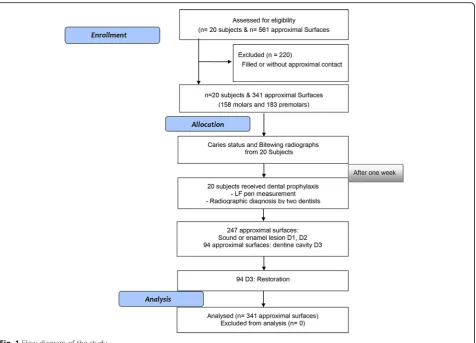
The patient drop-out-rate was 0 %. As the LF pen had failed to function in certain instances, 18 surfaces (5 %) were excluded from the analysis. The reason for this be-ing, that on the examination day no functioning probe was available to replace the defective one. BW revealed enamel caries on 247 surfaces and 94 surfaces with car-ies extending into dentine (Fig. 1).
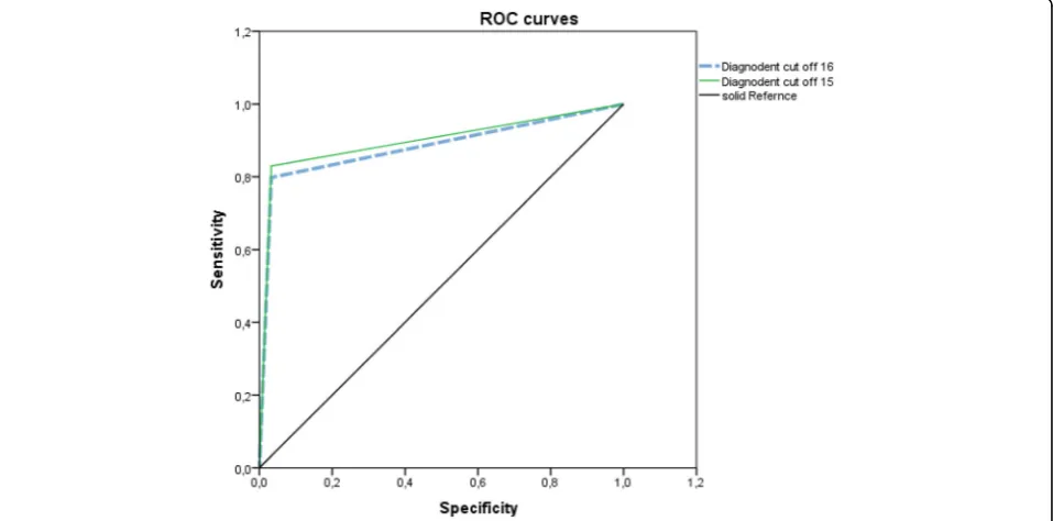
the two different cut-off limits at dentine caries levels on a graph. The optimal cut-off limits for the LF pen was 15 for dentine caries in comparison to the manufac-turer’s cut-off limits (16) (Table 1).
It was observed that the value of sensitivity was low for visual methods and high for the LF pen scores. Caries at the D3 level (dentinal lesion) showed values of specificity and sensitivity to be slightly higher for the LF pen at a cut-off value of 15 (Fig. 2). The Spearman rank correlation for approximal caries with radiography was 0.82 (LF pen 15), 0.79 (LF pen 16) and 0.12 (visual). The ROC (receiver operating characteristic) was significantly greater with LF Pen 15 and 16 compared to visual (Fig. 2).
Discussion
All studies assessing caries radiographically show a marked increase in cavitation when the radiolucency
reaches the outer half of dentine. In vitro studies have shown that the cavitation rate of inner enamel and outer dentine increases from 11 to 66 % and 65 to 100 % when the radiolucency has reached the outer half of dentine [27–29]. The use of BW as the gold standard in this study conformed with other studies having similar study designs [4, 8]. Furthermore, as several studies have shown LF to have a good reproducibility [8, 29, 30], it was not considered necessary to reconfirm the reprodu-cibility of the LF pen. The examiner was found to have a good reproducibility. Visual inspection after tooth separ-ation was not evaluated, due to non-acceptance on the subjects part, for reasons of discomfort [31] and time (two appointments). Moreover, in the daily dental prac-tice setting, the decision to treat approximal caries is made without separation of the teeth. Thus, not separat-ing the teeth prior to measurement with the LF pen also Fig. 1Flow diagram of the study
Table 1Area under the ROC curves for LF pen for detection of approximal surfaces for cut-off limits 16 and 15
Method Area Standard error p Confidence interval (95 % CI)
LF pen with cut off 16 0.883 0.026 0.002 0.833 0.933
LF pen with cut off 15 0.899 0.024 0.001 0.852 0.945
provided us with the knowledge of how functional the device is in a daily dental practice setting. Two authors have claimed that dental separation cannot be used as a validation method for permanent teeth because of low reliability [32, 33]. The study by Hintze et al. in 1998 showed, that two thirds of the surfaces under study were assessed as sound. After tooth separation, only a few (0.5–2.6 %) of these surfaces were found to be cavitated, thus showing a lack of reproducibility. The authors sug-gested that the tooth separation method cannot be used as a gold standard for validation of other diagnostic methods [33]. Ekstrand’s criteria for caries diagnosis is the most commonly used method in daily clinical prac-tice, which most dentists agree upon to decide on the treatment need of a carious lesion [34]. In general, the studies that used a ranked diagnostic system such as that proposed by Ekstrand et al., which is also the basis of the ICDAS II criteria, had higher values for diagnostic parameters [35]. Similar to other study, we used the Ekstrand’s criteria for the present study [36].
A recent systematic review and meta-analysis with 75 studies, demonstrated that the fluorescence-based method for caries diagnosis tends to have similar accuracy for all types of teeth, dental surfaces or settings [37].
To evaluate the accuracy of a diagnostic method it is necessary to determine the validation method that ex-presses the true state of the disease. Sensitivity and spe-cificity have been used in several in vitro and in vivo studies for the evaluation of the effectiveness of differ-ent caries diagnostic methods [19, 38–42]. Cut-off
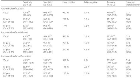
points for evaluation with the LF pen in primary teeth [11] and in permanent teeth [8] were obtained from an earlier study. Accordingly, readings higher than 5 were considered to be non-cavitated lesions, and measure-ments higher than 16, were considered to be cavitated. The results of this study revealed that the cut-off value of 15 had a slightly better validity than that of 16. In addition, the present study showed that the detection accuracy for approximal dentine caries was similar be-tween the bitewing radiographs and clinical caries exca-vation of the same teeth (Table 2). This result was congruent with the result of other studies [4, 43].
lesions of 72.4 %. Mendes et al. speculated that the com-bined strategies could perform better in permanent teeth with a higher prevalence of non-evident caries lesions [10]. Nevertheless, most of the studies do not recom-mend the use of the LF or LF pen for permanent or pri-mary teeth as the gold standard or sole diagnostic tool for occlusal caries detection [4, 39, 44, 45] or approxi-mal caries detection [4] diagnostic tool to be used in combination with visual diagnostic techniques and/or radiography to assist the clinician when making opera-tive decisions [46].
The first limitation of this study was that tooth separ-ation was not carried out before visual examinsepar-ation in order to evaluate the approximal surface and thus to make a decision regarding preventive or operative treat-ment. The visual diagnosis was for the approximal area used in our study was not perfect, due to difficulty in viewing non-cavitated enamel lesions in this area [47], however it is functional method and provided the best alternative. In the present study, radiolucency in dentine was used as an indication for operative treatment of the lesion. This method is also the method of choice in daily clinical practice for detecting carious lesions and as an indication for treatment with a filling [48].
Use of only one examiner for the study, could be seen as another limiting factor of this study. This examiner was, however, calibrated with another experienced den-tist prior to the commencement of the study on five
subjects, who later did not participate in the study. The intra- and interexaminer reproducibility was found to be good.
Conclusion
The results of this study show that the pen-type laser fluorescence device can be used for the detection of approximal carious lesions in permanent teeth similar to dental radiographs as the gold standard in a daily clinical practice setting.
Within the limitations of this study, the newly formu-lated cut-off limit of 15 instead of the cut-off limit of 16 for the pen-type laser fluorescence device provided a slightly higher validity, higher values of sensitivity and specificity, as compared to the gold standard BW. The high sensitivity and specificity of the LF pen make it suitable for the detection of approximal caries, in sub-jects with a similar caries experience, instead of radio-graphs for the clinician in a dental practice. However, further studies in larger sample size, with different caries experience, are needed to extend the standard usage of the LF pen for detection approximal caries.
Abbreviations
BW:Bitewing radiographs; D3 level: Dentin lesion; DMFS: The decayed, missing, filled surfaces; LF pen: Pen-type laser fluorescence device; LF: Laser fluorescence device
Acknowledgements
The authors are grateful to the 20 subjects who participated in this study.
Table 2Sensitivity–specificity of visual and LF pen of approximal surfaces as compared with bitewing radiographs
Sensitivity (95 % CI)
Specificity (95 % CI)
False positive False negative Accuracy (95 % CI)
Spearman (standard error)
Approximal surfaces (all)
Visual 4.3 %a,b
(1.0–8.6)
99.3 %a,b (98.3–100)
95.7 % 0.7 % 74.9 %a,b
(70.3–79.5)
0.15 (0.06)
LF pen (Cut-off 16)
79.8 %a (71.4–88,2)
96.8 %a (94.6–99,0)
20.2 % 3.2 % 92.1 %a
(89.2–95.0)
0.80 (0.04)
LF pen (Cut-off 15)
83 %b (75.2–90.9)
96.8 %b (94.6–99.0)
17 % 3.2 % 93.0 %b
(90.2–95.8)
0.82 (0.04)
Approximal surfaces (Molars)
Visual 4.3 %a,b
(2.0–10.6)
98.4 %a,b (96.1–100)
95.7 % 1.6 % 73.3 %a,b
(66.4–80.2)
0.13 (0.09)
LF pen (Cut-off 16)
73.9 %a (60.3.87.5)
95.5 %a (91.5–99.5)
26.1 % 4.5 % 89.2 %a
(84.1–94.3)
0.73 (0.06)
LF pen (Cut-off 15)
78.3 %b (61.9–88.7)
95.5 %b (91.5–99.5)
21.7 % 4.5 % 90.5 %b
(85.7–95.3)
0.75 (0.06)
Approximal surfaces (Premolars)
Visual 4.3 %a,b
(1.56–10.16)
100 %a,b (100–100)
95.7 % 0 % 76.5 %a,b
(70.4–82.6)
0.18 (0.06)
LF pen (Cut-off 16)
85.4 %a (75.4–95.4)
97.8 %a (95.3–100)
14.6 % 2.2 % 94.5 %a
(91.2–97.8)
0.86 (0.04)
LF pen (Cut-off 15)
87.5 %b (78.1–96.9)
97.8 %b (95.3–100)
12.5 % 2.2 % 95.1 %b
(92.0–98.2)
0.87 (0.04)
Funding
The University of Duesseldorf provided the funding for materials, etc. The authors were involved in the study design, the data collection and analysis, the decision to publish, or the preparation of the manuscript.
Availability of data and materials
The full trial protocol can be accessed by the ethical committee of University of Duesseldorf (No. 3081). The datasets are included as Excel data.
Authors’contributions
MB planned the study design, conducted the analysis and interpretation of the data and drafted the manuscript. NW carried out the study and performed the statistical analysis of the data. PS participated in drafting the paper. GD contributed to critical revision of the manuscript. SZ contributed in drafting the paper. All authors read and approved the final manuscript.
Competing interests
There are no non-financial competing interests (of a political, personal, reli-gious, ideological, academic, intellectual, commercial or other nature) to declare in relation to this manuscript.
Consent for publication
The 20 subjects, who were interested and agreed to participate, were asked to sign the consent form and enrolled in the study.
Ethics approval and consent to participate
The study was approved by the ethical committee of University of Duesseldorf (No. 3081), University of Duesseldorf, Germany, where the full trial protocol can be accessed. The Study was carried out in accordance with the Declaration of Helsinki. The study has the following German Clinical Trials Register number: DRKS00004817 on DRKS on 12thMarch 2013.
Author details
1Department of Operative and Preventive Dentistry, University Witten/ Herdecke, Alfred-Herrhausen-Str. 50, 58448 Witten, Germany.2Department of Operative and Preventive Dentistry and Periodontics, Heinrich-Hein University Duesseldorf, Moorenstr. 5, 40225 Duesseldorf, Germany. 3Department of Orthodontics, University Witten/Herdecke, Alfred-Herrhausen-Str. 50, 58448 Witten, Germany.
Received: 5 September 2016 Accepted: 26 October 2016
References
1. Bader JD, Shugars DA, Bonito AJ. A systematic review of the performance of methods for identifying carious lesions. J Public Health Dent. 2002;62(4): 201–13.
2. Smith NJ. Risk assessment: the philosophy underlying radiation protection. Int Dent J. 1987;37(1):43–51.
3. Gwinnett AJ. A comparison of proximal carious lesions as seen by clinical radiography, contact microradiography, and light microscopy. J Am Dent Assoc. 1971;83(5):1078–80.
4. Huth KC, Lussi A, Gygax M, Thum M, Crispin A, Paschos E, Hickel R, Neuhaus KW. In vivo performance of a laser fluorescence device for the approximal detection of caries in permanent molars. J Dent. 2010;38(12):1019–26. 5. Wenzel A. Bitewing and digital bitewing radiography for detection of caries
lesions. J Dent Res. 2004;83(Spec No C):C72–5.
6. Banerjee A, Gilmour A, Kidd E, Watson T. Relationship between S. mutans and the autofluorescence of carious dentin. Am J Dent. 2004;17(4):233–6. 7. König K, Flemming G, Hibst R. Laser-induced autofluorescence spectroscopy
of dental caries. Cell Mol Biol (Noisy-le-Grand). 1998;44(8):1293–300. 8. Lussi A, Hack A, Hug I, Heckenberger H, Megert B, Stich H. Detection of
approximal caries with a new laser fluorescence device. Caries Res. 2006; 40(2):97–103.
9. Kuhnisch J, Bucher K, Hickel R. The intra/inter-examiner reproducibility of the new DIAGNOdent Pen on occlusal sites. J Dent. 2007;35(6):509–12. 10. Mendes FM, Novaes TF, Matos R, Bittar DG, Piovesan C, Gimenez T,
Imparato JC, Raggio DP, Braga MM. Radiographic and laser fluorescence methods have no benefits for detecting caries in primary teeth. Caries Res. 2012;46(6):536–43.
11. Novaes TF, Matos R, Braga MM, Imparato JC, Raggio DP, Mendes FM. Performance of a pen-type laser fluorescence device and conventional methods in detecting approximal caries lesions in primary teeth–in vivo study. Caries Res. 2009;43(1):36–42.
12. Novaes TF, Matos R, Raggio DP, Braga MM, Mendes FM. Children’s discomfort in assessments using different methods for approximal caries detection. Braz Oral Res. 2012;26(2):93–9.
13. Novaes TF, Matos R, Gimenez T, Braga MM, DE Benedetto MS, Mendes FM. Performance of fluorescence-based and conventional methods of occlusal caries detection in primary molars - an in vitro study. Int J Paediatr Dent. 2012;22(6):459–66.
14. Shi XQ, Welander U, Angmar-Mansson B. Occlusal caries detection with KaVo DIAGNOdent and radiography: an in vitro comparison. Caries Res. 2000;34(2):151–8.
15. Neuhaus KW, Rodrigues JA, Hug I, Stich H, Lussi A. Performance of laser fluorescence devices, visual and radiographic examination for the detection of occlusal caries in primary molars. Clin Oral Investig. 2010;15 (5):635-41. 16. Matos R, Novaes TF, Braga MM, Siqueira WL, Duarte DA, Mendes FM. Clinical
performance of two fluorescence-based methods in detecting occlusal caries lesions in primary teeth. Caries Res. 2011;45(3):294–302. 17. Sinanoglu A, Ozturk E, Ozel E. Diagnosis of occlusal caries using laser
fluorescence versus conventional methods in permanent posterior teeth: a clinical study. Photomed Laser Surg. 2014;32(3):130–7.
18. Huth KC, Neuhaus KW, Gygax M, Bucher K, Crispin A, Paschos E, Hickel R, Lussi A. Clinical performance of a new laser fluorescence device for detection of occlusal caries lesions in permanent molars. J Dent. 2008; 36(12):1033–40.
19. Lussi A, Megert B, Longbottom C, Reich E, Francescut P. Clinical performance of a laser fluorescence device for detection of occlusal caries lesions. Eur J Oral Sci. 2001;109(1):14–9.
20. Shi XQ, Tranaeus S, Angmar-Mansson B. Validation of DIAGNOdent for quantification of smooth-surface caries: an in vitro study. Acta Odontol Scand. 2001;59(2):74–8.
21. Mendes FM, Nicolau J. Utilization of laser fluorescence to monitor caries lesions development in primary teeth. J Dent Child (Chic). 2004;71(2):139–42. 22. Mendes FM, Hissadomi M, Imparato JC. Effects of drying time and the
presence of plaque on the in vitro performance of laser fluorescence in occlusal caries of primary teeth. Caries Res. 2004;38(2):104–8. 23. Chawla N, Messer LB, Adams GG, Manton DJ. An in vitro comparison of
detection methods for approximal carious lesions in primary molars. Caries Res. 2012;46(2):161–9.
24. Braga MM, Morais CC, Nakama RC, Leamari VM, Siqueira WL, Mendes FM. In vitro performance of methods of approximal caries detection in primary molars. Oral Surg Oral Med Oral Pathol Oral Radiol Endod. 2009;108(4):e35–41. 25. Ekstrand KR, Ricketts DN, Kidd EA. Reproducibility and accuracy of three
methods for assessment of demineralization depth of the occlusal surface: an in vitro examination. Caries Res. 1997;31(3):224–31.
26. Hintze H, Wenzel A, Danielsen B. Behaviour of approximal carious lesions assessed by clinical examination after tooth separation and radiography: a 2. 5-year longitudinal study in young adults. Caries Res. 1999;33:415–22. 27. Mejare I, Grondahl HG, Carlstedt K, Grever AC, Ottosson E. Accuracy at
radiography and probing for the diagnosis of proximal caries. Scand J Dent Res. 1985;93(2):178–84.
28. Waggoner WF, Ashton JJ. Predictability of cavitation based upon radiographic appearance: comparison of two film types. Quintessence Int. 1989;20(1):55–60.
29. Pinelli C, Loffredo Lde C, Serra MC. Effect of drying on the reproducibility of DIAGNOdent to detect caries-like lesions. Braz Dent J. 2010;21(5):405–10. 30. Aljehani A, Yang L, Shi XQ. In vitro quantification of smooth surface caries
with DIAGNOdent and the DIAGNOdent pen. Acta Odontol Scand. 2007; 65(1):60–3.
31. Rimmer PA, Pitts NB. Temporary elective tooth separation as a diagnostic aid in general dental practice. Br Dent J. 1990;169(3–4):87–92.
32. De Araujo FB, Rosito DB, Toigo E, dos Santos CK. Diagnosis of approximal caries: radiographic versus clinical examination using tooth separation. Am J Dent. 1992;5(5):245–8.
34. Braga MM, Mendes FM, Ekstrand KR. Detection activity assessment and diagnosis of dental caries lesions. Dent Clin North Am. 2010;54(3):479–93. 35. Dunkley S, Ashley P. Use of a ranked scoring system to detect occlusal
caries in primary molars. Int J Paediatr Dent. 2007;17(4):267–73. 36. Mendes FM, Siqueira WL, Mazzitelli JF, Pinheiro SL, Bengtson AL.
Performance of DIAGNOdent for detection and quantification of smooth-surface caries in primary teeth. J Dent. 2005;33(1):79–84.
37. Gimenez T, Braga MM, Raggio DP, Deery C, Ricketts DN, Mendes FM. Fluorescence-based methods for detecting caries lesions: systematic review, meta-analysis and sources of heterogeneity. PLoS One. 2013;8(4):e60421. 38. Alwas-Danowska HM, Plasschaert AJ, Suliborski S, Verdonschot EH. Reliability
and validity issues of laser fluorescence measurements in occlusal caries diagnosis. J Dent. 2002;30(4):129–34.
39. Bamzahim M, Shi XQ, Angmar-Mansson B. Occlusal caries detection and quantification by DIAGNOdent and Electronic Caries Monitor: in vitro comparison. Acta Odontol Scand. 2002;60(6):360–4.
40. Akarsu S, Koprulu H. In vivo comparison of the efficacy of DIAGNOdent by visual inspection and radiographic diagnostic techniques in the diagnosis of occlusal caries. J Clin Dent. 2006;17(3):53–8.
41. Reis A, Zach Jr VL, de Lima AC, de Lima Navarro MF, Grande RH. Occlusal caries detection: a comparison of DIAGNOdent and two conventional diagnostic methods. J Clin Dent. 2004;15(3):76–82.
42. Sheehy EC, Brailsford SR, Kidd EA, Beighton D, Zoitopoulos L. Comparison between visual examination and a laser fluorescence system for in vivo diagnosis of occlusal caries. Caries Res. 2001;35(6):421–6.
43. Chen J, Qin M, Ma W, Ge L. A clinical study of a laser fluorescence device for the detection of approximal caries in primary molars. Int J Paediatr Dent. 2012;22(2):132–8.
44. Lussi A, Imwinkelried S, Pitts N, Longbottom C, Reich E. Performance and reproducibility of a laser fluorescence system for detection of occlusal caries in vitro. Caries Res. 1999;33(4):261–6.
45. Costa AM, Yamaguti PM, De Paula LM, Bezerra AC. In vitro study of laser diode 655 nm diagnosis of occlusal caries. ASDC J Dent Child. 2002;69(3): 249–53. 33.
46. Ribeiro AA, Purger F, Rodrigues JA, Oliveira PR, Lussi A, Monteiro AH, Alves HD, Assis JT, Vasconcellos AB. Influence of contact points on the performance of caries detection methods in approximal surfaces of primary molars: an in vivo study. Caries Res. 2015;49(2):99–108.
47. Shoaib L, Deery C, Ricketts DN, Nugent ZJ. Validity and reproducibility of ICDAS II in primary teeth. Caries Res. 2009;43(6):442–8.
48. Kidd EAM, Pitts NB. A reappraisal of the value of the bitewing radiograph in the diagnosis of posterior approximal caries. Br Dent J. 1990;169:195–200.
• We accept pre-submission inquiries
• Our selector tool helps you to find the most relevant journal
• We provide round the clock customer support
• Convenient online submission
• Thorough peer review
• Inclusion in PubMed and all major indexing services
• Maximum visibility for your research
Submit your manuscript at www.biomedcentral.com/submit