662
Development and Validation of HPTLC Method for
Quantitative Estimation of Nuciferine from Lotus leaves
NAFSIN A. PATEL AND ARUNA P. JADHAV*
Department of Quality Assurance, BharatiVidyapeeth’s College of Pharmacy, C.B.D. Belapur, Navi Mumbai-400 614, India Nafsin and Aruna: Detection and Quantification of Nuciferine by HPTLC
Nelumbo nucifera (Nelumbonaceae), commonly known as lotus, is an aquatic perennial plant. A simple, precise and accurate high-performance thin-layer chromatography method for quantitative estimation of an alkaloid, nuciferine from methanol extract of lotus leaf was developed. The method employed high-performance thin-layer chromatography aluminium plate precoated with silica gel 60F254 (10×10 cm) as stationary phase and separation was achieved by using a suitable mobile phase chloroform:ethylacetate:methanol:glacial acetic acid [6.5:1:2.5:0.025 v/v/v/v] and Dragendorff’s reagent was used for detection. Quantitation of the method was achieved by densitometric method at 272 nm using Camag TLC scanner 3. This method showed good separation of nuciferine in the extract with Rf value 0.44±0.02. The regression analysis data indicated good linear relationship for the calibration plots for nuciferine in the range of 100-450 ng/spot and regression coefficient was 0.998. The accuracy and reliability of the proposed method was ascertained by evaluating various validation parameters like linearity, precision, accuracy, robustness, specificity, limit of quantification and limit of detection according to the ICH guideline. Statistical analysis of the data showed that the method is reproducible and selective for the estimation of nuciferine. The developed high-performance thin-layer chromatography method can be used for quantification of nuciferine in different species of lotus and formulations containing nuciferine.
Key words: High performance thin layer chromatography, lotus leaf, nuciferine, quantification, alkaloids
*Address for correspondence
E-mail: [email protected]
Nelumbo nucifera is perennial, rhizomatous, aquatic plant belonging to the family of Nelumbonaceae, which has numerous common names (Indian lotus, Chinese water lily and sacred lotus)[1,2].It is widely grown and consumed over the world, mainly in China,
This is an open access article distributed under the terms of the Creative Commons Attribution-NonCommercial-ShareAlike 3.0 License, which allows others to remix, tweak, and build upon the work non-commercially, as long as the author is credited and the new creations are licensed under the identical terms
Accepted 20 June 2017 Revised 28 February 2017
July-August 2017 663
India, Japan, Korea, South East Asia, Russia and some countries in Africa[3]. Both types of lotus leaves,
aerial as well as floating are large, non-wettable and orbicular and 20-90 cm in diameter. The aerial leaves are cup-shaped whereas floating leaves are flat[4]. The leaf of N. nucifera is blue-green in colour, aromatic
in odour and bitter in taste[5]. Primarily they are used for resolving summer heat and to stop bleeding. It is used as food stuff and used as traditional medicine in China[6]. The leaves contain alkaloids, glycosides
and flavonoids[7]. The major pharmacological active
components of lotus leaves are aporphine alkaloids such as nuciferine, 2-hydroxy-1-methoxyaporphine,
pronuciferine and roemerine[8]. These alkaloids compounds have been reported to show biological
activities including antioxidant, antiHIV, antimicrobial,
antihyperlipidemic, antiobesity and antidiabetic[9].
The aim of present study was to develop a validated analytical method for detection and quantification of nuciferine. The critical factors needed to be considered
while developing a new method are physicochemical
properties of analyte (pH, solubility), extraction
process, sample preparation, selection of mobile phase
and stationary phase, solution and stability. The high performance thin layer chromatography (HPTLC)
method[10,11] developed was accurate, robust, precise
and specific for the determination of nuciferine in Lotus leaves. The developed method was found to
be superior to previously reported methods as it was less time consuming, less solvent required for mobile
phase as compared to HPLC and gas chromatography-mass spectrometry (GC-MS), sample preparation is
easy, low analytical requirement, visual detection is possible, less chances of cross contamination and low
cost required. The proposed method was validated as per International Conference on Harmonisation (ICH)
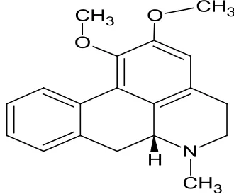
guidelines. Reference standard of nuciferine was
procured from Numex Chemical Corporation, Mumbai, India (fig. 1). Nuciferine is the aporphine alkaloid with
empirical formula C19H21NO2 and molecular weight
295.38. Melting point of nuciferine was found to be
165.5o. Solubility of nuciferine is in alcohol. Analytical grade reagents; chloroform, ethyl acetate, methanol
and glacial acetic acid (S. D. Fine-Chem Limited, Mumbai, India) were used. The fresh lotus leaves were collected from Seawoods-Darave, Navi Mumbai. The plant material was dried and authenticated for its correct botanical identity. The HPTLC system consisted of a Camag Linomat V sample applicator equipped with a 100 μl Hamilton syringe, Camag TLC
scanner 3 was used for densitometric analysis, and
these were supported with Camag WinCATS software. Camag twin-trough chamber (10×10 cm) was used.
Application of sample was done on precoated silica gel 60F254 (Merck, Germany).
The standard solution was prepared by dissolving
accurately weighed 10 mg of nuciferine in 10 ml methanol solution, which gives solution of
concentration 1000 µg/ml. The fresh lotus leaves were
dried in tray dryer at 55o temperature. The dried lotus
leaves were powdered using domestic mixer. About 1 g of powdered lotus leaves was extracted using 100 ml (4×25 ml) methanol by maceration process. To facilitate extraction, magnetic stirrer was used for 24 h and filtered through Whatmann filter paper. The final volume of 100 ml extract was used for nuciferine quantification.
The HPTLC analysis was performed on Merck silica
gel 60F254 plates (10×10 cm). The chromatographic
ascending development was performed using
a mixture of mobile phase chloroform:ethyl acetate:methanol:glacial acetic acid (6.5:1:2.5:0.025 v/v/v/v) to a migration distance of 80 mm. The chamber
was previously saturated for 25 min at temperature 25±2°. Sample and standard zones were applied as band of 6 mm wide at distance from the plate side edge 10 mm and distances from bottom of the plate 10 mm. After development the plates were dried and were sprayed with Dragendorff’s derivatization reagent
for visual detection. Quantification of nuciferine was achieved by scanning with Camag TLC Scanner 3 (slit dimension 5×0.33 mm) at wavelength of 272 nm using deuterium lamp. The chromatogram was plotted as
absorbance verses Rf values.
The developed method was validated as per ICH guidelines. The linearity of an analytical procedure
is its ability to obtain test results, which are directly proportional to the concentration of analyte in the
sample. Linearity was observed by plotting drug concentration against peak area for standard and was
N O
CH3
H
O CH3
664
validated by the correlation coefficients. The linearity
of method was carried out by using standard solution
of nuciferine in the range of 100-450 ng/spot. Limit of detection (LOD) and limit of quantification (LOQ) were defined as k×SD/s (k is constant, s is slope of calibration curve and SD is standard deviation). The
amount for which signal to noise ratio (S/N) were 3 and
10, respectively is defined as LOD and LOQ.
Precision provides an indication of random error. Determination of repeatability and intermediate precision was carried out by measuring intraday variation and
interday variation in triplicate. The precision performed
three times on same day termed as intraday precision while the precision performed over three different days
was known as interday precision. Determination of
nuciferine for three different days at three concentration levels 100, 300 and 450 was performed and the results
were expressed as percent relative standard deviation. The system precision was carried out with 6 replicates of
same concentration of 300 ng/spot for nuciferine and %
RSD of the replicate injection was calculated. Accuracy is also known as percent recovery. It was performed by applying known concentration and further spiking by 80,
100 and 120% w/v amount of analyte in triplicate and the accuracy is then calculated as percentage of analyte
recovered from assay. The robustness of the method was
studied in triplicate at 100 ng/spot and 300 ng/spot for
nuciferine by deliberately making small changes such
as chamber saturation time variation (25±5 min) and
variation in mobile phase composition (6.7:0.8:2.5:0.025 v/v/v/v) and (6.3:1.2:2.5:0.025 v/v/v/v). Specificity of
the method was performed by applying standard and
test solutions and by confirming their Rf values and differentiating standards from other closely related compounds, showing that the method was specific. The peak purity was evaluated by comparing the spectrum of
standard nuciferine with sample.
In this study, HPTLC method was developed for quantification of nuciferine in Lotus leaf. The solvent system chloroform:ethyl acetate:methanol:glacial acetic acid (6.5:1:2.5:0.025 v/v/v/v) showed good separation and resolution of nuciferine peak without interference from the other compounds present in extract. TLC
chamber was saturated for 25 min with mobile phase to show good resolution with reproducible retention factors (Rf). For quantitative determination, peak area was used. The Rf value of nuciferine was found to be 0.44±0.02
(fig. 2). Nuciferine showed good linear relationship over
concentrations range of 100-450 ng/spot (regression equation is, y=11.563x+255.07 and r2=0.998). The LOD
and LOQ were found to be 6.5 ng/spot and 19.8 ng/ spot. The percentage coefficient of variation (%RSD) of
intraday precision and interday precision of nuciferine was found to be less than 2%, which was within the
limits as shown in Table 1. The system precision results
showed that the repeatability of measurement of the area
Fig. 2: HPTLC chromatogram of standard nuciferine
July-August 2017 665
was precise with RSD 1.41% for nuciferine since their
coefficient of variation was less than 2% it is possible to
conclude that the instrument has good precision (n=3).
The recovery was found to be 100.55, 99.14 and 98.96% for nuciferine in lotus leaf as shown in Table 2. There
was no change in Rf values by varying mobile phase
composition (6.7:0.8:2.5:0.025 v/v/v/v) by +5% (1.01% RSD for nuciferine) and (6.3:1.2:2.5:0.025 v/v/v/v) –5%
(0.46% RSD for nuciferine) and by varying saturation
time by +5 min (0.47% RSD for nuciferine) and –5 min (0.98% RSD for nuciferine). Therefore the developed
method was robust. Derivatization of chromatogram with Dragendorff’s reagent provided orange colored spot at Rf value 0.44 corresponding to pure nuciferine, which differentiates closely related structures, indicating
the method was specific.
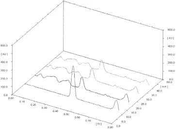
The quantification of nuciferine from lotus leaf extract was carried out in triplicate (fig. 3). The total amount of nuciferine in lotus leaf extract was found to be 0.308% w/w. The summary of all validated parameters are shown in Table 3. These above results revealed that
the developed method enables rapid, precise, sensitive,
Compound Level of percentage recovery of marker (ng)Total amount marker found Amount of (ng)
Recovery
(%) Percentage of RSD Mean recovery (%)
Nuciferine 100300 450
554 615 677
556.08 608.77 668.998
100.37 98.988 98.81
0.736 1.102
0.74 99.389
Each result was an average of three observations performed at 80, 100 and 120% levels. RSD is relative standard deviation
TABLE 2: RECOVERY STUDY OF NUCIFERINE
Fig. 3: 3D Chromatogram nuciferine with sample
3D chromatogram of standard nuciferine was observed at Rf 0.44 in track 1. 3D chromatogram of nuciferine present in sample
(lotus leaves methanolic extract) was observed at Rf 0.44 in track 2, 3 and 4 Compound Concentration (ng/spot)
Intraday Interday
Measured
concentration RSD % concentrationMeasured RSD %
(ng/spot) (ng/spot)
100 115.952 1.581 120.47 1.46
Nuciferine 300 310.452 0.84 321.15 0.768
450 464.417 1.28 465.824 0.254
Each result was an average of three observations. Concentration levels used for precision parameter were 100, 300, 450 ng/spot for nuciferine, RSD: relative standard deviation
666
and highly accurate quantification of nuciferine. The developed method was validated according to ICH
guideline[12]. The method was found to be simple,
sensitive, precise, accurate, and specific for estimation
of nuciferine from lotus leaf (N. nucifera).
Acknowledgements:
The authors thank Dr. H. M. Pandit, Botanist, Mumbai
for authenticating the plant material.
Financial assistance:
None.
Conflict of interests:
None declared.
REFERENCES
1. Jung HA, Jung YJ, Yoon NY, Jeong DM, Bae HJ, Kim DW, et al. Inhibitory effects of Nelumbo nucifera leaves on rat lens aldose reductase, advanced glycation end products formation, and oxidative stress. Food Chem Toxicol 2008;46:3818-26.
2. Moro CF, Yonekura M, Kouzuma Y, Agrawal GK, Rakwal R. Lotus – A source of food and medicine: Current status and future perspectives in context of the seed proteomics. Int J Life Sci 2013;7(1):1-5.
3. Wu SH, Sun CR, Cao XJ, Zhou H, Zhang H, Pan YJ. Preparative counter-current chromatography isolation of liensinine and its analogues from embryo of the seed of Nelumbo nucifera Gaertn using upright coil planet centrifuge with four multilayer coils connected in series. J Chromatogr A 2004;1041:153-62.
4. Raghunathan K, Mitra R. Pharmacognosy of Indigenous Drugs. Vol. 1. New Delhi: Central Council for Research in
Ayurveda and Siddha;1982. p. 806.
5. Shoji N, Umeyama A, Saito N, Iuchi A, Takemoto T, Kajiwara A, et al. Asimilobine and lirinidine, serotonergic receptor
antagonists, from Nelumbo nucifera. J Nat Prod
1987;50:773-4.
6. Kashiwada Y, Aoshima A, Ikeshiro Y, Chen YP, Furukawa H, Fujioka T, et al. Anti-HIV benzylisoquinoline alkaloids and flavonoids from the leaves of Nelumbo nucifera, and structure-activity correlations with related alkaloids. Bioorg Med Chem 2005;13:443-8.
7. Mukherjee PK, Mukherjee D, Maji AK, Rai S, Heinrich M. The sacred lotus (Nelumbo nucifera)-phytochemical and therapeutic profile. J Pharm Pharmacol 2009;61(4):407-22. 8. Xiao GQ, Lu XY, Tian Y, Zhou XM. The research advance of
alkaloids from lotus leaf. Chem Bioen 2006;23:1-2.
9. Xia X, Cui-Rong S, Xiao-Jing D, Rui-Lin H, Yuan-Jiang P, Zhang-Fa Y. LC/MS Guided Isolation of Alkaloids from Lotus Leaves by pH-Zone- Refining Counter-Current
Chromatography.Molecules 2011;16:2551-60.
10. Reich E, Schibli A. High-Performance Thin Layer Chromatography for the Analysis of Medicinal Plants. 1st ed. Switzerland: Thieme Medical Publishers; 2006. p. 193-14. 11. Sethi PD. HPTLC Quantitative Analysis of Pharmaceutical
Formulations. 1st ed. New Delhi: CBS Publishers; 1996. p. 4-28.
12. https://www.ich.org/fileadmin/Public_Web_Site/ICH_
Products/Guidelines/Quality/Q2_R1/Step4/Q2_R1__ Guideline.pdf.
Parameters Nuciferine
Linearity range (ng/spot) 100-450
Correlation coefficient 0.998±0.0001
LOD (ng/spot) 6.5
LOQ (ng/spot) 19.8
Percentage of recovery (n=3)
Lotus leaves (Nelumbo nucifera) 99.389
Precision (%RSD) Precise
Robustness Robust
Specificity Specific
n=3 represents each was an average of three observations, RSD: relative standard deviation, LOD: limit of detection, LOQ: limit of quantification