Article
High PD-1 Expression Level is Associated with an Unfavorable Prognosis in Patients with Cervical Adenocarcinoma
Masako Ishikawa1, Kentaro Nakayama1*, Kohei Nakamura1, Hitomi Yamashita1, Tomoka Ishibashi1, Toshiko Minamoto1, Kouji Iida1, Sultana Razia1, Noriyoshi Ishikawa2, Satoru Nakayama3, Yoshiro Otsuki4 and Satoru Kyo1
1Department of Obstetrics and Gynecology, Shimane University School of Medicine
2Department of Organ Pathology, Shimane University School of Medicine, 6938501, Izumo, Japan 3Department of Obstetrics and Gynecology, Seirei Hamamatsu Hospital, 4308558, Hamamatsu, Japan
4Department of Organ Pathology, Seirei Hamamatsu Hospital, 4308558, Hamamatsu, Japan *Correspondence to:Kentaro Nakayama, MD, PhD Shimane University School of Medicine
Enyacho 89-1, Izumo, Shimane and Japan 6938501
Keywords: programmed cell death-1(PD-1), programmed cell death-ligand 1(PD-L1), cervical adenocarcinoma (CA), immune-checkpoint inhibition (ICI)
Abstract: The effectiveness of immunotherapy for cervical adenocarcinoma (CA) has not been demonstrated yet. It may be possible for us to use programmed cell death 1 (PD-1), programmed cell death-ligand 1 (PD-L1), and CD8 as biomarkers of response to immune therapy in CA patients. In the present study, we aimed to investigate whether the expression levels of PD-1, PD-L1, and CD8 can predict the prognosis of CA patients and their response to ICI therapy. The levels of the PD-1, PD-L1, and CD8 proteins were analyzed by immunohistochemical analysis from formalin-fixed, paraffin-embedded tumor samples. The correlation between the expression levels and patient prognosis was analyzed by the Kaplan–Meier method and univariate and multivariate Cox proportional hazard regression model. We observed a significant inverse-correlation between the PD-1 and CD8 expression (p=0.001, chi square test). We also found a significant inverse-correlation between the PD-L1 and CD8 expression (p=0.027). The overall survival was significantly worse in patients with positive PD-1 expression (p=0.027). Similarly, the progression-free survival was also worse (p=0.087). Our results demonstrate that a high level of PD-1 expression is associated with a poor prognosis in CA patients. Further research is necessary to identify the molecular mechanisms that mediate this association.
Keywords: cervical adenocarcinoma; immune-checkpoint inhibitor; programmed death- 1(PD-1), programmed death-ligand 1(PD-L1), CD8 expression, lymphocyte, survival analysis
1. Introduction
Cervical cancer is the fourth most common cancer in women according to the World Health Organization. In Japan, there are 13,000 new cases of cervical cancer and 3,500 cervical cancer-related deaths every year [1, 2]. Thus, cervical adenocarcinoma has become a major health concern among women because of its high prevalence and poor prognosis. Adenocarcinoma accounts for approximately 10%-25% of uterine cervical cancer cases [1-4]. In Japan, cervical adenocarcinoma is one of the primary health problems in young women because the morbidity and mortality associated with it have increased in the past two decades [5].
Recently, several studies have reported new immunotherapy options for cancer that effectively stimulate the host immune response to eliminate tumor cells. For example, treatment with
nivolumab or pembrolizumab, antibodies against programmed cell death-1 (PD-1), has markedly improved the overall survival of patients suffering from malignant tumors with poor prognoses. However, as these immune checkpoint inhibitors are effective only against a few types of cancers, only a small percentage of cancer patients benefit from their use. Therefore, there is a crucial need to identify potential markers than can predict the success of these new immune therapies. To achieve this goal, a large number of studies have investigated the tumor microenvironment and clarified the mechanisms of immunoediting and tumor immunogenicity, as well as the composition of the tumor-infiltrating cells, such as lymphocytes and macrophages.
Furthermore, several studies have investigated whether PD-1 can be used as a predictive biomarker of disease treatment success or as a predictor of poor prognosis. The effectiveness of immunotherapy alone or as a combination therapy in cervical adenocarcinoma has not been demonstrated yet. Thus, in this study, we aimed to investigate whether the expression levels of PD-L1, PD-1, and CD8 can predict the prognosis of cervical adenocarcinoma patients and their
response to immune-checkpoint inhibition therapy.
2. Materials and methods
2.1. Tissue samples
Tissue samples were obtained from the Department of Obstetrics and Gynecology at Shimane University School of Medicine (Shimane, Japan) and Seirei Hamamatsu Hospital (Shizuoka, Japan) between 2003 and 2017. A total of 82 samples (obtained during surgery or biopsy) were collected from patients with uterine cervical adenocarcinoma. The diagnoses were confirmed by an expert gynecopathologist in our institution. Most patients were primarily treated with surgery and received adjuvant therapy, such as chemotherapy, radiotherapy, or concurrent chemoradiotherapy (CCRT) with cisplatin (weekly dose of 40 mg/m2). Clinical information was obtained
retrospectively from electronic medical records. The acquisition of tumor tissues was approved by the Shimane University Institutional Review Board (IRB No. 20070305-1)
2.2. Immunohistochemical analysis
The expression levels of PD-1, PD-L1, and CD8 were evaluated by immunohistochemical analysis (IHC). Formalin-fixed and paraffin-embedded (FFPE) sections (4-μm thick) were dewaxed in xylene and hydrated in graded alcohol solutions. After antigen retrieval in a sodium citrate buffer, the slides were incubated overnight at 4°C with antibodies against CD-8 (1:100; Roche, Basel, Switzerland), PD-L1 (ab205921, Abcam, Cambridge, United Kingdom), and PD-1 (Roche). One pathologist blinded to the clinicopathologic factors evaluated the samples under a light microscope. PD-1 and CD8 levels on the surface of lymphocytes were detected in the microenvironment of the tumor. PD-L1 expression was detected on the tumor cell plasma membrane or in the cytoplasm.
2.3. IHC of PD-1, PD-L1, and CD8
The density of tumor infiltrating lymphocytes was stratified into two categories by CD8: 0; undetectable, 1+; low density (0-30%), 2+; moderate-high density (≥ 30%). Cases that were 2+ were counted as positive in our analysis.
2.4. Statistical analyses
Analysis of the correlation between PD-1, PD-L1, and CD8 expression and clinicopathological characteristics was performed using the chi-squared test. Furthermore, correlations between the expression levels of each of these 3 proteins were determined using the chi-squared test. The progression-free survival (PFS) and overall survival (OS) rates were analyzed with the Kaplan–Meier method using the log–rank test.
Univariate and multivariate Cox proportional hazard regression models were used with binomial logistic regression for ordered categorical variables. Statistical calculations were performed using IBM SPSS (IBM, Armonk, NY, USA), version 23. A P-value <0.05 was considered statistically significant.
3. Results
3.1 Clinical and pathological features
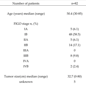
The patients’ clinicopathological characteristics are summarized in Table 1. In the present study, the clinical stages were determined according to guidelines of the International Federation of Gynecology and Obstetrics (FIGO): stages ⅠA, ⅠB1, ⅠB2, ⅡA, ⅡB, ⅢB, and ⅣB were observed in 5, 36, 12, 5, 15, 7, and 2 cases, respectively. The patients were initially treated as follows: 72 patients underwent radical hysterectomy followed by adjuvant therapy such as concurrent chemoradiotherapy (CCRT). Nine patients with advanced stage cancer were initially treated with CCRT. One patient underwent chemotherapy without surgery because of multiple distant metastases. Radiotherapy (whole pelvic irradiation) or chemotherapy (paclitaxel 175 mg/m2 and carboplatin area under the curve = 5 mg/m2) was performed postoperatively in patients with a high recurrence risk (locally advanced stage, non-SCC histology type, bulky tumor(≧4 cm), deep infiltration depth of the cervical tumor (grade 2 or 3), lymph node metastasis, or lymphovascular space invasion).
Table 1. Patients characteristics with cervical adenocarinoma
Number of patients n=82
Age (years) median (range) 50.4 (30-85)
FIGO stage n, (%)
ⅠA 5 (6.1)
ⅠB 48 (58.5)
ⅡA 5 (6.1)
ⅡB 14 (17.1)
ⅢA 0
ⅢB 8 (9.8)
ⅣA 0
ⅣB 2 (2.4)
Tumor size(cm) median (range) 32.7 (0-80)
Tumor size
< 4cm 49 (59.6)
≧4cm 33 (40.4)
LSI
Yes 39(47.6)
No 29 (35.4)
unknown 14 (17.0)
Metastases paraaortic LN
Yes 17 (20.7)
No 65 (79.3)
Metastases pelvic LN
Yes 3 (3.7)
No 79 (92.3)
Metastases distance
Yes 1(1.2)
No 81 (98.8)
Treament
Operation only 24 (29.2)
Operation+adjuvant(RT or CCRT or CT) 48 (58.5)
Rdiotherapy(RT, CCRT) 8 (9.8)
Chemotherapy 2 (2.5)
Recurrence within 5 years
Yes 20 (24.4)
No 60 (75.6)
Death within 5 years
Yes 19 (23.2)
No 63(76.8)
The chemotherapy regimens adopted were paclitaxel plus carboplatin, paclitaxel plus cisplatin, docetaxel plus carboplatin, irrinotecan plus cisplatin, and gemsitabine. In the CCRT regimen, cisplatin was administered weekly in 5-6 courses of 40 mg/m2.
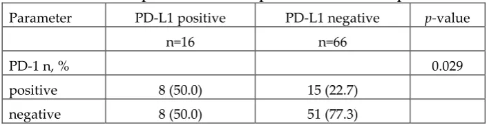
3.2. Correlation between the expression levels of PD-1, PD-L1, and CD8 in cervical adenocarcinoma.
We observed a significant inverse correlation between PD-1 expression and CD8 expression on the tumor-infiltrating lymphocytes (p=0.001, chi square test; Table 2A). We also found a significant inverse correlation between PD-L1 expression and CD8 expression in the tumor-infiltrating lymphocytes (p=0.027, chi square test; Table 2B). Furthermore, PD-1 expression was significantly positively correlated with PD-L1 expression in the tumor-infiltrating lymphocytes (p=0.029, chi square test; Table 2C).
Table2A. Relationship between PD-1 expression and CD8 expression Parameter PD-1 positive PD-1 negative p-value
n=23 n=59
CD8 n, % 0.001
positive 9 (39.1) 46 (78.0)
negative 14 (60.9) 13 (22.0)
Table2B. Relationship between PD-L1 expression and CD8 expression Parameter PD-L1 positive PD-L1 negative p-value
n=16 n=66
CD8 n, % 0.027
positive 7 (43.8) 48 (72.7)
Table2C. Relationship between PD-1 expression and PD-L1expression Parameter PD-L1 positive PD-L1 negative p-value
n=16 n=66
PD-1 n, % 0.029
positive 8 (50.0) 15 (22.7)
negative 8 (50.0) 51 (77.3)
There was no significant correlation between the expression levels of PD-1, PD-L1, and CD8 and the clinicopathological factors examined (Supplementary Table 1A-C).
3.3. Correlation between the prognosis of cervical adenocarcinoma and the expression levels of PD-1, PD-L1, and CD8.
3.4. Univariate analysis of prognostic factors in patients with cervical adenocarcinoma.
There were no significant differences in the progression free survival (PFS) or overall survival (OS) between the positive and negative PD-1 cases. Similarly, there were no differences in PFS and OS between positive or negative CD8 and PD-1 cases. Tumor size and metastatic invasion of the lymph-vascular space were the only parameters significantly correlated with PFS and OS of the patients (Table 3, Table 4).
Table 3. Univariate and multivariate analysis of progression-free survival using a Cox proportional hazards model in patients with cervical adenocarcinoma
Factors Patients Univariate analysis Mutivariate analysis
n HR 95% CI p-value HR 95% CI
p-value
<60 58 0.552 0.236-1.293 0.171
≧60 24 ref.
FIGO stage
<ⅡB 58 0.194 0.081-0.464 <0.0001 ref.
≧ⅡB 24 ref. 1.101 0.320-3.787 0.879
histology
non-gastric
type 69 0.819 0.277-2.422 0.819
gastric
type 13
tumor size (mm)
<40 50 0.135 0.050-0.369 0.0001 0.18 0.046-0.702 0.013
≧40 32 ref. ref.
Metastasis pelvic
lymphnode
Negative 65 0.379 0.162-.0889 0.026 0.583 0.147-2.307 0.442
Positive 17 ref. ref.
Metastasis paraaortic
lymphnode
Negative 79 0.209. 0.061-0.707 0.012 0.235 0.012-4.687 0.343
Positive 3 ref. ref.
Metastasis distance
Negative 81 0.078 0.009-0.644 0.018 0.454 0.042-4.941 0.517
Positive 1 ref. ref.
Metastasis
LSI
Negative 29 0.453 0.087-2.353 0.004 0.035 0.003-0.387 0.005
Positive 39 ref. ref.
PD-1
Negative 59 ref. ref.
Positive 23 2.756 0.815-9.319 0.103 1.791 0.408-7.854 0.44
Negative 66 ref. ref. Positive 16 1.153 0.391-3.420 0.792 6.251 0.610-64.077 0.123
CD8
Negative 27 ref. 0.507 0.118-2.170 0.36
Positive 55 1.512 0.591-3.872 0.388 ref.
Table 4.Univariate and multivariate analysis of overall survival using a Cox proportional hazards model in patients with cervical adenocarcinoma
Factors Patients Univariate analysis Mutivariate analysis
n HR 95% CI p-value HR 95% CI p-value
Age (y)
<60 58 0.58 0.233-1.446 0.243
≧60 24 ref.
FIGO stage
<ⅡB 58 0.222 0.087-0.565 0.002 0.968 0.284-3030. 0.958
≧ⅡB 24 ref. ref.
histology non-gastric
type 69 0.67 0.222-2.021 0.477
gastric type 13 ref. tumor size
(mm)
<40 50 0.168 0.060-0.468 0.001 0.171 0.036-0.812 0.026
≧40 32 ref. ref.
Metastasis pelvic
lymphnode
Negative 65 0.515 0.202-1.309 0.163
Positive 17 ref.
Metastasis paraaortic
lymphnode
Negative 79 0.321 0.074-1.405 0.131
Positive 3 ref.
Metastasis distance
Negative 81 0.026 0.002-0.283 0.003 0.102 0.009-1.205 0.07
Positive 1 ref. ref.
Metastasis
LSI
Negative 29 0.053 0.007-0.416 0.005 0.034 0.002-0.521 0.015
Positive 39 ref. ref.
PD-1
Negative 59 ref. ref.
Positive 23 6.765 0.903-50.705 0.063 5.311 0.549-51.377 0.149 PD-L1
Negative 66 ref. ref.
Positive 16 1.331 0.382-2.196 0.667 2.414 0.439-13.264 0.311
CD8
Negative 27 ref. 0.286 0.055-1.498 0.139
Positive 55 1.694 0.609-4.710 0.313 ref.
4. Discussion
The present study reports two major findings: First, we revealed a significant inverse correlation between PD-1 and CD8 expression levels of tumor-infiltrating lymphocytes . We also found a significant inverse correlation between PD-L1 expression and CD8 expression on tumor-infiltrating lymphocytes. These findings have already been demonstrated in previous studies on ovarian cancer [6] and other malignant tumors [7,8]. In cervical adenocarcinoma, tumor cells expressing PD-L1 may be protected from the destructive activity of CD8+ lymphocytes. The reduction of CD8 expression levels may not be the only mechanism by which PD-L1 promotes tumor immune escape. It may be possible that PD-L1 on tumor cells induces functional impairment of tumor-specific T cells without reducing their CD8 levels, as reported for antiviral T cells [9,10]. A recent meta-analysis study concluded that the correlations between the survival of cancer patients and the expression of PD-L1 vary among different tumor types [11].
Second, we showed that the high expression level of PD-1 is associated with a poor prognosis in cervical adenocarcinoma patients (Figure 2a). Some previous studies showed no significant difference between the expression levels of immune-checkpoint associated proteins and the prognosis of cervical adenocarcinoma patients [12]. However, the results of these studies were controversial [13]. In the current study, the level of CD8 on tumor-infiltrating lymphocytes was not found to be associated with a favorable prognosis. A similar result was reported in a previous study on cervical squamous cell carcinoma and adenocarcinoma [14-16].
We also observed a significant correlation between high PD-1 expression levels and worse overall survival of cervical adenocarcinoma patients. The expression levels of PD-L1 and CD8 were not significantly correlated with the patients’ prognosis. The overexpression of PD-L1 on tumor cells might inhibit the activity of CD8-expressing lymphocytes in the microenvironment of the tumor. Previous studies suggested that CD8+ lymphocytes could not engage with PD-L1-positive tumor cells. Therefore, the expression levels CD8 and PD-L1 are expected to be inversely correlated [6] [8].
If we use PD-1 inhibitor for cervical adenocarcinoma patients with high PD-1 expression, their immune activity against tumor cells would recover and may decrease the size of the tumor.
Future research on biomarkers that predict response to checkpoint blockade immunotherapies could increase their durable responses.
malignant tumors, such as ovarian carcinoma and cervical carcinoma. Thus, it was difficult to determine the expression levels of proteins in these lymphocytes.
Another limitation was that we did not count lymphocytes separately in the tumor epithelium and tumor stroma as was done in previous studies [12] [13]. As we speculated that the function of tumor-infiltrating lymphocytes is not different in the epithelium and stroma, in the current study, we counted together the lymphocytes in both sections. In future studies, it is necessary to count lymphocytes individually in the stroma and epithelial sections.
Furthermore, the use of PD-L2 antibodies has recently been suggested as a new strategy of immune-checkpoint therapy [17]. The expression level of PD-L2 has already been examined in some tumors. Furthermore, some studies demonstrated that anti-PD-L1 plus anti-PD-L2 therapy has improved antitumor effects [18].
Finally, as many cervical adenocarcinoma patients still have a poor prognosis, it is essential to develop new immunotherapies for the treatment of this cancer.
Author Contributions:
Masako Ishikawa and Kentaro Nakayama drafted the manuscript. Kohei Nakamura, Hitomi Yamashita, Tomoka Ishibashi, Toshiko Minamoto, Koji Iida, Noriyoshi Ishikawa, and Sultana Razia carried out the IHC studies. Kentaro Nakayama participated in the design of the study. Satoru Nakayama, Yoshiro Otuski, Noriyoshi Ishikawa carried out the pathological diagnosis. Satoru Kyo conceived of the study, participated in its design and coordination, and helped in drafting the manuscript. All authors have read and approved the final manuscript.
conception and design of the study; K.N., M.I.
acquisition and analysis of data; K.N., T.I., H.Y., T.M., K.I., S.R. carrying out the pathological diagnosis; S.N., Y.O., N.I. drafting the manuscript or figures; M.I., S.R.
conceived of the study; S.K.
Figure legends
Figure1. A-F. HE staining and immunohistochemical analysis of the specimens from patients with cervical adenocarcinoma.
A,B: Immunostaining of PD-1. A, positive expression of PD-1; B, no expression of PD-1. C,D: Immunostaining of PD-L1. C, positive expression of PD-L1; D, no expression of PD-L1. E,F: Immunostaining of CD8: E, CD8 expression score of +2; F, CD8 expression scores of 0 and +1.
Figure 2 A.B.C. Kaplan-Meier analysis of progression-free and overall survival associated with each immune-checkpoint-related molecular. A: Kaplan-Meier analysis of progression-free survival and overall survival between the PD-1(+) group and PD-1(-) group. B: Kaplan-Meier analysis of progression-free survival and overall survival between the PD-L1(+) group and PD-L1(-) group. C: Kaplan-Meier analysis of the progression-free survival and overall survival between the CD8(+) group and CD8(-) group.
Supplementary Table 1 A.B.C. Relationship between the patients’ clinicopathological factors and immune-checkpoint-related molecules according to the Chi-square test.
A: Relationship between the patients’ clinicopathological factors and PD-1. B: Relationship between the patients’ clinicopathological factors and PD-L1. C: Relationship between the patients’ clinicopathological factors and CD8.
There was no significant correlation between these factors and immune-checkpoint-related molecules.
Author Contributions:Masako Ishikawa and Kentaro Nakayama drafted the manuscript. Kohei Nakamura, Hitomi Yamashita, Tomoka Ishibashi, Toshiko Minamoto, Kiyoka Sawada, Yuki Yoshimura, Koji Iida, Noriyoshi Ishikawa, and Sultana Razia carried out the IHC studies. Kentaro Nakayama participated in the design of the study. Satoru Nakayama, Yoshiro Otuski, Noriyoshi Ishikawa carried out the pathological diagnosis. Satoru Kyo conceived of the study, participated in its design and coordination, and helped in drafting the manuscript. All authors have read and approved the final manuscript.
CONFLICTS OF INTEREST:The authors declare no conflicts of interest.
References
1. Vital Statistics Japan (Ministry of Health, Labour and Welfare, Cancer Registry and Statistics. Cancer Information Service, National Cancer Center, Japan.
2. Hori, M.; Matsuda, T.; Shibata, A.; Katanoda, K.; Sobue, T.; Nishimoto, H.; Cancer incidence and incidence rates in Japan in 2009: a study of 32 population-based cancer registries for the Monitoring of Cancer Incidence in Japan (MCIJ) project. Jpn. J. Clin. Oncol. 2015, 45, 884-891.
3. Vinh-Hung, V.; Bourgain, C.; Vlastos, G.; Cserni, G.; De Ridder, M.; Storme, G.; Vlastos, A.T.; Prognostic value ofhistopathology and trends in cervical cancer: a SEER populationstudy. BMC Cancer 2007, 7, 164.
4. Wilbur, D.C.; Colgan, T.J.; Ferenczy, A.S.; Kurman, R.J.; Carcangiu, M.L.; Herrington, C.S.; Glandular tumorus and precursors. World Health Organization Classification of Tumours Pathology &Genetics. Tumours of Female Reproductive Organs, 4th 2014, 183-189.
5. Japanese Journal of Gynecologic Oncology, Statement for the Year 2012. 5-year results. Acta Obstetricaet Gynecologica Japonica, 2019, 727-747.
6. Hamanishi, J.; Mandai, M.; Iwasaki, M.; Okazaki, T.; Tanaka, Y.; Yamaguchi, K.; Higuchi, T.; Yagi, H.; Takakura, K.; Minato, N.; Honjo, T.; Fujii, S.; Programmed cell death 1 ligand 1 and tumor-infiltrating CD8+ T lymphocytes are prognostic factors of human ovarian cancer. Proc. Natl. Acad. Sci. U S A 2007, 104, 3360-3365.
7. Dong, H.; Strome, S.E.; Salomao, D.R.; Tamura, H.; Hirano, F.; Flies, D.B.; Roche, P.C.; Lu, J.; Zhu, G.; Tamada, K.; Lennon, V.A.; Celis, E.; Chen, L.; Tumor-associated B7-H1 promotes T-cell apoptosis: a potential mechanism of immune evasion. Nat. Med. 2002, 8, 793-800.
8. Iwai, Y.; Ishida, M.; Tanaka, Y.; Okazaki, T.; Honjo, T.; Minato, N.; Involvement of PD-L1 on tumor cells in the escape from host immune system and tumor immunotherapy by PD-L1 blockade. Proc. Natl. Acad. Sci. U S A 2002, 99, 12293-12297.
9. Barber, D.L.; Wherry, E.J.; Masopust, D.; Zhu, B.; Allison, J.P.; Sharpe, A.H.; Freeman, G.J.; Ahmed, R.; Restoring function in exhausted CD8 T cells during chronic viral infection. Nature 2006, 439, 682-687.
10. Hirano, F.; Kaneko, K.; Tamura, H.; Dong, H.; Wang, S.; Ichikawa, M.; Rietz, C.; Flies, D.B.; Lau, J.S.; Zhu, G.; Tamada, K.; Chen, L.; Blockade of B7-H1 and PD-1 by monoclonal antibodies potentiates cancer therapeutic immunity. Cancer Res. 2005, 65, 1089-1096.
11. Wu, P.; Wu, D.; Li, L.; Chai, Y.; Huang, J.; PD-L1 and Survival in Solid Tumors: A Meta-Analysis. PLoS. One. 2015, 10: e0131403.
13. 13. Heeren, A.M.; Punt, S.; Bleeker, M.C.; Gaarenstroom, K.N.; van der Velden, J.; Kenter, G.G.; de Gruijl, T.D.; Jordanova, E.S.; Prognostic effect of different PD-1 expression patterns in squamous cell carcinoma and adenocarcinoma of the cervix. Mod. Pathol. 2016, 29, 753-763.
14. 14. Punt, S.; van Vliet, M.E.; Spaans, V.M.; de Kroon, C.D.; Fleuren, G.J.; Gorter, A.; Jordanova, E.S.; FoxP3(+) and IL-17(+) cells are correlated with improved prognosis in cervical adenocarcinoma. Cancer Immunol. Immunother. 2015, 64, 745-753.
15. 15. Karim, R.; Jordanova, E.S.; Piersma, S.J.; Kenter, G.G.; Chen, L.; Boer, J.M.; Melief, C.J.; van der Burg, S.H.; Tumor-expressed B7-H1 and B7-DC in relation to PD-1+ T-cell infiltration and survival of patients with cervical carcinoma. Clin. Cancer Res. 2009, 15, 6341-6347.
16. 16. Spaans, V.M.; Peters, A.A.; Fleuren, G.J.; Jordanova, E.S.; HLA-E expression in cervical adenocarcinomas: association with improved long-term survival. J. Transl. Med. 2012, 10, 184.
17. 17. Yearley, J.H.; Gibson, C.; Yu, N.; Moon, C.; Murphy, E.; Juco, J.; Lunceford, J.; Cheng, J.; Chow, L.Q.M.; Seiwert, T.Y.; Handa, M.; Tomassini, J.E.; McClanahan, T.; PD-L2 Expression in Human Tumors: Relevance to Anti-PD-1 Therapy in Cancer. Clin. Cancer Res. 2017, 23, 3158-3167.