Sensory Neuromodulation
Robert D. Black*, Ph.D.; Lesco L. Rogers, MD Scion NeuroStim, Raleigh, NC, USA
*Corresponding author: [email protected]
Abstract: We describe a model of neurological disease based on dysfunctional brain oscillators. This is not a new model, but it is not one that is generally appreciated by clinicians. The value of this model lies in the predictions it makes and the utility it provides in translational applications, in particular for neuromodulation devices. We provide a perspective on the difference between neuromodulation devices that enforce an externally administered stimulus with devices that provide input to sensory receptors and thus stimulate endogenous sensory networks. Current forms of clinically applied neuromodulation are of the former type, including devices such as (implanted) deep brain stimulators (DBS) and various, noninvasive methods such as transcranial magnetic stimulation (TMS) and transcranial current methods (tACS, tDCS). The challenge with these methods is that they are not sensitive to underlying neuronal dynamics and work by applying an empirically derived electrical current waveform to affect dynamical patterns. Neuromodulation of a sensory organ accesses the same pathways that natural environmental stimuli do and, importantly, the modulatory signal will be transformed as it travels through the brain, allowing the modulation input to be consistent with regional dynamics. We present specific examples of devices that rely on sensory neuromodulation and evaluate the translational potential of these approaches. We argue that sensory neuromodulation is well suited to probe and, ideally, repair dysfunctional brain oscillators, thus providing a novel therapeutic approach for neurological diseases.
Keywords: neuromodulation, noninvasive, sensory networks
Graphical Abstract:
Part 1: Oscillatory Dynamics
There is now growing evidence that brain dynamics are underpinned by collective oscillatory states. In this monograph, we explore the proposal that neurological disease can be modeled as dysfunctional brain oscillators. We further consider how artificial neurostimulation methods might alter brain oscillators and the potential for improving functional deficits resulting from disease. Most particularly, we examine neurostimulation introduced into an endogenous sensory network and examine how this approach is categorically different from current clinical methods of neuromodulation.
Neuronal oscillations:
The observation of neuronal oscillations has been documented in relatively simple animals such as aplysia (Elmariah, 2007) and jelly fish (Nath, 2017). EEG recordings have been reported in eels (Barthelemy, 1977), fish (Robb, 2003) and reptiles (De Vera, 1994). De Vera et al. (1994) suggested a homology between the waking state of reptiles and slow-wave sleep in mammals. This is an evocative suggestion, but the primary finding is that oscillations enable behavior in animals in a manner that cannot be deduced based simply on the static architecture of the connectome.
Though EEG recordings have been studied for decades since the original work by Berger in the 1920’s, their significance was not immediately understood. Until recently many researchers were unsure of whether the oscillations recorded in EEG time series were meaningful or simply epi-phenomena of Hebbian firing patterns. Fries (2005) proposed that oscillatory activity in the brain is actually central to function, enabling a means by which transient pathways can form and fade, based on demand. His communication through coherence model provides an elegant answer to the question of how dynamic organization of the cortex occurs. More recently, McLelland & VanRullen (2016) reviewed communication-through-coherence and refinements.
Buzsaki’s (2006) comprehensive book takes a consistent, oscillation-centric perspective on the primacy of oscillations to brain function. He starts with a general review of periodic, nonlinear and chaotic phenomena in nature and from that develops a framework for understanding coupled oscillators and details results from invasive neuroscience studies before turning to non-invasive methods used in human research, including EEG, MEG and functional imaging. He paints a story of continuity, both evolutionarily and architecturally, from small clusters of neurons to the whole brain. Voytek & Knight (2015) suggested that dynamic network communication relies on coordination via neuronal oscillations, the disruption of which can result in clinical disorders. Assenza et al. (2017) and Fox et al. (2014) reiterated the view that dysfunctional brain oscillators are associated with disease and they review how neuromodulation may be helpful in altering and improving brain oscillator function.
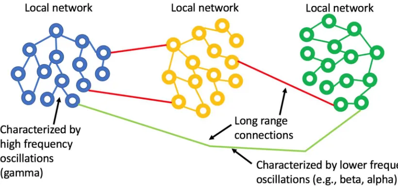
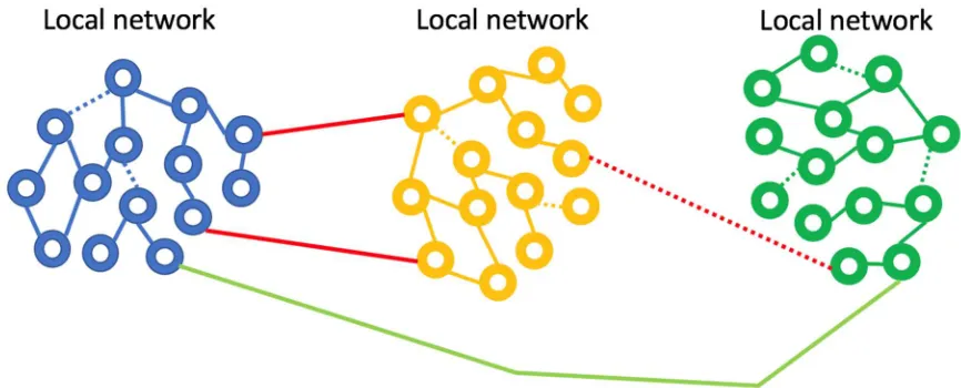
seems very well matched to the observation that gamma oscillations enable localized neuronal dynamics and slower (alpha, beta, etc.) EEG frequency bands are indeed associated with longer-range dynamics. The small-world topology is an optimal configuration for the efficient use of space (synaptic density) while still enabling global communication.
Figure 1: Small world network topology. The highest density of synaptic connections is local and longer-range connections are sparse. This topology is consistent with high frequency, short-range oscillations (gamma) being local and longer range, slower oscillations being regional or global.
CFC - Cross frequency correlation:
Figure 2: The behavior of a simple, undamped oscillator as a function of excitation frequency. The amplitude of the response of the oscillator (blue) to the driving force peaks when the driver reaches the natural resonance of the oscillator. The phase of the oscillator with respect to the driver (orange) is in phase when the driving frequency is lower than the resonant frequency and anti-phase when the driving frequency exceeds resonance.
The formation of dynamic small-world ensembles is partially facilitated by developmentally established neuronal pathways, but the transient selection of certain subsets of all possible pathways must rely on an encoding scheme. How might encoding and recall be enabled in an oscillator-based model? Hoppensteadt & Izhikevich (1998) provide a simple yet general schema that models thalamocortical interactions via weakly coupled oscillators and they develop an intuitive framework in analogy to FM radio principles. A primary insight from their model is that a cortical oscillator may participate in different ensembles by changing its frequency without changing the strengths of synaptic connections. The authors also clarify the point that the terms “inter-spike intervals,” “frequency modulation,” and “phase modulation” all describe the same thing, in the neuroscience, electrical engineering, and mathematical physics literature respectively, even though that unity of description is not generally appreciated (figure 4). The crucial observation is that frequency and phase encoding are both biologically plausible as means for creating the sort of transient neuronal ensembles that are consistent with oscillator-based theories of brain function. The particulars of how frequency and phase encoding are instantiated in real brain networks are not yet fully understood, but established analytical methods from past studies of oscillatory networks provide a fertile basis for model generation.
Figure 4: Spiking of a relaxation oscillator (the basis for the Hodgkin-Huxley model), where an applied perturbation has shifted the phase of the spike train. This shift can be viewed as a change in spike timing, phase modulation, or frequency modulation: they are all equivalent mathematically. Adapted from Hoppensteadt & Izhikevich (1998).
well matched to the challenge of interfacing with brain oscillators. Additionally, we will look at some evidence that suggests that taking an oscillator-centric view of neurological disease might also provide insights into innate protective pathways in the brain that orchestrate underlying biochemical processes. That is, it may be that the brain is able to marshal a biochemical response to repair and maintain neurons in response to the aberrant firing patterns of damaged brain oscillators.
Part 2: Methods of Neuromodulation
There are several recent and excellent reviews of clinical neuromodulation (see the Workshop summary under Bain et al., 2015, and references therein). Deep brain stimulation (DBS) describes a category of interventions seeking to alter regional neuronal activity via a surgically implanted electrode powered by an implanted pulse generator. Non-invasive brain stimulation (NIBS) describes a class of devices that use externally placed electrodes to direct electrical current into brain (usually cortical) tissue, that current being direct or alternating. Transcranial magnetic stimulation (TMS) makes use of a rapidly changing magnetic field to induce current flow in the cortex and as such is a method that does not require direct contact to the head. These methods have been used extensively in a research context and there has been translation to clinical medicine as well. DBS, especially for movement disorders associated with Parkinson’s disease, is a well-established approach, but it is typically reserved for late-stage disease because of the invasive nature of the placement procedure and the concommitant high cost (Umemura et al., 2016). TMS has been cleared by the FDA for major depressive disorder (Connolly et al., 2012) and for migraine headache (Conforto et al., 2014). Two other alternating current methods have been cleared for the treatment of migraine headache, one using an applicator on the neck for stimulation of the vagus nerve (Silberstein, 2016) and another applying current to the forehead (over a branch of the trigeminal nerve; Schoenen et al., 2013). Despite these clinical successes, there is a general concern about the design of NIBS studies and the need for a comprehensive approach that includes attention to the mechanism of action for a given intervention (Frohlich & Schmidt, 2013).
et al. (2013) summarized the challenge of matching an exogenously generated stimulus to the target brain oscillator: “An important implication of this finding is that the frequency of applied stimulation should be matched to the frequency of the endogenous oscillatory state.…[and] the choice of stimulation frequency could represent a serious challenge as there is no clear preferred resonance or peak frequency.” We propose that neuromodulation of a sensory network addresses this matching challenge. If the brain target is accessed by endogenous neural (sensory) pathways and the modulation signal is applied to the sensory organ, the modulation is transformed in a way that it is matched to the native dynamics of the target region.
That a sensory system processes and transforms incoming stimuli is perhaps most intuitively understood by considering vision. The light pattern on the retina changes rapidly as the result of saccadic eye movements and these signals follow the optic nerve to the first visual cortical region (V1). The visual scene is broken into constituent elements, as was described in the seminal work by Hubel & Wiesel (1959) and further processing proceeds in a hierarchical fashion. Near the top of the visual hierarchy, the brain is able to maintain a persistent representation of a viewed object even though the input activity patterns from the retina itself do not match the activity patterns in that part of the cortex representing the viewed object (Wurtz et al., 2011). The retinal signals were transformed and combined with input from other brain regions with the net effect that a new dynamic oscillatory state consistent with the represented object emerged. It is this sort of transformation of raw sensory inputs into meaningful patterns at the targeted brain region that marks the difference between sensory neuromodulation and other current formats. There is no way to emulate these detailed, regional signal transformations with exogenously applied stimulation methods (NIBS) with just one or a limited number of frequencies.
Visual, auditory, somatosensory and vestibular neurostimulation methods are described in the literature and we now evaluate examples of clinical studies with translational intent to better illustrate sensory neuromodulation.
A well-described method for modulating visual perception makes use of an optokinetic drum, a rotating cylinder with light and dark stripes that is viewed by a subject. Most commonly an optokinetic experiment aims to induce perceptions of self-motion and nystagmus. Kikuchi et al. (2009) performed optokinetic stimulation while acquiring BOLD MRI data and demonstrated activation of cortical areas related to visual movement processing and deactivation of the parieto-insular cortex, which is primary in vestibular processing. Chokron et al. (2007) described studies using optokinetic methods for mitigating unilateral spatial neglect. The general aim was to correct spatial bias by realigning perception of spatial coordinates. The authors review a number of such studies and conclude that the effects were transient when single sessions were used. Kerkhoff et al. (2006) performed longitudinal optokinetic stimulations and reported persistent improvement over a 2-week follow up period, which may imply a neuroplastic change as the basis for durability. Iaccarino et al. (2016) presented evidence in support of the reduction of plaque formation in a murine model of Alzheimer’s disease via optogenetically driven modulation of interneurons in the gamma frequency range. This is an interesting case wherein changes in oscillatory stimulation trigger a protective biochemical response. Using neuromodulation to activate neuroprotection is a topic to which we will return later.
stimulation as a treatment for tinnitus. Starting with a guinea pig model, the authors found that fusiform cells exhibited increased spontaneous activity and cross-unit synchrony, which are physiological correlates of tinnitus in a majority of patients. Through empirical means, they found that bimodal (but not unimodal) stimulation produced long-term depression in the dorsal cochlear nucleus in guinea pigs. The stimulus method consists of sound stimuli, delivered by insert earphones, and somatosensory stimuli, delivered by electrodes placed on the skin of the cervical spine or cheek. The auditory stimulus was based on the individual subject’s tinnitus spectrum and audiogram. The electrical (somatosensory) signal was timed to have a specific temporal relationship with the auditory signals (again, based on empirical findings in the guinea pig model). The device was designed for home use, thus facilitating daily, 30-minute treatment sessions for two, 4-week sessions (with a gap) before being crossed over to the opposite treatment arm (active or placebo). Ten of the twenty human subjects had clinically significant reductions in their tinnitus scores. This study is a fascinating example of the use of two forms of sensory neuromodulation in an interactive protocol. The availability of a well-developed animal model allowed for specific hypotheses about the mechanism of action to be developed. Since tinnitus is a sensory processing disorder, its alignment with a sensory neuromodulation approach is logical.
Bellesi et al. (2014) evaluated the possibility of enhancing slow-wave sleep (during non-REM sleep) using acoustic stimulation. The authors first evaluate the literature addressing the use of transcranial direct current stimulation (tDCS) and TMS, concluding that these methods are, at present, impractical. They target modulation of a peripheral evoked slow wave (K-complex) using entrainment via auditory stimulation. They list parameters of relevance for acoustic stimulation: intensity, frequency, timing (with respect to the onset of slow wave sleep) and entrainment. The latter parameter is not independent of the others and instead speaks to the goal of matching the acoustic stimulus frequency to that of an endogenous oscillator. Acoustic stimulation at 0.8 Hz fits with the EEG power band of 0.5-1.0 Hz associated with slow-wave sleep and has resulted in higher intensity, suggesting entrainment of what is thought to be a spontaneous thalamocortical oscillation. A remaining challenge for this approach is to time the administration of the acoustic stimulus, since stimulation at the wrong time in the sleep cycle can actually have an arousal effect. The authors suggest the use of ambulatory EEG to assess the proper time for administration, but this requirement calls into question the practicality of the method.
The PONS device has been used in an RCT to assess efficacy for chronic balance deficits resulting from mild-to-moderate TBI (traumatic brain injury). Though not yet in peer-reviewed, published form, reported results are positive for a high-frequency stimulation arm (75.4% of subjects met the targeted improvement in SOT [sensory organization test] scores). The study sham, delivering a low-frequency stimulation, also proved to be efficacious and thus the primary endpoint was not met. This is an ongoing challenge with non-invasive neuromodulation studies in general: reproduction of a comparable procedural experience for sham-arm subjects as compared with active-arm subjects. Wildenberg at al. (2013) hypothesized that the afferent output from tongue stimulation enters the brainstem in proximity to the vestibular and trigeminal nuclei, moving upwards to the cortex, and is able to influence cortical processing of visual motion. Leonard et al. (2017) undertook an imaging study with multiple sclerosis subjects to assess effects of PONS stimulation over a 14-week treatment period. They found evidence of improved motor performance in active-arm subjects, as judged by BOLD MRI data localized to the motor cortex, suggestive of a neuroplastic change (specifically, the changes were durable enough to be subsequently recorded with fMRI). Of relevance here is that the mechanism of action of the PONS device seems to involve extensive pathways, accessed via a sensory input channel, albeit an indirect one via the tongue, and that there is evidence of durable neuroplastic change with beneficial effects for subjects (without significant device-related adverse events). A crucial observation is that this approach to sensory neuromodulation enables stimulation of brainstem regions. But the modulatory effects progress up from the brainstem to areas including the visual cortex and parieto-insular vestibular cortex (Wildenberg, 2013). And, importantly, the modulation signal follows endogenous sensory pathways.
We now turn to vestibular neuromodulation (VNM) and argue that this often-neglected sensory channel presents what is perhaps the best conduit for sensory neuromodulation. Caloric vestibular stimulation (CVS) is a widely used diagnostic technique, in particular for the study of balance disorders, and was initially explained by Barany (1911). The Fitzgerald-Hallpike (Fitzgerald & Hallpike, 1942) protocol for CVS is used routinely in the diagnosis of vestibular disorders. Galvanic vestibular stimulation (GVS) is a transcranial current method with the specific intent of creating a voltage bias across the two sets of vestibular organs by placing electrodes on the mastoid bone behind the ears (Fitzpatrick & Day, 2004). CVS and GVS, along with rotational methods, will be collectively referred to as vestibular neuromodulation (VNM).
The neuroscience of the vestibular system has been illuminated in extensive studies in animals and humans, but this rich literature is generally underappreciated by neurologists outside of audiological and balance related specialties (figure 5). Ayres (1972) book on the multi-sensory and integrative aspects of the vestibular system is a particularly cogent reference that illuminates the higher order cognitive elements of vestibular processing, going well beyond the study of balance.
hippocampus and the cerebellum. Three components were mainly characterized by negative deflections of the BOLD signal: the pre- and postcentral gyrus, the anterior cingulate gyrus, the precuneus, the occipital lobe, and the supplementary motor area.
Lopez & Blanke (2011) reviewed the extensive literature around the structures and pathways comprising the thalamocortical vestibular system. The authors concluded that there is no unique and well-defined primary vestibular cortex comparable to primary sensory cortex for vision, somatosensation, and audition. The vestibular system is often referred to as a multi-sensory sense since it has direct or indirect projections into all cortical regions and vestibular sensory flow impacts the interpretation of other sensory modalities in the brain. And thus in addition to mediating balance, emerging evidence suggests that the vestibular network expands into dimensions of emotional processing, mental health and social cognition (Lopez, 2016).
Hitier et al. (2014) addressed the role of vestibular pathways in cognition, focusing on five major pathways that transmit vestibular sensory information to the distributed vestibular cortex: 1) vestibulo-thalamocortical; 2) dorsal tegmental nucleus via the lateral mammillary nucleus; 3) nucleus reticularis pontis oralis; 4) via the cerebellum; and 5) (hypothesized) via the basal ganglia. The cerebellum evolved from the vestibular and trigeminal nuclei (Bishop, 1959) and thus has a key role in processing vestibular sensory flow.
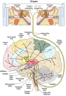
Figure 5: A schematic view of some of the widespread connectivity of the vestibular system. The top panel illustrates the induction of CVS using warm/cool ear inserts (see figure 8 for example thermal waveforms). The 8th cranial nerve conveys vestibular stimulation to the vestibular nuclei
in the brainstem.
words, modulation of the firing rate of the vestibular system was transformed and resulted in a wholly different firing pattern in the LC.
Part 3: Models of disease of oscillators (clinical examples)
Let us look at some specific examples where a neurological disease may be viewed as resulting from the dysfunction of oscillatory states and evaluate whether that perspective generates any new insights.
Epilepsy is often viewed simply as being characterized by hypersynchronous seizures, but the ictal and inter-ictal patterns of synchronization are more complicated than that. Muller et al. (2014) described varying degrees of synchronization prior to and during a seizure event. They described the peri-ictal evolution of brain network function as transitioning from a predominantly random topology to a more regular network and back again. They suggested that high synchronization at the end of a seizure is in fact a signature of the termination of the seizure. Yet throughout, they stated that there is a consistent trace of topology associated with the default mode network. In other words, the default organization is not lost during a seizure and ultimately reasserts itself. Perhaps counter-intuitively, epilepsy seems to be characterized by cortical regions that have poor connectivity and the hypersynchronous activity of a seizure works to re-integrate disconnected regions (Rummel et al., 2013; Schindler, private communication). Kusmierczak et al. (2105) addressed changes in local and long-range connectivity between cortical neurons as a component of the epileptogenic process after deafferentiation in an animal model. They found evidence that as axons started to reconnect the transected regions, the balance of long and short range excitatory connections altered neuronal excitability. It may be that a similar process occurs during post-traumatic epilepsy or that similar imbalances occur with idiopathic epilepsy. Is a seizure the brain’s attempt to re-establish normal cortical connectivity?
Finding a biomarker for migraine onset is a current aim in headache research. Coppola et al. (2012) provided evidence that, interictally, migrainuers exhibit poor sensory habituation. Goadsby et al. (2017) argued that migraine headache is a disorder of sensory processing, which cycles based on development (genetics) and environment. They focused on abnormal brainstem function in the premonitory phase of a migraine, when the well-known sensory phobias emerge. Brighina et al. (2009) provided evidence that cerebellar inhibition is reduced in migraineurs and it is known that the cerebellum exerts inhibitory control on the cortex. Since the brainstem, cerebellum and cortex are all involved in sensory processing, a failure of habituation would seem to be a result of widespread network dysfunction. Interestingly, as the migraine develops, sensory habituation normalizes (Coppola et al., 2013). Is a migraine the brain’s attempt to re-establish normal sensory sensitivity?
severity of disease. Indeed, de Hemptinne et al. found that CFC increased with the DBS device turned off, and decreased when the device was active. How the neurodegenerative effects of PD altered the normal amount of CFC is not wholly clear, but this is a clear case whereby changes in normal brain oscillator function have a clear and measureable clinical consequence. As a consequence of neurodegeneration, the brain is not able to re-establish normal connectivity, but through artificial stimulation it is possible to reduce pathological CFC.
In these three examples, epilepsy, migraine and PD, we see that the diseases are characterized by widespread network dysfunction, which implies collective action of brain oscillators, if one accepts that healthy brain function is underpinned by collective oscillations. Taking an oscillator-centric viewpoint leads to a generalized way of understanding neurological diseases that are typically not considered together (though migraine and epilepsy are sometimes discussed jointly in terms of cortical spreading depression). Further, there is evidence for migraine and epilepsy that the brain acts to re-establish a more stable network configuration, but that allostatic drive seems to be associated with a seizure (in the case of epilepsy) or a migraine and therefore the brain’s response seems associated with the signal pathologies of the diseases. The use of DBS for motor dysfunction in PD suggests that direct alteration of aberrant oscillatory states can be therapeutic and we shall argue that sensory neuromodulation is particularly well-suited as a therapeutic methodology in this regard.
Part 4: What keeps oscillators functional?
Adult neurogenesis may occur to a limited degree in the hippocampal complex (Anacker & Hen, 2017), but changes in existing neuronal structures in the adult brain are largely the result of neuroplastic alterations in networks; in synaptic connectivity. Adult learned behavior and memory formation can only occur through synaptic modification. Alterations in connectivity as a result of stroke have historically provided a significant source of understanding about how function is enabled by specific brain regions (Hallett, 2005). One particular observation with stroke patients provides an interesting conceptual model for functional loss more generally. An idling neuron (Neubauer et al., 1992) is a term that was coined to describe a neuron found in an ischemic penumbra with a living soma, but with a reduced dendritic arbor or overall reduced metabolism. The idling neuron is not dead, but it is not part of a network and therefore not functional. Within this model, early stroke intervention improves the likelihood of re-integration of the idling neuron into a functionally useful configuration. One can see an analogy with neurodegenerative disease, where a neuron may be disconnected, but still alive. More generally, what innate processes maintain neural networks that underpin oscillatory brain states?
neuroplasticity in specific targeted regions. They found the benefits of aerobic exercise were indeed widespread, suggesting a primary causal relationship, but the study was not designed to speak directly to mechanism of action. The role of sleep in maintaining stable brain networks is a subject of intensive study. Larson-Prior et al. (2009) concluded that the spontaneous BOLD MRI activity seen in the descent to sleep reflect processes that maintain functional integrity of the brain. They went on to note that even under general anesthesia, default functional connectivity remains intact. (If this were not the case, how would the patient re-emerge from anesthesia with an intact mind?) The synaptic homeostasis hypothesis (SHY; Tononi & Cirelli, 2006) posits that synaptic strength weakens during sleep in order to, conceptually, preserve important neuronal connections and winnow out unimportant ones. This must obviously occur while maintaining the DMN and in fact evidence supports the importance of sleep for proper DMN function (Gujar et al., 2010).
Chan et al. (2016) considered correlates between neuronal oscillations and mitochondrial dysfunction. In this case, the authors focused on the disruption of cortical oscillators due to weakness of the neurons comprising those oscillators, resulting from metabolic stress via mitochondrial damage. The authors looked at a number of examples of diseases for which this linkage could have diagnostic and therapeutic import. We now return to the idea that changes in baseline oscillator function may trigger innate neuroprotective responses. Might the brain use changes in oscillators to trigger biochemical responses that work to fix dysfunctional oscillators?
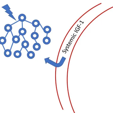
Figure 6: Systemic IGF-1 is transported through the blood-brain-barrier to the site of neuronal activity. IGF-1 binding protein is cleaved from the IGF-1 molecule, allowing passage through the barrier. Once in the CNS, IGF-1 has a limited half-life because it no longer has its binding protein chaperone. See Nishijima et al., 2016 for a more detailed figure.
We therefore see that the maintenance of stable oscillatory states in the brain is essential to proper function over time and this reality suggests that there are innate response mechanisms that support this stability. That neuronal dynamics regulate the transport of IGF-1 through the BBB provides a specific example of how a neurotrophic biochemical response acts to support neuronal health and function. Toth et al. (2015) reported on the disruption of neurovascular coupling in a murine model resulting from IGF-1 deficiency, emphasizing the integrative role of neuronal activity and the metabolic function mediated by the vasculature.
Part 5: Details for One Form of Sensory Neuromodulation
risks. It is interesting to note that NREM was once called synchronous sleep, reflective of the dominant activity seen on EEG recordings.
Sensory neuromodulation delivers a signal, even if it is non-physiological, via an innate network that processes the artificial signal just as it would naturally occurring sensory stimuli. We propose, therefore, that sensory neuromodulation may act to encourage developmentally established network behavior and thus may act to strengthen endogenous oscillators that couple to the sensory network. In other words, the sensory traffic drives target oscillators in a manner consistent with innate function. We hypothesize that sensory neuromodulation might then act to rehabilitate dysfunctional neural networks, bringing them back closer to a developmental state via neuroplastic modification (figure 7). Evidence for such rehabilitative potential for sensory neuromodulation is not definitive, but we now review some clinical results that support this hypothesis.
Figure 7: Neurological disease represented as weak or broken network connections. Sensory neuromodulation is hypothesized to force activity in weak networks, strengthening synapses and improving coupling. The IGF-1 mechanism may be a key player in this network repair process.
VNM:
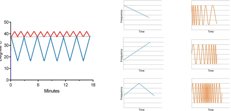
Rogers & Smith (US 8,262,717) conceived of a solid-state CVS device that would warm and cool the ear canal, thus enabling longitudinal therapy in the home. In order to extend treatment times with CVS, a time-varying thermal stimulus is needed to avoid adaptation of the vestibular hair cells (Bock et al., 1979) and so the solid-state CVS device was equipped with the means to deliver time-varying thermal waveforms, independently to both ears (figure 8). CVS alters the tonic firing rate, of ~100 Hz, of the regularly firing vestibular hair cells. Applying a triangular temperature waveform (fig. 8) results in a time-varying firing pattern in the vestibular hair cells around the 100 Hz tonic rate. Additionally, the envelope of the triangular waveform establishes a slower modulation and when both ears are stimulated simultaneously, at different frequencies, the resulting afferent firing pattern reaching the vestibular nuclei can be quite complex and spans frequencies from 0.01 Hz to 100+ Hz. Therefore, even though the time course of thermal transfer to the inner ear during CVS is slow, at least on the order of seconds, it is important to recognize that modulation of the equilibrium firing rate means that frequencies centered on 100 Hz are also present.
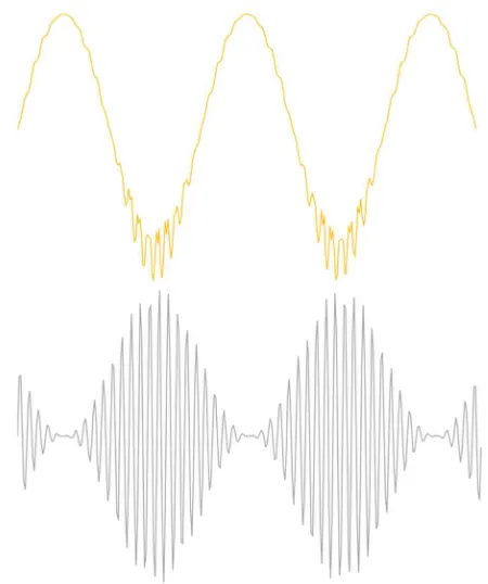
Figure 8: An example of time varying waveforms used with the solid-state CVS device (left). One ear receives a warm triangle wave and the other a cold triangle wave. As seen in figure 5, warming increases the afferent firing rate and cooling decreases it. On the right, the effect of a time-varying temperature on the hair cell firing rate is shown graphically. The response (orange) frequency shifts in time, a phenomenon called a “chirp.” Thus a complex range of firing rate modulations can be achieved.
episodic migraines.
The migraine study involved slow modulations in temperature, in the range of 0.008 – 0.033 Hz. The temperature changes then induce alterations in the firing rate of vestibular hair cells at the in the same range of frequencies. Is there any evidence that such slow oscillations might be important, on top of the basic need to avoid adaptation of the vestibular response? Black et al. (2016) provided evidence of entrainment of a pontine pacing center, which engendered oscillations in cerebral blood flow velocity, using time-varying CVS. In that study, the oscillations appeared to sharpen to what was presumably a natural resonance, at about 0.025 Hz, and persisted when CVS was stopped, a strong indicator of entrainment. Sliwka et al. (2001) reported that migraineurs had abnormal B wave activity (interictal), spontaneous oscillations in blood flow velocity in a range from 0.008 – 0.05 Hz. B waves are thought to originate in the pons and may be part of a the autoregulatory response (the B wave period is roughly the transit time of blood from the heart to the brain and back). Swlika et al. suggested that abnormal B wave activity in migraineurs may stem from a dysfunction in the monoaminergic/serotonergic system in the brainstem, a hypothesis that overlaps independent models of migraine pathogenesis (Coppola et al., 2009). Therefore, evidence for entrainment of B waves with time-varying CVS could be a biomarker of utility when seeking to titrate therapy for individual migraineurs.
Oscillations in the B wave frequency range have also been observed in BOLD MRI studies aimed at measuring cortical functional connectivity (Cordes et al., 2001; Bharath et al., 2017; Leopold et al., 2003; Schmidt, 2009) and appear in slow-wave sleep (Dang-Vu et al., 2008). Dang-Vu et al. suggested a possible relationship between the observed oscillations in slow-wave sleep and waking DMN, implying a restorative role of sleep on large-scale cortical functional organization. Oscillations in the B waves range during sleep have also been seen with EEG (Terzano et al., 1985) and with transcranial Dopper sonography in newborns (Ferrarri et al., 1994). Are the oscillations seen in functional connectivity studies related to those seen in cerebrovascular studies or is there just a coincidental overlap in frequency ranges? Even if the two phenomena don’t share a common pontine pacing center, entraining one sets up the possibility of entraining the other. Measuring the onset and strength of entrainment of targeted brain oscillators presents a tangible method for the titration of therapy. No functional brain oscillator can remain isolated (uncoupled) from other oscillators (this is the essence of the small-world network concept) and time-varying CVS presents a powerful method for exciting complete networks, innervated by the vestibular system. Coupling occurs not just for oscillators that have the same resonant frequency, but cross-frequency coupling (CFC) allows for interactions between oscillators having different fundamental frequencies.
on cognition.
Time-varying CVS has also been used in a case study with two subjects in a minimally conscious state (Vanzan et al., 2017) with the aim of increasing awareness. Spontaneous recovery for these subjects generally occurs within a time window after emergence from coma and both of the subjects were past the time when natural recovery was expected. As measured with coma recovery scales (WHIM & CRS-R), improvement seemed to show a time-locked association with CVS treatment epochs, which consisted of 1-month of daily treatment, followed by 1-month of sham treatment, and ending with a second month of active treatment. Improvements were recorded during active treatment and no gains were seen during sham treatment. Note that the subjects were likely unaware of the active/sham changes. One of the subjects in particular demonstrated durable gains and progressed from an inability to move his gaze to a person in the room to initiating conversation and answering simple questions. The authors noted that the apparent gains imply improved function across a number of brain regions, but most particularly the thalamocortical projection system, and neuroplastic modification of neuronal pathways. They further suggested that vestibular pathways go well beyond a role in autonomic motor control and underlie higher cognitive states, a viewpoint consistent with the literature reviewed in Part 2.
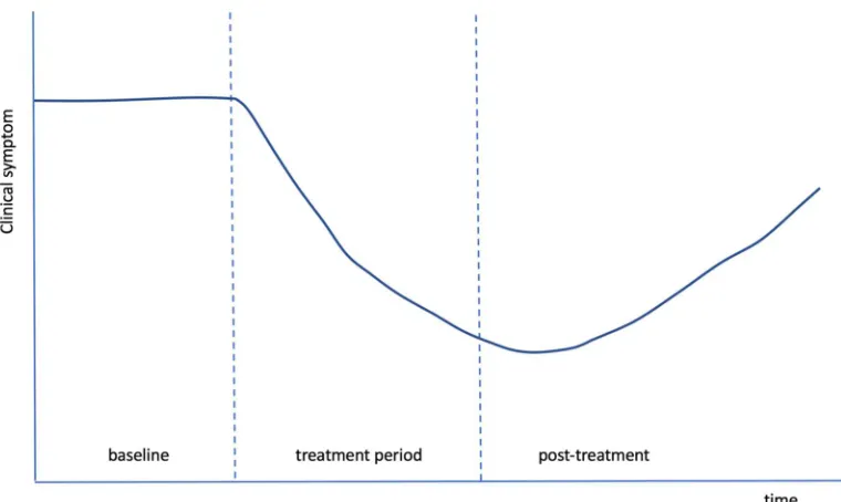
Figure 9: A composite graph of the response, over time, of multiple, different symptoms in Wilkinson et al. (accepted). The same characteristic shape and time course was seen across independent measures, suggesting a common effect, most likely neuroplastic modification of the dysfunctional pathways associated with the various symptoms.
Part 6: Discussion and future questions
but also because the vestibular sensory network is so well-suited to the task of delivering neuromodulation throughout the brain.
At present, the way in which sensory neuromodulation works, and how well it works, is not fully established and more clinical studies and mechanism of action studies are required. Yet it is reasonable to hypothesize, using the oscillator model of neurological disease, about what to expect.
Why it helps and doesn’t hurt
An oscillator-centric model clarifies how sensory neuromodulation can in principal work broadly and may mitigate multiple disease symptoms. This is because the nature of networked oscillators is to enable cross coupling between individual oscillators and networks. As an illustrative example, consider the cardiac oscillator. Heart rate variability (HRV) is a measure of the ability of the heart to respond to changes in demand from the body. High HRV means that the heart is very adaptive and is associated with good health. Low HRV means that the heart is not adaptive and means that the cardiac oscillator system is too weakly coupled to other brain-driven oscillator networks. If an oscillator is too weakly coupled, it is ineffective and thus the interventional goal is to improve and increase coupling to a normal level. In the case of the cardiac oscillator, exercise and a healthful lifestyle are well accepted as improving cardiac function, improving HRV (Kiviniemi et al., 2010). Aerobic exercise encourages the heart to be rate adaptive and we submit that forcing the cardiac oscillator to be responsive to the demands of the body is achieved by exposing it to an appropriately stressful stimulus. Conversely, inactivity places low demands on HRV and the cardiac oscillator will become more weakly coupled to the body. The cardiac oscillator has a developmentally established level of coupling to the body, but the strength of coupling can change: it can improve via exercise or it can decline via inactivity. We assert that beneficial coupling is encouraged by forcing a target oscillator to work with other network oscillators and coupling is altered (figure 7) through synaptic modification (neuroplasticity). This realization has important consequences for therapeutic intervention. Firstly, it’s not possible to harm the function of a target oscillator by entraining it with other oscillatory networks for, indeed, entrainment is fundamental to its function. Thus oscillator entrainment by sensory neuromodulation will not interfere with normal function. The stimulus does not need to be focused only on a damaged region, for instance. Secondly, entrainment works to reinforce the integrity of an oscillatory network’s developmentally derived form. Sensory neuromodulation provides input through endogenous channels and thereby forces functional responses, which reinforce synaptic coupling, in those areas innervated by the sensory pathways. It is for this reason that the extensive innervation of the vestibular network recommends it as a preferred conduit for sensory neuromodulation therapy. Understanding how sensory neuromodulation should work to maintain and improve coupling between oscillators would lead to the expectation of extensive effects on function (and thus potentially the mitigation of multiple disease symptoms) and durability of gains (because the effect is to create neuroplastic modification of the coupling between oscillators).
component may not be amenable to therapy alone without some form of physical or cognitive behavioral therapy. Let us look at each of these three examples in turn.
A neurodegenerative disease (like PD) or disease that is trauma-induced (like TBI or stroke) results in an alteration in pre-disease oscillatory networks. Early evidence from Wilkinson et al. (accepted) suggested that there may be a plastic response to time-varying CVS that works to return function broadly. It may be that concommitant physical training could help with motor dysfunction, in particular, and this would result in a ratchet effect whereby neuromodulation improves function and physical therapy improves function further. Studies with the PONS device typically included a physical training component (Chisholm et al., 2014). An interesting question for future research is whether sensory neuromodulation creates neuroplastic facilitation that augments conventional physical therapy.
Diseases like schizophrenia and idiopathic migraine are developmental. If there is no disease-free baseline to which to return, what does this mean for the applicability of sensory neuromodulation? Gerretsen et al. (2017) used irrigation-based CVS with schizophrenia subjects and found evidence of transiently improved insight to illness. The authors concluded that, similar to hemispatial neglect, CVS acted to balance activity in the two hemispheres, making use of the potential of unilateral CVS to preferentially activate one hemisphere. This is an interesting example as it points out the spectral nature of diseases like schizophrenia where improvement in one symptom may still have an impact on patient outcomes. Wilkinson et al. (2017) offered clear evidence of benefit from time-varying CVS for idiopathic episodic migraine. Why this is so is unknown, but we noted earlier that the brain actually does try to re-establish normal sensory habituation during an attack. And so perhaps a helpful viewpoint would be to think of the migraine brain as having two states, habituating and non-habituating, and neuromodulation increases the prevalence of habituation, thus reducing the sensory dysfunction that may be the source of the disease (Goadsby et al., 2017).
For behaviorally driven diseases like type-II diabetes and addiction, it seems unlikely that neuromodulation alone can’t be effective since habits and lifestyle must also be adjusted. The aim, rather, for neuromodulation could be to make the adoption of lifestyle changes easier and to help to establish new habitual patterns.
Part 7: Conclusion
The advent of home-use, low risk devices for delivering sensory neuromodulation creates a distinct advantage in that such therapies can be used early in the progress of a disease, versus implanted devices that are relegated to late-stage disease. This potential meshes with current efforts to detect neurodegenerative disease at an early stage, via the use of biomarkers. Taking therapeutic action simply on the basis of genetic profiling is a challenging decision, ethically, however a low-risk, low side-effect therapy mitigates the downside of acting early.
The brain, viewed from an engineer’s context, is energy intensive (consuming some 20% of metabolic output in about 2% of body mass) and is comprised of irreplaceable parts (no extensive adult neurogenesis). That leads to a poor prognosis for long-term function, unless innate support and repair mechanisms are in place. The challenge is different for organs that do generate new cells. Autoregulation is clearly one mechanism that supports brain function. The clearance of waste products, by the circulatory system and lymphatic and glymphatic routes, is also important. We find that the IGF-1 model proposed by Nishijima et al. (2010) presents a new avenue for model building, linking brain oscillators to underlying biochemical pathways that help to facilitate neuroprotection.
Acknowledgement: The authors thank Lanty L. Smith for discussions and support.
Appendix:
References:
Aberg, N. D., K. G. Brywe and J. Isgaard (2006). "Aspects of growth hormone and insulin-like growth factor-I related to neuroprotection, regeneration, and functional plasticity in the adult brain." ScientificWorldJournal 6: 53-80
Akundi, R. S., L. Zhi and H. Bueler (2012). "PINK1 enhances insulin-like growth factor-1-dependent Akt signaling and protection against apoptosis." Neurobiol Dis 45(1): 469-478 PMC3225697.
Ali, M. M., K. K. Sellers and F. Frohlich (2013). "Transcranial alternating current stimulation modulates large-scale cortical network activity by network resonance." J Neurosci 33(27): 11262-11275
Anacker, C. and R. Hen (2017). "Adult hippocampal neurogenesis and cognitive flexibility - linking memory and mood." Nat Rev Neurosci
Aru, J., J. Aru, V. Priesemann, M. Wibral, L. Lana, G. Pipa, W. Singer and R. Vicente (2015). "Untangling cross-frequency coupling in neuroscience." Curr Opin Neurobiol 31: 51-61
Assenza, G., F. Capone, L. di Biase, F. Ferreri, L. Florio, A. Guerra, M. Marano, M. Paolucci, F. Ranieri, G. Salomone, M. Tombini, G. Thut and V. Di Lazzaro (2017). "Corrigendum:
Oscillatory Activities in Neurological Disorders of Elderly: Biomarkers to Target for Neuromodulation." Front Aging Neurosci 9: 252 PMC5539606.
Aviles-Olmos, I., T. Foltynie, J. Panicker, D. Cowie, P. Limousin, M. Hariz, C. J. Fowler and L. Zrinzo (2011). "Urinary incontinence following deep brain stimulation of the pedunculopontine nucleus." Acta Neurochir (Wien) 153(12): 2357-2360
Ayadi, A. E., M. J. Zigmond and A. D. Smith (2016). "IGF-1 protects dopamine neurons against oxidative stress: association with changes in phosphokinases." Exp Brain Res 234(7): 1863-1873 PMC4893922.
Ayres, A. J. (1972). Sensory integration and learning disorders. Los Angeles, Calif.,, Western Psychological Services.
Bain, L. and e. al. (2015). Non-Invasive Neuromodulation of the Central Nervous System: Opportunities and Challenges: Workshop Summary, Institute of Medicine.
Barany, R. and K. Wittmaack (1911). "Funktionelle Prufung des Vestibularapparates (functional testing of the vestibular apparatus)." Otol. Ges. 20: 184-238
Barthelemy, L., D. Mabin, A. Belaud and C. Peyraud (1977). "[Electrical activity of the brain of the eel (Anguilla anguilla L.) subjected to hypoxia and hypercapnia]." J Physiol (Paris) 73(8): 1035-1044
Barton, R. A. and C. Venditti (2014). "Rapid Evolution of the Cerebellum in Humans and Other Great Apes." Current Biology 24: 1-5
Bellesi, M., B. A. Riedner, G. N. Garcia-Molina, C. Cirelli and G. Tononi (2014). "Enhancement of sleep slow waves: underlying mechanisms and practical consequences." Front Syst Neurosci 8: 208 4211398.
Bharath, R. D., R. Panda, J. Saini, K. Sriganesh and G. S. U. Rao (2017). "Dynamic local connectivity uncovers altered brain synchrony during propofol sedation." Sci Rep 7(1): 8501 PMC5561230.
Bishop, G. H. (1959). "The relation between nerve fiber size and sensory modality: phylogenetic implications of the afferent innervation of cortex." J Nerv Ment Dis 128(2): 89-114
Bitto, A., C. Lerner, C. Torres, M. Roell, M. Malaguti, V. Perez, A. Lorenzini, S. Hrelia, Y. Ikeno, M. E. Matzko, R. McCarter and C. Sell (2010). "Long-term IGF-I exposure decreases autophagy and cell viability." PLoS ONE 5(9): e12592 2935370.
Black, R.D., Balaban, C.D., Silberstein, S.D. (2018). “Vestibular Neuroscience for the Headache Neurologist.” Invited review to appear in Headache Currents.
Black, R. D., L. L. Rogers, K. K. Ade, H. A. Nicoletto, H. D. Adkins and D. T. Laskowitz (2016). "Non-Invasive Neuromodulation Using Time-Varying Caloric Vestibular Stimulation." IEEE J Transl Eng Health Med 4: 2000310
Bock, O., H. von Koschitzky and W. H. Zangemeister (1979). "Vestibular adaptation to long-term stimuli." Biol Cybern 33(2): 77-79
Brighina, F., A. Palermo, M. L. Panetta, O. Daniele, A. Aloisio, G. Cosentino and B. Fierro (2009). "Reduced cerebellar inhibition in migraine with aura: a TMS study." Cerebellum 8(3): 260-266
Buzsáki, G. (2006). Rhythms of the brain. Oxford ; New York, Oxford University Press.
Chan, F., N. Z. Lax, C. H. Davies, D. M. Turnbull and M. O. Cunningham (2016). "Neuronal oscillations: A physiological correlate for targeting mitochondrial dysfunction in
neurodegenerative diseases?" Neuropharmacology 102: 48-58
Chisholm, A. E., R. N. Malik, J. S. Blouin, J. Borisoff, S. Forwell and T. Lam (2014).
"Feasibility of sensory tongue stimulation combined with task-specific therapy in people with spinal cord injury: a case study." J Neuroeng Rehabil 11: 96 PMC4057581.
Chokron, S., E. Dupierrix, M. Tabert and P. Bartolomeo (2007). "Experimental remission of unilateral spatial neglect." Neuropsychologia 45(14): 3127-3148
Conforto, A. B., E. Amaro, Jr., A. L. Goncalves, J. P. Mercante, V. Z. Guendler, J. R. Ferreira, C. C. Kirschner and M. F. Peres (2014). "Randomized, proof-of-principle clinical trial of active transcranial magnetic stimulation in chronic migraine." Cephalalgia 34(6): 464-472
Connolly, K. R., A. Helmer, M. A. Cristancho, P. Cristancho and J. P. O'Reardon (2012).
"Effectiveness of transcranial magnetic stimulation in clinical practice post-FDA approval in the United States: results observed with the first 100 consecutive cases of depression at an academic medical center." J Clin Psychiatry 73(4): e567-573
Coppola, G., V. De Pasqua, F. Pierelli and J. Schoenen (2012). "Effects of repetitive transcranial magnetic stimulation on somatosensory evoked potentials and high frequency oscillations in migraine." Cephalalgia 32(9): 700-709
Coppola, G., C. Di Lorenzo, J. Schoenen and F. Pierelli (2013). "Habituation and sensitization in primary headaches." J Headache Pain 14: 65 3733593.
Coppola, G., F. Pierelli and J. Schoenen (2009). "Habituation and migraine." Neurobiol Learn Mem 92(2): 249-259
Cordes, D., V. M. Haughton, K. Arfanakis, J. D. Carew, P. A. Turski, C. H. Moritz, M. A. Quigley and M. E. Meyerand (2001). "Frequencies contributing to functional connectivity in the cerebral cortex in "resting-state" data." AJNR Am J Neuroradiol 22(7): 1326-1333
D'Mello, S. R., K. Borodezt and S. P. Soltoff (1997). "Insulin-like growth factor and potassium depolarization maintain neuronal survival by distinct pathways: possible involvement of PI 3-kinase in IGF-1 signaling." J Neurosci 17(5): 1548-1560
Dang-Vu, T. T., M. Schabus, M. Desseilles, G. Albouy, M. Boly, A. Darsaud, S. Gais, G. Rauchs, V. Sterpenich, G. Vandewalle, J. Carrier, G. Moonen, E. Balteau, C. Degueldre, A. Luxen, C. Phillips and P. Maquet (2008). "Spontaneous neural activity during human slow wave sleep." Proc Natl Acad Sci U S A 105(39): 15160-15165 PMC2567508.
De Vera, L., J. Gonzalez and R. V. Rial (1994). "Reptilian waking EEG: slow waves, spindles and evoked potentials." Electroencephalogr Clin Neurophysiol 90(4): 298-303
Dosenbach, N. U., D. A. Fair, F. M. Miezin, A. L. Cohen, K. K. Wenger, R. A. Dosenbach, M. D. Fox, A. Z. Snyder, J. L. Vincent, M. E. Raichle, B. L. Schlaggar and S. E. Petersen (2007). "Distinct brain networks for adaptive and stable task control in humans." Proc Natl Acad Sci U S A 104(26): 11073-11078 PMC1904171.
Drews, J. (2000). "Drug discovery: a historical perspective." Science 287(5460): 1960-1964
Ebert, A. D., A. J. Beres, A. E. Barber and C. N. Svendsen (2008). "Human neural progenitor cells over-expressing IGF-1 protect dopamine neurons and restore function in a rat model of Parkinson's disease." Exp Neurol 209(1): 213-223
Elmariah, H. (2007). IDENTIFICATION OF NEURAL ACTIVITY IN APLYSIA
ABDOMINAL GANGLIA USING IMPEDANCE MEASUREMENT. MS, U. of Florida.
Ferrarri, F., A. W. Kelsall, J. M. Rennie and D. H. Evans (1994). "The relationship between cerebral blood flow velocity fluctuations and sleep state in normal newborns." Pediatr Res 35(1): 50-54
Fitzgerald, G. and C. S. Hallpike (1942). "Studies in human vestibular function: observations on the directional preponderance of caloric nystagmus resulting from cerebral lesions. Part 1." Brain 65: 115-137
Fitzpatrick, R. C. and B. L. Day (2004). "Probing the human vestibular system with galvanic stimulation." J Appl Physiol 96(6): 2301-2316
Fox, M. D., R. L. Buckner, H. Liu, M. M. Chakravarty, A. M. Lozano and A. Pascual-Leone (2014). "Resting-state networks link invasive and noninvasive brain stimulation across diverse psychiatric and neurological diseases." Proc Natl Acad Sci U S A 111(41): E4367-4375 PMC4205651.
Fox, M. D. and M. E. Raichle (2007). "Spontaneous fluctuations in brain activity observed with functional magnetic resonance imaging." Nat Rev Neurosci 8(9): 700-711
Fries, P. (2005). "A mechanism for cognitive dynamics: neuronal communication through neuronal coherence." Trends Cogn Sci 9(10): 474-480
Gerretsen, P., D. D. Pothier, C. Falls, M. Armstrong, T. Balakumar, H. Uchida, D. C. Mamo, B. G. Pollock and A. Graff-Guerrero (2017). "Vestibular stimulation improves insight into illness in schizophrenia spectrum disorders." Psychiatry Research 251: 333-341
Goadsby, P. J., P. R. Holland, M. Martins-Oliveira, J. Hoffmann, C. Schankin and S. Akerman (2017). "Pathophysiology of Migraine: A Disorder of Sensory Processing." Physiol Rev 97(2): 553-622
Gontier, G., C. George, Z. Chaker, M. Holzenberger and S. Aid (2015). "Blocking IGF Signaling in Adult Neurons Alleviates Alzheimer's Disease Pathology through Amyloid-beta Clearance." J Neurosci 35(33): 11500-11513
Grabherr, L., G. Macauda and B. Lenggenhager (2015). "The Moving History of Vestibular Stimulation as a Therapeutic Intervention." Multisens Res 28(5-6): 653-687
Grinberg, Y. Y., M. E. Dibbern, V. A. Levasseur and R. P. Kraig (2013). "Insulin-like growth factor-1 abrogates microglial oxidative stress and TNF-alpha responses to spreading depression." J Neurochem 126(5): 662-672 3752330.
Grinberg, Y. Y., L. A. Zitzow and R. P. Kraig (2017). "Intranasally administered IGF-1 inhibits spreading depression in vivo." Brain Res 1677: 47-57 PMC5993215.
Gu, Y., C. Wang and A. Cohen (2004). "Effect of IGF-1 on the balance between autophagy of dysfunctional mitochondria and apoptosis." FEBS Lett 577(3): 357-360
Gujar, N., S. S. Yoo, P. Hu and M. P. Walker (2010). "The unrested resting brain: sleep
deprivation alters activity within the default-mode network." J Cogn Neurosci 22(8): 1637-1648 PMC2883887.
Hallett, M. (2005). "Neuroplasticity and rehabilitation." J Rehabil Res Dev 42(4): xvii-xxii Hariz, M. (2017). "My 25 Stimulating Years with DBS in Parkinson's Disease." J Parkinsons Dis 7(s1): S35-S43 PMC5345632.
Hitier, M., S. Besnard and P. F. Smith (2014). "Vestibular pathways involved in cognition." Front Integr Neurosci 8: 59 4107830.
Hoppensteadt, F. C. and E. M. Izhikevich (1998). "Thalamo-cortical interactions modeled by weakly connected oscillators: could the brain use FM radio principles?" Biosystems 48(1-3): 85-94
Hubel, D. H. and T. N. Wiesel (1959). "Receptive fields of single neurones in the cat's striate cortex." J Physiol 148: 574-591 PMC1363130.
Iaccarino, H. F., A. C. Singer, A. J. Martorell, A. Rudenko, F. Gao, T. Z. Gillingham, H. Mathys, J. Seo, O. Kritskiy, F. Abdurrob, C. Adaikkan, R. G. Canter, R. Rueda, E. N. Brown, E. S.
Boyden and L. H. Tsai (2016). "Gamma frequency entrainment attenuates amyloid load and modifies microglia." Nature 540(7632): 230-235 PMC5656389.
Iadecola, C. (2017). "The Neurovascular Unit Coming of Age: A Journey through Neurovascular Coupling in Health and Disease." Neuron 96(1): 17-42 PMC5657612.
Kang, B. P., A. Urbonas, A. Baddoo, S. Baskin, A. Malhotra and L. G. Meggs (2003). "IGF-1 inhibits the mitochondrial apoptosis program in mesangial cells exposed to high glucose." Am J Physiol Renal Physiol 285(5): F1013-1024
Kataoka, H., Y. Okada, T. Kiriyama, Y. Kita, J. Nakamura, S. Morioka, K. Shomoto and S. Ueno (2016). "Can Postural Instability Respond to Galvanic Vestibular Stimulation in Patients with Parkinson's Disease?" J Mov Disord 9(1): 40-43 4734983.
Kerkhoff, G., H. Hildebrandt, S. Reinhart, M. Kardinal, V. Dimova and K. S. Utz (2011). "A long-lasting improvement of tactile extinction after galvanic vestibular stimulation: two Sham-stimulation controlled case studies." Neuropsychologia 49(2): 186-195
Kerkhoff, G., I. Keller, V. Ritter and C. Marquardt (2006). "Repetitive optokinetic stimulation induces lasting recovery from visual neglect." Restor Neurol Neurosci 24(4-6): 357-369
Kikuchi, M., Y. Naito, M. Senda, T. Okada, S. Shinohara, K. Fujiwara, S. Y. Hori, Y. Tona and H. Yamazaki (2009). "Cortical activation during optokinetic stimulation - an fMRI study." Acta Otolaryngol 129(4): 440-443
Kim, D. J., V. Yogendrakumar, J. Chiang, E. Ty, Z. J. Wang and M. J. McKeown (2013). "Noisy galvanic vestibular stimulation modulates the amplitude of EEG synchrony patterns." PLoS One 8(7): e69055 3715484.
Kiviniemi, A. M., A. J. Hautala, H. Kinnunen, J. Nissila, P. Virtanen, J. Karjalainen and M. P. Tulppo (2010). "Daily exercise prescription on the basis of HR variability among men and women." Med Sci Sports Exerc 42(7): 1355-1363
Klingner, C. M., G. F. Volk, C. Flatz, S. Brodoehl, M. Dieterich, O. W. Witte and O. Guntinas-Lichius (2012). "Components of vestibular cortical function." Behav Brain Res 236C: 194-199 Kolev, O. (1990). "How caloric vestibular irritation influences migraine attacks." Cephalalgia 10(4): 167-169
Kuramoto, Y. (1984). "Cooperative Dynamics of Oscillator Community: A Study Based on Lattice of Rings." Prog Theor Phys Suppl 79: 223-240
Kusmierczak, M., F. Lajeunesse, L. Grand and I. Timofeev (2015). "Changes in long-range connectivity and neuronal reorganization in partial cortical deafferentation model of
Larson-Prior, L. J., J. M. Zempel, T. S. Nolan, F. W. Prior, A. Z. Snyder and M. E. Raichle (2009). "Cortical network functional connectivity in the descent to sleep." Proc Natl Acad Sci U S A 106(11): 4489-4494 PMC2657465.
Lecrux, C. and E. Hamel (2011). "The neurovascular unit in brain function and disease." Acta Physiol (Oxf) 203(1): 47-59
Leonard, G., Y. Lapierre, J. K. Chen, R. Wardini, J. Crane and A. Ptito (2017). "Noninvasive tongue stimulation combined with intensive cognitive and physical rehabilitation induces neuroplastic changes in patients with multiple sclerosis: A multimodal neuroimaging study." Mult Scler J Exp Transl Clin 3(1): 2055217317690561 PMC5466147.
Leopold, D. A., Y. Murayama and N. K. Logothetis (2003). "Very slow activity fluctuations in monkey visual cortex: implications for functional brain imaging." Cereb Cortex 13(4): 422-433
Levine, A. J., C. R. Harris and A. M. Puzio-Kuter (2012). "The interfaces between signal
transduction pathways: IGF-1/mTor, p53 and the Parkinson Disease pathway." Oncotarget 3(11): 1301-1307 PMC3717794.
Lopez, C. (2016). "The vestibular system: balancing more than just the body." Curr Opin Neurol 29(1): 74-83
Lopez, C. and O. Blanke (2011). "The thalamocortical vestibular system in animals and humans." Brain Res Rev 67(1-2): 119-146
Manto, M., J. M. Bower, A. B. Conforto, J. M. Delgado-Garcia, S. N. da Guarda, M. Gerwig, C. Habas, N. Hagura, R. B. Ivry, P. Marien, M. Molinari, E. Naito, D. A. Nowak, N. Oulad Ben Taib, D. Pelisson, C. D. Tesche, C. Tilikete and D. Timmann (2012). "Consensus paper: roles of the cerebellum in motor control--the diversity of ideas on cerebellar involvement in movement." Cerebellum 11(2): 457-487
Major, R. J. and A. A. J. Valdivia (2018). "Mechanism of Synaptic Dysfunction and How This Disruption in IGF-1 homeostasis Leads to Neurodegenerative Diseases: A Theory." Biology and Medicine 10(4)
Marks, K. L., D. T. Martel, C. Wu, G. J. Basura, L. E. Roberts, K. C. Schvartz-Leyzac and S. E. Shore (2018). "Auditory-somatosensory bimodal stimulation desynchronizes brain circuitry to reduce tinnitus in guinea pigs and humans." Sci Transl Med 10(422)
Mason, J. L., K. Suzuki, D. D. Chaplin and G. K. Matsushima (2001). "Interleukin-1beta promotes repair of the CNS." J Neurosci 21(18): 7046-7052
McGeoch, P. D., L. E. Williams, T. Song, R. R. Lee, M. Huang and V. S. Ramachandran (2009). "Post-stroke tactile allodynia and its modulation by vestibular stimulation: a MEG case study." Acta Neurol Scand 119(6): 404-409
McLelland, D. and R. VanRullen (2016). "Theta-Gamma Coding Meets Communication-through-Coherence: Neuronal Oscillatory Multiplexing Theories Reconciled." PLoS Comput Biol 12(10): e1005162 PMC5065198.
Muller, M. F., C. Rummel, M. Goodfellow and K. Schindler (2014). "Standing waves as an explanation for generic stationary correlation patterns in noninvasive EEG of focal onset seizures." Brain Connect 4(2): 131-144
Nath, R. D., C. N. Bedbrook, M. J. Abrams, T. Basinger, J. S. Bois, D. A. Prober, P. W.
Sternberg, V. Gradinaru and L. Goentoro (2017). "The Jellyfish Cassiopea Exhibits a Sleep-like State." Current Biology
Neubauer, R. A., S. F. Gottlieb and A. Miale, Jr. (1992). "Identification of hypometabolic areas in the brain using brain imaging and hyperbaric oxygen." Clin Nucl Med 17(6): 477-481
Ng, F. and B. L. Tang (2013). "When is Sirt1 activity bad for dying neurons?" Front Cell Neurosci 7: 186 3807049.
Nieto-Estevez, V., C. Defterali and C. Vicario-Abejon (2016). "IGF-I: A Key Growth Factor that Regulates Neurogenesis and Synaptogenesis from Embryonic to Adult Stages of the Brain." Front Neurosci 10: 52 PMC4763060.
Nishiike, S., S. Nakamura, S. Arakawa, N. Takeda and T. Kubo (1996). "GABAergic inhibitory response of locus coeruleus neurons to caloric vestibular stimulation in rats." Brain Res 712(1): 84-94
Nishijima, T., J. Piriz, S. Duflot, A. M. Fernandez, G. Gaitan, U. Gomez-Pinedo, J. M. Verdugo, F. Leroy, H. Soya, A. Nunez and I. Torres-Aleman (2010). "Neuronal activity drives localized blood-brain-barrier transport of serum insulin-like growth factor-I into the CNS." Neuron 67(5): 834-846
Nishijima, T., I. Torres-Aleman and H. Soya (2016). "Exercise and cerebrovascular plasticity." Prog Brain Res 225: 243-268
Okada, Y., Y. Kita, J. Nakamura, H. Kataoka, T. Kiriyama, S. Ueno, M. Hiyamizu, S. Morioka and K. Shomoto (2015). "Galvanic vestibular stimulation may improve anterior bending posture in Parkinson's disease." Neuroreport 26(7): 405-410
Pan, W., R. Soma, S. Kwak and Y. Yamamoto (2008). "Improvement of motor functions by noisy vestibular stimulation in central neurodegenerative disorders." J Neurol 255(11): 1657-1661
Pinal, D., M. Zurron, F. Diaz and P. Sauseng (2015). "Stuck in default mode: inefficient cross-frequency synchronization may lead to age-related short-term memory decline." Neurobiol Aging 36(4): 1611-1618
Pitt, J., K. C. Wilcox, V. Tortelli, L. P. Diniz, M. S. Oliveira, C. Dobbins, X. W. Yu, S.
Nandamuri, F. C. A. Gomes, N. DiNunno, K. L. Viola, F. G. De Felice, S. T. Ferreira and W. L. Klein (2017). "Neuroprotective astrocyte-derived insulin/insulin-like growth factor 1 stimulates endocytic processing and extracellular release of neuron-bound Abeta oligomers." Mol Biol Cell 28(20): 2623-2636 PMC5620371.
Raichle, M. E. (2015). "The brain's default mode network." Annu Rev Neurosci 38: 433-447
Robb, D. H. F. and B. Roth (2003). "Brain activity of Atlantic salmon (Salmo salar) following electrical stunning using various field strengths and pulse durations." Aquaculture 216: 363-369
Robida-Stubbs, S., K. Glover-Cutter, D. W. Lamming, M. Mizunuma, S. D. Narasimhan, E. Neumann-Haefelin, D. M. Sabatini and T. K. Blackwell (2012). "TOR signaling and rapamycin influence longevity by regulating SKN-1/Nrf and DAF-16/FoxO." Cell Metab 15(5): 713-724 3348514.
Rode, G., C. Tilikete, J. Luaute, Y. Rossetti, A. Vighetto and D. Boisson (2002). "Bilateral vestibular stimulation does not improve visual hemineglect." Neuropsychologia 40(7): 1104-1106
Rogers & Smith (US patent 8,262,717): “Vestibular stimulation apparatus and associated methods of use.”
Rummel, C., M. Goodfellow, H. Gast, M. Hauf, F. Amor, A. Stibal, L. Mariani, R. Wiest and K. Schindler (2013). "A systems-level approach to human epileptic seizures." Neuroinformatics 11(2): 159-173
Samoudi, G., M. Jivegard, A. P. Mulavara and F. Bergquist (2014). "Effects of Stochastic Vestibular Galvanic Stimulation and LDOPA on Balance and Motor Symptoms in Patients With Parkinson's Disease." Brain Stimul
Schmidt, S. H. (2009). Bandwidth-Specific Functional Connectivity of Physiological Low Frequency Oscillations in fMRI. Dr. Med., Aus der Klinik für Neurologie
der Medizinischen Fakultät Charité – Universitätsmedizin Berlin.
Schott, G. D. (1993). "Penfield's homunculus: a note on cerebral cartography." J Neurol Neurosurg Psychiatry 56(4): 329-333 PMC1014945.
Seppi, K., D. Weintraub, M. Coelho, S. Perez-Lloret, S. H. Fox, R. Katzenschlager, E. M. Hametner, W. Poewe, O. Rascol, C. G. Goetz and C. Sampaio (2011). "The Movement Disorder Society Evidence-Based Medicine Review Update: Treatments for the non-motor symptoms of Parkinson's disease." Mov Disord 26 Suppl 3: S42-80 PMC4020145.
Sharma, A. N., E. S. B. F. da Costa, J. C. Soares, A. F. Carvalho and J. Quevedo (2016). "Role of trophic factors GDNF, IGF-1 and VEGF in major depressive disorder: A comprehensive review of human studies." J Affect Disord 197: 9-20 4837031.
Silberstein, S. D., A. H. Calhoun, R. B. Lipton, B. M. Grosberg, R. K. Cady, S. Dorlas, K. A. Simmons, C. Mullin, E. J. Liebler, P. J. Goadsby, J. R. Saper and E. S. Group (2016). "Chronic migraine headache prevention with noninvasive vagus nerve stimulation: The EVENT study." Neurology 87(5): 529-538 4970666.
Sliwka, U., S. Harscher, R. R. Diehl, R. van Schayck, W. D. Niesen and C. Weiller (2001). "Spontaneous oscillations in cerebral blood flow velocity give evidence of different autonomic dysfunctions in various types of headache." Headache 41(2): 157-163
Sonntag, W. E., F. Deak, N. Ashpole, P. Toth, A. Csiszar, W. Freeman and Z. Ungvari (2013). "Insulin-like growth factor-1 in CNS and cerebrovascular aging." Front Aging Neurosci 5: 27 3698444.
Sultan, F., M. Augath, S. Hamodeh, Y. Murayama, A. Oeltermann, A. Rauch and P. Thier (2012). "Unravelling cerebellar pathways with high temporal precision targeting motor and extensive sensory and parietal networks." Nat Commun 3: 924
Sung, H. Y., E. N. Choi, D. Lyu, I. Mook-Jung and J. H. Ahn (2014). "Amyloid beta-mediated epigenetic alteration of insulin-like growth factor binding protein 3 controls cell survival in Alzheimer's disease." PLoS One 9(6): e99047 PMC4070895.
Suzuki, J., Y. Kaziro and H. Koide (1998). "Synergistic action of R-Ras and IGF-1 on Bcl-xL expression and caspase-3 inhibition in BaF3 cells: R-Ras and IGF-1 control distinct anti-apoptotic kinase pathways." FEBS Lett 437(1-2): 112-116
Talukdar, T., A. Nikolaidis, C. E. Zwilling, E. J. Paul, C. H. Hillman, N. J. Cohen, A. F. Kramer and A. K. Barbey (2017). "Aerobic Fitness Explains Individual Differences in the Functional Brain Connectome of Healthy Young Adults." Cereb Cortex: 1-10