Original Research Article
To Analyze the Impact of
18F-FDG PET/CT in Nodal Staging and Overall
Staging in Recently Diagnosed Non-Treated Cases of Squamous Cell
Carcinoma of Head and Neck: A Prospective Study
Bhavay Sonik
1*, Padma S. Sundaram
2, P. Shanmuga Sundaram
31*Assistant Professor, Department of Nuclear Medicine,
Guru Gobind Singh Medical College and Hospital, Faridkot, Punjab, India. 2Professor, 3Professor and Head, Department of Nuclear Medicine & PET/CT, Amrita Institute of Medical Sciences, Cochin, Kerala, India.
ABSTRACT
Background: 85% to 95% neoplasms of the head and neck are squamous cell carcinomas with most common sites being oral cavity, pharynx and larynx. The preservation of function, especially as it relates to speech, swallowing, and mastication, as well as cosmetic considerations, are considered essentials for determining the most effective management paradigm for squamous cell carcinomas of head and neck (SCCHN). The aim of this present study is to analyze the impact of 18F-FDG PET/CT in nodal staging and overall staging of recently diagnosed non-treated cases of SCCHN when compared with clinical and / or conventional imaging techniques such as CECT and MRI as and when available.
Materials and Methods: The present study was conducted in the Department of Nuclear Medicine and PET CT, Amrita Institute of Medical Sciences, Cochin, Kerala from November 2011 to May 2013. The study group comprised of non-treated patients of both sexes with age ranging from 20 to 81 years referred to our department with an established tissue diagnosis of SCCHN for 18F-FDG-PET/CT Whole Body scan for evaluation of disease status. Data including age, sex, primary site, levels of neck lymph node involvement at presentation, scopy (direct / indirect) findings, histopathology / FNAC report of primary site & lymph nodes and conventional imaging findings were recorded. AJCC 7th edition, 2010 was used for staging.
Results: 37 patients were included in the study, male: female – 33: 4, mean age of presentation for males was 57.5 years and for the females was 55 years. Oropharynx was the most common site of primary tumour in 40% of patients (n=15). At presentation no nodes palpable, ipsilateral and bilateral neck
lymph nodes were present in 32.4% (n=12), 48.7% (n=18) and 18.9% (n=7) of patients respectively. Most common pre PET/CT nodal staging, according to 7th edition of AJCC in 32 patients (excluding 5 patients with nasopharyngeal malignancy), was N0 in 12 patients (37.5%). Post-PET/CT upstaging, downstaging and no change in nodal staging was seen in 11 (32.3%), 3 (8.1%) and 22 (59.5%) patients respectively. Overall change in staging was noted in 13% of patients when compared to conventional imaging techniques. Conclusion: 18F-FDG PET/CT accurately predicts nodal stage better than clinical and/or conventional imaging techniques leading to overall stage change in patients with SCCHN. Thus, it acts as a valuable tool in determining the exact nodal spread establishing the exact treatment plan.
Keywords: Squamous Cell Carcinoma, PET, CT.
*Correspondence to:
Dr. Bhavay Sonik, Assistant Professor,
Department of Nuclear Medicine,
Guru Gobind Singh Medical College and Hospital, Faridkot, Punjab, India.
Article History:
Received: 28-02-2017, Revised: 13-03-2017, Accepted: 30-03-2017
Access this article online Website:
www.ijmrp.com
Quick Response code
DOI:
10.21276/ijmrp.2017.3.2.082
INTRODUCTION
Head and neck cancer is a broad term that encompasses epithelial malignancies that arise in the paranasal sinuses, nasal cavity, oral cavity, pharynx and larynx. 85% to 95% of these epithelial malignancies are squamous cell carcinomas of the head and neck (SCCHN) and most common sites are the oral cavity, pharynx and larynx.1,2 The most important risk factors are tobacco and alcohol consumption1 and are implicated in 75% of all SCCHN and have a multiplicative combined effect.3 Treatment decision in
considerations are considered essentials for determining the most effective management paradigm for SCCHN. This is particularly true for oral cavity, oropharyngeal, and laryngeal carcinomas.5 Thus, precise prediction of the extent of the primary tumours, cervical node status and distant metastatic spread is important for treatment planning and prognosis. Patients with known distant metastases can possibly be spared the toxicities of aggressive and often unnecessary loco-regional therapy.6
The most common known imaging modalities in clinical use are Contrast Enhanced Computed Tomography (CECT) and Magnetic Resonance Imaging (MRI), which rely on criteria of contrast-enhancement patterns and size for the detection of lymph nodal metastases, which are not specific and have suboptimal sensitivity and specificity for the detection of metastatic disease.5,6 Role of 18F-Flouro-Deoxy-Glucose (18F-FDG) PET and 18F-FDG PET/CT
has been evaluated in the initial staging of head and neck carcinomas in a number of studies and is extremely useful in the situations where anatomic imaging is equivocal and the disease cannot be assessed by direct visualization.7-11 The aim of this present study is to analyse the impact of 18F-FDG PET/CT in nodal staging and overall staging of recently diagnosed non-treated cases of SCCHN when compared with clinical and/or conventional imaging techniques such as CECT and MRI as and when available.
MATERIALS AND METHODS
The present study was conducted in the Department of Nuclear Medicine and PET CT, Amrita Institute of Medical Sciences, Cochin, Kerala over a period of about one and half years in association with the Departments of Head and Neck Surgery, Medical Oncology, Surgical Oncology and Radiation Oncology beginning in November 2011 and till May 2013. Institutional ethical committee clearance was obtained. The study group comprised of non-treated patients of both sexes with age ranging from 20 to 81
years referred to our department with an established tissue diagnosis of SCCHN for 18F-FDG-PET/CT Whole Body scan for evaluation of disease status. Patients who had any treatment history of previous head / neck malignancy or any other site malignancy were excluded from the study. Informed consent was obtained from patient after informing about the entire procedure. A set of instructions were given to the patient a day before the appointment and on the day of appointment they were repeated. 8-10 mCi of 18F-FDG was injected intravenously in euglycemic status. Time of injection was noted along with pre-injection and post-injection counts. Whole body PET/CT images (head to mid thigh) were acquired after 45 min to 60 min post injection. Oral & IV contrast was given for CT part of the study in patients with normal renal function status. As and when required, dual point imaging (2-3 hours post injection of 18F-FDG) or imaging with hands down was also performed.
Data sets were reconstructed using iterative reconstruction techniques. Image fusion was performed using co-ordinate based fusion software and subsequently reviewed on ADWPET work station. Both PET and CT data were evaluated separately as well as after image fusion. Semi-quantitative analysis was done in form of standard uptake value (SUV max in gms/ml).
Detailed history was obtained before the 18F-FDG PET/CT scan and used along with patient data extracted from our hospital information system prior the 18F-FDG PET/CT study. Data included age, sex, primary site, levels of neck lymph nodal involvement at presentation, scopy (direct / indirect) findings, histopathology / FNAC report of primary site & lymph nodes and conventional imaging findings were recorded. AJCC 7th edition, 2010 was used for staging.
Study population was divided into two groups i.e. Group A and B, depending whether conventional imaging was done or not prior to the PET/CT scan respectively to see the impact of 18F-FDG PET/CT on nodal staging.
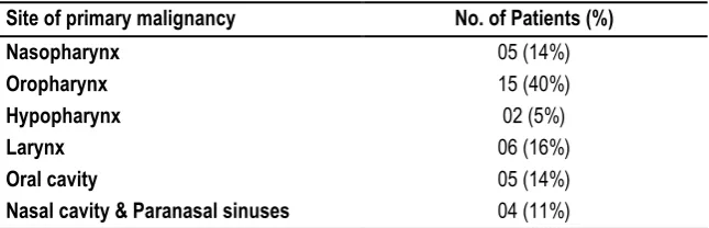
Table 1: Site of Primary Tumour at presentation
Table 2: Distribution of lymph nodal metastases at presentation
RESULTS
A total of 37 patients were included in this study. Of the 37 patients included in the study, there were 33 males (89.2%) and 4 females (10.8%). The mean age of presentation for males was 57.5 years and for the females was 55 years. The overall mean and median age of presentation for the study population was 57.2 and 63 years respectively. Oropharynx was the most common site
of primary tumour, seen in 15 cases (40%), in our study group. Other sites of primary tumour and their %age incidence in our study are summarized in Table 1.
Table 2 summarizes the distribution of lymph nodal metastases at presentation. At presentation ipsilateral (single / multiple levels) lymph nodes were present in majority of the patients (48.7%). Table 3 elaborates the pre 18F-FDG PET/CT Nodal staging at
Site of primary malignancy No. of Patients (%)
Nasopharynx 05 (14%)
Oropharynx 15 (40%)
Hypopharynx 02 (5%)
Larynx 06 (16%)
Oral cavity 05 (14%)
Nasal cavity & Paranasal sinuses 04 (11%)
Pattern No. of Patients (%)
Ipsilateral 18 (48.7 %)
Bilateral 07 (18.9%)
presentation (excluding Nasopharynx) according to 7th edition of AJCC staging guidelines 2010 for 32 patients. Most common pre 18F-FDG PET/CT nodal staging, by the way of clinical examination
and conventional imaging of neck in these 32 patients was N0 (n-12, 37.5%). Only 6.2% patients (n-2) were staged to N3 category. Respective nodal stage for each primary site (excluding nasopharynx) at presentation is shown in Graph 1.
For 5 patients with nasopharyngeal primary, N1, N2 and N3 nodal staging was seen in n-2 (40%), n-1 (20%) and n-2 (40%) patients respectively. All patients with N3 lymph nodes (n-4) had ipsilateral neck node distribution.
We divided our study population into two groups i.e. Group A and B, depending whether conventional imaging was done or not prior to the 18F-FDG PET/CT scan. Group A (n-25) consisted of those patients who had undergone CECT and / or MRI of the head and/or neck region (n-23) and metastatic workup i.e. CECT thorax
(n-1) and CECT abdomen (n-1). Group B (n-12) consisted of those patients who underwent directly 18F-FDG PET/CT imaging after clinical examination.
For all 37 patients, impact of 18F-FDG PET/CT on nodal staging (N) and overall staging according to the 7th edition of AJCC 2010 is shown in Table 4 and Table 5. Nodal upstaging and downstaging was seen in a total of 11 and 3 patients respectively. 22 patients demonstrated no change in nodal staging. Overall change in staging i.e. upstaging and downstaging was noted in 5 and 2 patients respectively amongst the 14 patients with change in nodal staging.
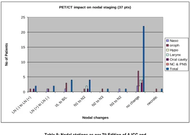
Table 6, 7 and graph 2 show changes in various nodal stations after 18F-FDG PET/CT. Also changes in nodal stations are divided among the group A and group B patients in Table 6.
Table 8 shows the Nodal stations as per 7th Edition of AJCC and number of patients in each station after 18F-FDG PET/CT.
Table3: Pre 18F-FDG PET/CT Nodal staging at presentation (excluding Nasopharynx) according to 7th edition of AJCC staging guidelines 2010 for 32 patients
Graph 1: Nodal staging for each primary site (excluding Nasopharynx)
Table 4: Post 18F-FDG PET/CT overall N stage change in 37 pts
Primary Tumour site (n-37) No change
(n-22)
Upstage (n-11)
Downstage (n-3)
Nasopharynx (n – 5) 2 1 2
Oropharynx (n – 14 + 1*) 7 6 1
Hypopharynx (n – 2) 2 - -
Larynx (n – 6) 4 2 -
Oral cavity (n – 5) 3 2 -
Nasal Cavity & Paranasal sinuses (n – 4) 4 - -
(*) 1 pt had FDG non avid necrotic lymph node.
Pre PET/CT Nodal stage
5
2
3
2
1
3
1 6
1 3
1
2
1 1
0 1 2 3 4 5 6 7
Oropharynx Hypopharynx Larynx Oral cavity NC & PNS
Site of Primary Tumor
N
o
o
f
Pa
ti
e
n
ts
N0
N1
N2a
N2b
N2c
N3
NODAL STAGING No. of Patients (%)
N0 12 (37.5 %)
N1 05 (15.6 %)
N2a 00 (0.0 %)
N2b 07 (21.9 %)
N2c 06 (18.8 %)
Table 5: Nodal stations as per AJCC and impact on staging after 18F-FDG PET/CT for n-14 patients Pt S
No
Primary Site Pre PET/CT
Stage
Post PET/CT Stage
Impact on N stage Overall Impact on
Staging
1 Nasopharynx N1 ; IVA N2; IV A Upstage None
2 Nasopharynx N3; IV B N2; III Downstage Yes
5 Nasopharynx N1; II B N0; II Downstage None
7 Oropharynx N2b; IVA N2c; IVA Upstage None
9 Oropharynx N2b; IVA N2c; IVA Upstage None
11 Oropharynx N2b; IVA N2c; IVA Upstage None
12 Oropharynx N1; III N2b; IVA Upstage Yes
14 Oropharynx N0; II N1; III Upstage Yes
16 Oropharynx N2b; IVA N3; IVB Upstage Yes
19 Oropharynx N2b; IVA N0; II Downstage Yes
23 Larynx N1; III N2a; IVA Upstage Yes
24 Larynx N1; III N2b; IVA Upstage Yes
29 Oral cavity N0; III N1; III Upstage None
32 Oral cavity N1; IVA N2b; IVA Upstage None
Table 6: Post 18F-FDG PET/CTchange invarious nodal stations in 37 pts
Parameters Nasopharynx
(n-5)
Oropharynx (n-15)
Hypopharynx (n-2)
Larynx (n-6)
Oral Cavity (n-5)
NC & PNS (n-4)
▪ LN (-) to LN (+) - 1
Group B
- - 1
Group B
-
▪ LN (+) to LN (-) 1
Group A
1 Group B
- - - -
▪ I/L to B/L (includes Nasopharynx N2 & N2c)
1 Group A
3 GroupA,A,B
- - - -
▪ N1 to N2 (excluding Nasopharynx N2)
NA 1
Group B
- 2
Group A,A
1 Group A
-
▪ N2 to N3 - 1
Group A
- - - -
▪ N3 to N2 1
Group B
- - - - -
▪ No change 2 7 2 4 3 4
▪ Necrotic - 1 - - - -
[LN(-): Lymph node negative, LN(+): Lymph node positive, I/L: Ipsilateral, B/L: Bilateral, NC: nasal cavity; PNS: Paranasal sinuses.]
Table 7: Post 18F-FDG PET/CT change in various nodal stations in 37 pts
CHANGE No of Patients
UPSTAGING 11
N0 to N1 2
N1 to N2 (nasopharynx) 1
N1 to N2a 1
N1 to N2b 3
N2b to N2c 3
N2b to N3 1
DOWNSTAGE 03
N1 to N0 1
N2b to N0 1
N3 to N2 1
NO CHANGE 22
Graph 2:PET/CT impact on various nodal stations for all 37 pts
Table 8: Nodal stations as per 7th Edition of AJCC and number of patients in each station after PET/CT
NODAL STATION No of patients (n-37)
N0* 13
N1 (Nasopharynx) --
N1 (all other tumours) 03
N2a 01
N2b 05
Bilateral (N2 for Nasopharynx and N2c for all others) 11
Nasopharynx – 3 Others – 8
N3 (Nasopharynx & all others) 04
(*) includes 1 patient with necrotic lymph node.
DISCUSSION
Our study population comprised of 37 patients, 33 males and 4 females. The overall mean and median age of our study group at presentation was 57.2 and 63 years respectively. Male predominance with median age of presentation in early 60s has been reported in the cancer statistics review done by Ries LAG et al.12 and Parkin DM et al.13 More than 50% of patients of SCCHN at presentation have regional lymph nodal metastases.14 In our study, 25 patients (67.6%) presented with lymph nodal metastases, inclusive of ipsilateral and bilateral nodal distribution. Presentation with ipsilateral lymph nodal metastases was more common than bilateral lymph nodal metastases (48.7% v/s 18.9%), similar to the pattern reported by Prestwich RJ et al (63.6% v/s 25.5%). However, incidence of bilateral lymph nodal metastases was higher when compared to the reported incidence of 10% by Jereeczek-Fossa BA et al.15 This may be due to smaller sample size of our population or late presentation.
According to the 7th edition of AJCC 2010 (excluding 5 patients with nasopharyngeal malignancy which are staged separately), N0 was the most common nodal station in 12 out of 32 patients (37.5%) at presentation. However, if patients with only lymph
nodal metastases were to be considered in these 32 patients, N2b was the most common nodal station in our study population seen in 7 (21.9%) patients, similar to the pattern reported by Prestwich RJ et al. In their study N2b nodal station was present in 21 out of 55 patients.
Lymph nodal metastases in our study were seen in patients having primary malignancy in nasopharynx, oropharynx, hypopharynx, larynx, oral cavity and nasal cavity and paranasal sinuses. Madison MT et al.16 had reported that primary tumours in these sites (excluding nasal cavity and paranasal sinuses), are most likely to have metastatic nodal spread. After 18F-FDG PET/CT scan, as per AJCC 2010, lymph nodal metastases were seen in 24 patients of our study population and 13 had N0 disease. Discordance in the level of nodal involvement between 18F-FDG PET/CT and clinical examination and/or conventional
imaging was observed in 14 patients (37.8%), similar to the incidence reported by Prestwich RJ et al.17 who had an incidence of 40% (n-22/55). Of these 14 patients, 11 were upstaged (78.6%) and 3 were downstaged (21.4%) patients. Similar results were also reported by Prestwich RJ et al. i.e. upstage of 72.7% patients (n-16) and downstage of 27.3% patients (n-6).
PET/CT impact on nodal staging (37 pts)
0 5 10 15 20 25
LN (-) to
LN (+ )
LN (+ ) to
LN (-)
I/L to B/L
N1 to N
2
N2 to N
3
N3 to N
2
no ch ange
necr otic
Nodal changes
N
o
o
f
P
a
ti
e
n
ts
In our study amongst the 14 patients with change in nodal staging, overall final staging was upstaged in 5 patients and downstaged in 2 patients. Out of these 5 patients, conventional imaging had been done in 3 patients. Thus, 18F-FDG PET/CT accurately predicted N staging better than the conventional imaging techniques in 13% of patients (a total of 23 procedures were done for nodal staging). Similar results have also been mentioned by the numerous studies in the literature18-20 that N staging is improved by 15% to 20% with the use of 18F-FDG PET/CT as it identifies nodal disease not otherwise detected on conventional imaging.
The maximum impact on nodal changes was seen in patients with oropharyngeal malignancy (n-7), where nodal staging was upstaged in 6 patients and downstaged in 1. Of these 7 patients, there was an overall stage in 4 patients – upstaging in 3 and downstaging in 1.
In our study, 18F-FDG PET/CT detected bilateral FDG avid lymph nodes in 4 out of 37 patients (10.8%) when clinically and/or on conventional imaging there was no evidence to suggest involvement of contralateral group of lymph nodes. Prestwich RJ et al. also had almost same results (9.1%). 18F-FDG PET/CT detected lymph nodal metastases in 2 out of 37 patients (5.4%) with no palpable lymph nodes on clinical examination; an observation similar to Prestwich RJ et al. However, Jeong HS et al21 reported that PET/CT had 20% more accuracy over physical examination for predicting pathological nodal involvement. This discordance may be due to different population under study, time of clinical presentation or sample size.
The major limitation of our study was the smaller sample size. Also, conventional imaging investigations were not available in all the patients to make head to head comparison with 18F-FDG PET/CT.
CONCLUSION
18F-FDG PET/CT accurately predicts nodal stage better than
clinical and/or conventional imaging techniques leading to overall stage change in patients with SCCHN. Thus, it acts as a valuable tool in determining the exact nodal spread establishing the exact treatment plan.
REFERENCES
1. Argiris A, Eng C. Epidemiology, staging, and screening of head and neck cancer. Cancer Treat Res 2003; 114: 15-60.
2. Blot WJ, McLaughlin JK, Winn DM et al. Smoking and drinking in relation to oral and pharyngeal cancer. Cancer Res 1988; 48:3282-87. 3. Vineis P, Aavanja M, Buffler P et al. Tobacco and cancer: recent epidemiological evidence. J Natl Cancer Inst 2004;96:99-106. 4. Argiris A, Karamouzis MV, Raben D, Ferris RL. Head and neck cancer. Lancet 2008; 371: 1695-709.
5. Lale Kostakoglu. Chapter 45 PET/CT Imaging. Head and Neck Imaging, Fifth Edition. Peter M. Som and Hugh D. Curtin.
6. de Bree R, Haigentz M Jr, Silver CE, et al. Oral Oncol 2012; 48 (9): 780- 786.
7. Laubenbacher C, Saumweber D, Wagner-Manslau C, et al. Comparison of 18F-FDG-PET, MRI and endoscopy for staging head and neck squamous cell carcinomas. J Nucl Med 1995; 36: 1747-57. 8. Wong WL, Chevertton E, McGurk M, et al. A prospective study of PET-FDG imaging for the assessment of head and neck squamous cell carcinoma. Clin Otolaryngol 1997; 22: 209-214.
9. Kim MR, Roh JL, Kim JS, et al. 18F-FDG-PET and bone scintigraphy for detection of metastases in patients with malignancies of the upper aerodigestive tract. Oral Oncol 2008; 44: 148-15. 10. Hannah A, Scott AM, Tochon-Danguy H, et al. Evaluation of 18F-FDG PET/CT with histopathological correlation in the initial staging of head and neck cancer. Ann Surg 2002; 236: 208-217.
11. Adam S, Baum RP, Stuckensen T, et al. Prospective comparison of 18F-FDG PET with conventional imaging modalities (CT, MRI, US) in lymph node staging of head and neck cancer. Eur J Nucl Med 1998; 25: 1255-1260.
12. Ries LAG, Melbert D, Krapcho M, et al. SEER Cancer Statistics Review, 1975-2004. Bethesda, MD: National Cancer Institute, 2006. 13. Parkin DM, Bray F, Ferlay J, Pisani P. Global cancer statistics, 2002. CA Cancer J Clin 2005; 55: 74-108.
14. Layland MK, Sessions DG, Lenox J. The influence of lymph node metastases in the treatment of squamous cell carcinoma of the oral cavity, oropharynx, larynx and hypopharynx: N0 versus N+. Laryngoscope 2005; 115: 629-639.
15. Jereczek-Fossa BA, Jassem J, Orecchia R. Cervical lymph node metastases of squamous cell carcinoma from an unknown primary. Cancer Treat Rev 2004; 30: 153-64.
16. Madison MT, Remley KB, Latchaw RE, et al. Radiological diagnosis: Staging of head and neck squamous cell carcinoma. Radiol Clin North Am 1994; 32: 163-181.
17. Prestwich RJ, Bhatnagar P, Chowdhury FU, et al. The impact of 18F-FDG PET CT prior to chemoradiotherapy for stage III/IV head and neck squamous cell carcinoma. ISRN Ocol 2012;2012:636379. doi: 10.5402/2012/636379. Epub 2012 Mar 24.
18. Yamazaki Y, Saitoh M, Notani K, et al. Assessment of cervical lymph node metastases using FDG-PET in patients with head and neck cancer. Ann Nucl Med 2008; 22: 177-184
19. Hafidh MA, Lacy PD, Hughes JP et al. Evaluation of the impact of addition of PET to CT and MR scanning in the staging of patients with head and neck carcinomas.Eur Arch Otorhinolaryngol 2006; 263: 853-59.
20. Krabbe CA, Dijkstra PU, Pruim J, et al. FDG PET in oral and oropharyngeal cancer. Value for confirmation of N0 neck and detection of occult metastases. Orol Oncol 2008; 44: 31-36.
21. Jeong HS, Baek CH, Son YI, et al. Use of integrated 18F-FDG PET/CT to improve the accuracy of initial cervical nodal evaluation in patients with head and neck squamous cell carcinoma. Head Neck 2007; 29: 203-210.
[
Source of Support: Nil.
Conflict of Interest: None Declared.
Copyright: © the author(s) and publisher. IJMRP is an official publication ofIbn Sina Academy of Medieval Medicine & Sciences, registered in 2001 under Indian Trusts Act, 1882. This is an open access article distributed under the terms of the Creative Commons Attribution Non-commercial License, which permits unrestricted non-commercial use, distribution, and reproduction in any medium, provided the original work is properly cited.
Cite this article as: Bhavay Sonik, Padma S. Sundaram, P. Shanmuga Sundaram. To Analyze the Impact of 18F-FDG PET/CT in
Nodal Staging and Overall Staging in Recently Diagnosed Non-Treated Cases of Squamous Cell Carcinoma of Head and Neck: A Prospective Study. Int J Med Res Prof. 2017; 3(2):395-400.