Review Article
Women and Oral Health
Cimona L. Saldanha
1*, Mohammad Mushtaq
21*
Associate Professor, Postgraduate Department of Obstetrics and Gynaecology,
Sher-I-Kashmir Institute of Medical Sciences, Srinagar, J&K, INDIA.,
2Head, Department of Orthodontics and Dentofacial Orthopedics,
Government Dental College and Hospital, Srinagar, J&K, INDIA.
Article History
Received: 12 Oct 2015 Revised: 19 Oct 2015 Accepted: 22 Oct 2015
*Correspondence to:
Dr. Cimona L. Saldanha Associate Professor, Postgraduate
Department of OBGYN Sher-I-Kashmir
Institute of Medical Sciences, Srinagar
ABSTRACT
Women go through several hormonal changes during their lifetime vis-à-vis puberty, menstruation, pregnancy and menopause. These kind of influences can be seen in women taking hormonal contraceptives and also hormonal imbalances caused in women who have eating disorders. There's definitely a gender-specific connection between women's hormones, gum disease, and specific health issues involving women. Although women tend to take better care of their oral health than men, the main message is women need to be even more vigilant about maintaining healthy teeth and gums to prevent or lessen the severity of some of women-specific health issues. A whole array of factors come into play when we discuss the oral health in women viz., type of oral hygiene practices, systemic illness and health, nutritional deficiencies, differences in metabolism, hormonal variations, aggravated tissue response to local irritants, impaired immunity, increased dietary intakes (refined carbohydrates) etc. A clear understanding of hormonal changes and its role in oral health and disease is needed for all the health care providers. Women in the reproductive age and who are pregnant should have thorough oral health screening and treatment. It is necessary to screen pregnant women for oral conditions during and after pregnancy to have better oral health related quality of life. Health care providers are recommended to advise all the women for regular dental visits.
KEYWORDS:
MRI, Knee pain, Menisci, Osteoarthritis.INTRODUCTION
Women go through several hormonal changes during their lifetime vis-à-vis puberty, menstruation, pregnancy and menopause. These kind of influences can be seen in women taking hormonal contraceptives and also hormonal imbalances caused in women who have eating disorders.1 During these periods, there is an increased
susceptibility to bacterial infection and its consequences like preterm births, fetal growth restriction and osteoporosis.
There's definitely a gender-specific connection between women's hormones, gum disease, and specific health issues involving women. Although women tend to take better care of their oral health than men, the main message is women need to be even more vigilant about maintaining healthy teeth and gums to prevent or lessen the severity of some of women-specific health issues." Krejci, a Case Western Reserve University periodontist, said, “Female gum disease related to hormonal fluctuations is a reality. It is recommended that even though a woman may brush teeth and floss also, she
should regularly visit her dentist every six months and even more so if she has more signs and symptoms of gum disease.”2
Bacteria build up on the gums forming bacterial plaque. Untreated, it leads to irritation and inflammation, which prompts the release of damaging and toxic byproducts that erode the bone that anchors teeth, causing broken and bleeding gums.
STAGES IN A WOMAN’S LIFE AND
IMPACT ON ORAL HEALTH
PUBERTY
PREGNANCY
Due to the high circulating hormone levels, particularly progesterone, there is an increased susceptibility of the gums to build up of bacterial plaque and ensuing gingivitis due to the swelling up and inflammation of the gums. This condition is called pregnancy gingivitis. Left untreated this condition causes pregnancy complications like preterm labour and birth, and low birth weight babies.
ORAL CONTRACEPTIVES
Like pregnancy, the circulating hormones do have an impact on the gingiva of the woman causing gingivitis and its sequelae. Newer hormonal preparations though have brought down this problem because their formulations are prepared with a much lower dose. Never the less it is always prudent to discuss the type and strength of the hormonal contraceptive tablets a woman is taking with her dental specialist in order for him/her to devise the optimal treatment plan.
Another reason why oral contraceptive usage has to be specifically discussed with the dentist is because some antibiotics can interfere by lowering the efficacy of the contraceptive pill.
Recently new evidence has surfaced that synthetic estrogen (of OC origin) plays a role in the malfunction of the temporomandibular joint (TMJ). It has been associated with osteoarthritis of that joint.4
In any case the dental physician and woman patient should discuss the situation with the physician counseling her to continue with the pill and also consider the option of using another added back up non hormonal
contraceptive modality during the course of her antibiotic treatment; as per a report from the ADA Council on Scientific affairs.5
EATING DISORDERS AND ORAL HEALTH Interestingly, as has been pointed out by a number of authors, it is the dental surgeon specialist who is the first professional healthcare provider to come in contact with such Eating Disorder Patients (EDPs).6,7
Enamel erosion is the most common apparent presentation in patients with ED.8,9,10 Tooth erosion takes
six months to two years to become clinically apparent.10,11,12,13
In one study done by Valena and Young, they have shown that erosive lesions are found on the lingual aspects of mandibular anterior teeth, and as such can differentiate thus between patients of Bulimia Nervosa or chronic gastroesophageal reflux and those with dental erosion due to extrinsic acids.14
Data on the increased incidence of dental caries in EDPs is conflicting with several studies proving for the rise in the condition and some stating against. In some of the studies the methodologies were not precise and in another few of them it was seen that the antisialogogue effects of the antidepressant treatment which was afforded to these patients was not taken into account and that this could be a confounding factor as to these conflicting results. However it there is consensus that there is a significant rise in the cariogenic
microorganisms in the EDPs like Streptococcus mutans
andlactobacillus.15,16
(A). Healthy gums holding teeth in place. (B) Periodontitis: Plaque left on teeth hardens to form tartar, which when
accumulated, pulls the gums away from the teeth to form pockets. This predisposes to tooth and underlying bone
infection. (C).Advanced periodontitis: Due to further receding gum line, the bone and ligamentous structures around
the tooth weaken and get infected resulting in increased tooth mobility which may even need extraction.18
EDPs are at an increased risk obviously for osteopenia and osteoporosis, a condition that increases their propensity for periodontal disease that tends to start early and advance faster too. In these patients, soft tissues are also affected. Periodontal conditions that are
these patients. It is pertinent to note that gingivitis can be seen in all age groups due to the response to supra-gingival plaque but periodontitis is a disease of adults.17
This fast progression of disease is postulated to be due to hypovitaminosis C in these patients. Many of the oral
nutritional deficiencies like iron and folic acid and the B-Complex group of vitamins. The resultant conditions caused are angular cheilitis, glossitis, oral ulceration, infections like candidiasis and staphylococcal infections.
Sialadenosis and necrotizing sialometaplasia are
conditions which are associated with BN; the former being due to probable cholinergic stimulation by induced vomiting and the latter due to trauma caused by introducing fingers or foreign objects to induce vomiting.19,20
PERINATAL PERIOD
This period during pregnancy, specifically commences at 22 completed weeks (154 days) of gestation and ends seven completed days after birth.21
The first report suggesting maternal periodontal infection as a possible risk factor for preterm low birth weight was published in 1996.22
Whatever scientific evidence on the association between maternal periodontal disease and risk of preterm birth and low birth weight is available is mixed or
confounding,22,23 but generally points to a positive
association. However, recent meta-analyses and other large trials have not shown any benefit of periodontal therapy during pregnancy in the reduction of preterm birth and infant low birth weight25.
Similarly, there have been conflicting results but an associated possibility with respect to the effect of periodontal disease on preeclampsia26. More research is
needed in these areas. More randomized controlled trials of periodontal treatment during the preconception period or non pregnant state may better define whether pre-pregnancy treatment could reduce adverse pre-pregnancy outcomes.
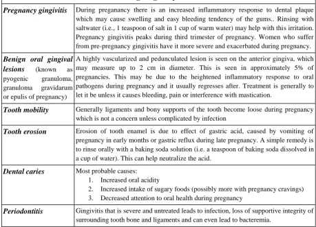
Physiologic changes during pregnancy may result in noticeable changes in the oral cavity27. These changes
include pregnancy gingivitis, benign oral gingival lesions, tooth mobility, tooth erosion, dental caries, and periodontitis (Table 1). It is important to reassure women about these various changes to the gums and teeth during pregnancy and to reinforce good oral health habits to
keep the gums and teeth healthy.
Table 1. Common Oral Conditions During Pregnancy
Pregnancy gingivitis
During preganancy there is an increased inflammatory response to dental plaquewhich may cause swelling and easy bleeding tendency of the gums.. Rinsing with saltwater (i.e., 1 teaspoon of salt in 1 cup of warm water) may help with this irritation. Pregnancy gingivitis peaks during third trimester of pregnancy. Women who suffer from pre-pregnancy gingivitis have it more severe and exacerbated during pregnancy.
Benign oral gingival
lesions
(known aspyogenic granuloma,
granuloma gravidarum
or epulis of pregnancy)
A highly vascularized and pedunculated lesion is seen on the anterior gingiva, which may measure up to 2 cm in diameter. This is seen in approximately 5% of pregnancies. This may be due to the heightened inflammatory response to oral pathogens during pregnancy and it usually regresses after. Treatment is generally to let it be unless it causes bleeding, pain or interference with mastication.
Tooth mobility
Generally ligaments and bony supports of the tooth become loose during pregnancywhich is not a concern unless complicated by infection
Tooth erosion
Erosion of tooth enamel is due to effect of gastric acid, caused by vomiting ofpregnancy in early months or gastric reflux during late pregnancy. A simple remedy is to rinse orally with a baking soda solution (i.e. a teaspoon of baking soda dissolved in a cup of water). This can help neutralize the acid.
Dental caries
Most probable causes:1. Increased oral acidity
2. Increased intake of sugary foods (possibly more with pregnancy cravings)
3. Decreased attention to oral health during pregnancy
Periodontitis
Gingivitis that is severe and untreated leads to infection, loss of supportive integrity ofsurrounding tooth bone and ligaments and can even lead to bacteremia.
Improved training in the importance of oral health, recognition of oral health problems, and knowledge of procedure safety during pregnancy may make health care providers more comfortable with assessing oral health and more likely to address it with patients.
The first prenatal visit should ensure that the woman be seen by a dentist if she has not has her visit in the past six months or has any oral health problems. Patients often need reassurance that prevention, diagnosis, and treatment of oral conditions, including dental X-rays (with shielding of the abdomen and thyroid) and local anesthesia (lidocaine with or without epinephrine), are safe during pregnancy. Conditions that require immediate treatment, such as extractions, root canals, and restoration (amalgam or composite) of untreated caries, may be managed at any time during pregnancy. Delaying treatment may result in more complex problems.
Counseling focuses on reinforcement of routine oral health maintenance, such as:
1. Limiting sugary foods and drinks
2. Brushing twice a day with fluoridated toothpaste 3. Flossing once daily, and
4. Dental visits twice a year.
Dental providers often recommend the use of chlorhexidine and fluoridated mouth rinses, and xylitol-containing chewing gum to decrease oral bacteria. No adverse effects have been reported with these products during pregnancy but they have not been studied extensively. For patients with vomiting secondary to morning sickness, hyperemesis gravidarum, or gastric reflux during late pregnancy, the use of antacids or rinsing with a baking soda solution (i.e., 1 teaspoon of baking soda dissolved in 1 cup of water) may help neutralize the associated acid. For additional information on oral health during the perinatal period, refer to Oral Health During Pregnancy: A National Consensus Statement, developed by the Health Resources and Services Administration’s Maternal and Child Health Bureau in collaboration with the American College of Obstetricians and Gynecologists and the American
Dental Association.28
Evidence has surfaced recently that prenatal oral health care improves pregnancy outcomes, ample evidence shows that oral health care during pregnancy is safe and should be recommended to improve the oral and general health of the woman. Improved oral health of the woman may decrease transmission of potentially cariogenic bacteria to infants and reduce children’s future risk of caries. For many women, obstetrician–gynecologists are the most frequently accessed health care professional, which creates a unique opportunity to educate women throughout their lifespan, including during pregnancy, about the importance of dental care and good oral
RECOMMENDATIONS
To spread awareness and timely management of oral disease, knowledge should be disseminated at patient level, healthcare provider level and public administration level as well for the proper implementation and dispersal of services and their utilization.
At the patient level:
Education is the key as has been elaborated earlier. Through word of mouth and tutelage to her children, a mother dispenses this knowledge to her family members and children and the trend percolates to the future generation.
At the Health care Provider level:
CMEs play a significant role in spreading awareness and information especially to core caregivers namely the obstetrician and the dentist. Camps and pubic campaigns play a positive role in prevention and early treatment of oral disease before complications can take place. There are websites and links on the internet that can be accessed by professionals to keep themselves updated and well informed, so as do deliver standards of care using evidence based medicine and guidelines.
eg. California Dental Association Foundation, 1201 K Street, Suite 1511, Sacramento, CA 95814. Telephone: (800) 232-7645, Ext.4916;
e-mail:[email protected]; website:http:/www.cdafoundation.org. Available.at
http://www.cdafoundation.org/Portals/0/pdfs/poh_guidel ines.pdf (guidelines) or
http://www.cdafoundation.org/Portals/0/pdfs/poh_policy _brief.pdf (policy brief).
These guidelines are designed to help health professionals deliver oral health services to pregnant women and young children. Topics include the importance of oral health; maternal physiological considerations related to oral health; pregnancy, oral conditions, and oral health care; oral health and early childhood; access to care; and systems improvement and public policy changes. Sample forms and websites for parents are included as appendices. A policy brief is also available
For improving perinatal oral health in the community: Government and NGOs to,
1) Assess and monitor perinatal oral health, 2) Enhance infrastructure and build partnerships, 3) Inform and empower the public to mobilize support, 4) Ensure adequate oral health workforce and systems for perinatal oral health,
5) Promote and support research and evidence-based practices24, 30
CONCLUSION
deficiencies, differences in metabolism, hormonal variations, aggravated tissue response to local irritants, impaired immunity, increased dietary intakes (refined carbohydrates) etc. Good oral health practices leading to optimal oral health before pregnancy, may be beneficial during gestation as well as the same may allow a continuing favorable oral health milieu. Pregnancy induced changes in immunity and local tissue response may add to the problems. One must understand the fact that the periodontal and dental caries status is generally poor in expectant mothers, and appropriate and timely care will go a long way in preventing more complex problems.
A clear understanding of hormonal changes and its role in oral health and disease is needed for all the health care providers. Women in the reproductive age and who are pregnant should have thorough oral health screening and treatment. It is necessary to screen pregnant women for oral conditions during and after pregnancy to have better oral health related quality of life. Health care providers are recommended to advise all the women for regular dental visits.
REFERENCES
1. Yadav R, Kachewar SG. Role of MRI in evaluation of painful knee. Int J Med Res Health Sci. 2014;3(1):84-7. 2. Guermazi A, Burstein D, Conaghan P, Eckstein F, Hellio Le Graverand-Gastineau MP, Keen H, et al. Imaging in osteoarthritis. Rheum Dis Clin North Am 2008;34:645e87.
3. Hunter D, Hellio Le Graverand M, Eckstein F. Radiologic markers of osteoarthritis progression. Curr Opin Rheumatol. 2009;21:110-7.
4. Guermazi A, Roemer FW, Hayashi D. Imaging of osteoarthritis: update from a radiological perspective. Curr Opin Rheumatol. 2011;23:484-91.
5. Pompan DC. Reassessing the role of MRI in evaluation of knee pain. American Family physician. 2012;85(3):224.
6. Mackenzie R, Dixon AK, Keene GS, Hollingworth W, Lomas DJ, Villar RN. Magnetic resonance imaging of the knee: assessment of effectiveness. Clin Radiol 1996;51:245–50.
7. Maurer EJ, Kaplan PA, Dussault RG, et al. Acutely injured knee: effect of MR imaging on diagnostic
thinking and therapeutic decisions.
Radiology1997;204:799–805.
8. Mansour MAM, Ahmed RA, Ibrahim A, Elhussein N, Aljuaid SA. Magnetic resonance imaging diagnostic procedures for knee joint injures at taif hospital, Saudi
Arabia. IOSR Journal of Nursing and Health Science. 2015;4(2):37-46.
9. Nasir A. The role of magnetic resonance imaging in the knee joint injuries, international research journal of medical sciences. 2013;1(5):1-7.
10. Yusuf E, Kortekaas MC, Watt I, Huizinga TW, Kloppenburg M. Do knee abnormalities visualised on MRI explain knee pain in knee osteoarthritis? A systematic review. Ann Rheum Dis. 2011;70:60-7. 11. Boden SD, Davis DO, Dina TS, et al. A prospective and blinded investigation of magnetic resonance imaging of the knee: abnormal findings in asymptomatic subjects. Clin Orthop. 1992;282:177–85.
12. Nikolaou VS, Chronopoulos E, Savvidou C et al. MRI efficacy in diagnosing internal lesions of the knee: a retrospective analysis. J Trauma Manag Outcomes. 2008;2:4.
13. Khanda GE, Akhtar W, Ahsan H, Ahmad N. Assessment of menisci and ligamentous injuries of the knee on magnetic resonance imaging: correlation with arthroscopy. J Pak Med Assoc. 2008;58:537–40.
14. Stoller DW, Martin C, Crues JV 3rd, Kaplan L, Mink JH. Menisci tears: pathologic correlation with MR imaging. Radiology 1987;163(3):731-5.
15. Mackenzie R, Palmer CR, Lomas DJ, Dixon AK. Magnetic resonance imaging of the knee: diagnostic performance studies. Clin Radiol 1996;51:251–7. 16. Litwic A, Edwards M, Dennison E, Cooper C. Epidemiology and Burden of Osteoarthritis. Br Med Bull. 2013;105:185–99.
17. Heidari B. Knee osteoarthritis prevalence, risk factors, pathogenesis and features: Part I. Caspian J Intern Med. 2011; 2(2): 205–12.
18. Kachewar SG, Kulkarni DS. Distant perijoint calcifications: sequel of non-traumatic brain injury-a review and case report. Journal of Clinical and Diagnostic Research 2013;7:2606-9.
19. Kachewar SG, Singh H. Perigenicular Heterotopic Ossification: A rare sequelae of non-traumatic brain injury. Nepal Journal of Neurosciences. 2010;1:21-3.
Copyright: © the author(s) and publisher IJMRP. This is an open access article distributed under the terms of the Creative Commons Attribution Non-commercial License, which permits unrestricted non-commercial use, distribution, and reproduction in any medium, provided the original work is properly cited.