REVIEW
A prospect of cell immortalization
combined with matrix microenvironmental
optimization strategy for tissue engineering
and regeneration
Yiming Wang
1,2, Song Chen
3, Zuoqin Yan
2*and Ming Pei
1,4*Abstract
Cellular senescence is a major hurdle for primary cell-based tissue engineering and regenerative medicine. Telomere
erosion, oxidative stress, the expression of oncogenes and the loss of tumor suppressor genes all may account for
the cellular senescence process with the involvement of various signaling pathways. To establish immortalized cell
lines for research and clinical use, strategies have been applied including internal genomic or external matrix
micro-environment modification. Considering the potential risks of malignant transformation and tumorigenesis of genetic
manipulation, environmental modification methods, especially the decellularized cell-deposited extracellular matrix
(dECM)-based preconditioning strategy, appear to be promising for tissue engineering-aimed cell immortalization.
Due to few review articles focusing on this topic, this review provides a summary of cell senescence and
immortaliza-tion and discusses advantages and limitaimmortaliza-tions of tissue engineering and regeneraimmortaliza-tion with the use of immortalized
cells as well as a potential rejuvenation strategy through combination with the dECM approach.
Keywords:
Cell senescence, Decellularized cell-deposited extracellular matrix, Differentiation, Immortalization,
Proliferation, SV40, Tissue engineering
© The Author(s) 2019. This article is distributed under the terms of the Creative Commons Attribution 4.0 International License (http://creat iveco mmons .org/licen ses/by/4.0/), which permits unrestricted use, distribution, and reproduction in any medium, provided you give appropriate credit to the original author(s) and the source, provide a link to the Creative Commons license, and indicate if changes were made. The Creative Commons Public Domain Dedication waiver (http://creat iveco mmons .org/ publi cdoma in/zero/1.0/) applies to the data made available in this article, unless otherwise stated.
Background
Tissue and organ failure is a prominent health issue that
cannot be ignored. Surgical intervention, organ
trans-plantation, artificial substitutes and mechanical devices
are methods to address this issue but all have
undesir-able short- and long-term consequences [
1
]. Tissue
engi-neering is an attractive method that enables fabrication
of functional tissue for tissue regeneration as well as the
establishment of physiological and pathological
mod-els for mechanistic studies [
2
]. This technique can
har-ness the intrinsic regenerative potential of primary cells
and expand them in a controlled environment before
reintroduction into the patient’s body. These natural,
synthetic or semisynthetic tissue and organ mimics are
expected to function normally in a tissue-specific
pat-tern as required [
1
,
3
]. However, primary cells derived
from non-cancerous tissues have a finite lifespan and
decreased proliferation ability when cultured in vitro.
After a limited number of divisions, cells enter a viable
state of permanent quiescence, termed cellular
senes-cence [
4
]. Cellular senescence, regulated by both intrinsic
and extrinsic factors, is characterized as two key
pheno-types, a stable proliferation arrest and altered secretory
pathway, the senescence-associated secretory phenotype
(SASP) [
5
].
In order to acquire an abundant number of cells for
functional tissue engineering, cellular senescence is
the major obstacle that needs to be overcome.
Numer-ous attempts have been made in past decades to deal
with cellular senescence in order to achieve
success-ful immortalization of primary cells. To establish
Open Access
*Correspondence: [email protected]; [email protected]
1 Stem Cell and Tissue Engineering Laboratory, Department
of Orthopaedics, West Virginia University, PO Box 9196, 64 Medical Center Drive, Morgantown, WV 26506-9196, USA
2 Department of Orthopaedics, Zhongshan Hospital of Fudan University,
180 Fenglin Road, Shanghai 200032, China
immortalized cell lines for research and clinical use,
strategies have been applied including internal genomic
or external matrix microenvironment modification.
Considering the potential risks of malignant
trans-formation and tumorigenesis of genetic
manipula-tion, environmental modification methods, especially
the decellularized cell-deposited extracellular matrix
(dECM)-based preconditioning strategy, appear to be
promising for tissue engineering-aimed cell
immor-talization. Due to few review articles focusing on this
topic, this review provides a summary of cell
senes-cence and immortalization and discusses advantages
and limitations of tissue engineering and regeneration
with the use of immortalized cells as well as a potential
rejuvenation strategy when combined with the dECM
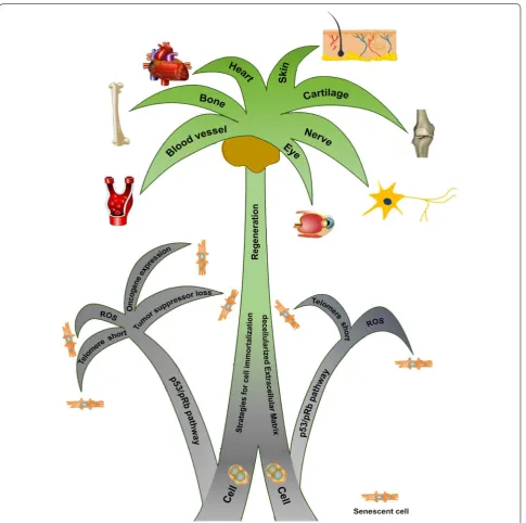
approach (Fig.
1
).
Cellular senescence
Cellular senescence is a process that imposes
irrevers-ible proliferative arrest on cells in response to internal
and external environmental changes. Various
stress-ors, including progressive telomere erosion,
oxida-tive stress, the expression of oncogenes and the loss of
tumor suppressors, contribute to the occurrence of
cel-lular senescence.
Replicative senescence
As vital structures that cap and protect the ends of
lin-ear chromosomes [
6
], shortening of telomeres happens
at every fission which eventually causes cells to reach
their “Hayflick limit” which halts growth after
approxi-mately 60 population doublings [
4
,
7
]. When
telom-eres are too short to function normally for capping,
replicative senescence (M1 stage, a cellular growth
arrest) occurs [
8
–
10
]. The critically short telomeres
are detected by cells as double-strand breaks and
trig-ger a deoxyribonucleic acid (DNA) damage response
(DDR) that consists of a series of signaling events
cen-tered on two anti-proliferative mechanisms, the p53/
p21 and p16/tumor suppressor retinoblastoma protein
(Rb) pathways. This cessation allows cells to repair the
DNA damage, but if the damage continues to exceed a
certain limit, apoptosis or senescence may occur [
11
].
Regulated by its upstream partner p16, one of the
cyc-lin-dependent kinase inhibitors (CKIs), Rb controls cell
cycle progression from G
1into S phase by binding to
and suppressing the activity of E2F transcription factor
1 (E2F1) [
10
,
12
,
13
].
Many researchers have verified the importance of both
pathways in directing senescence as the suppression of
either p53 or Rb alone failed to achieve cell
immortaliza-tion [
14
–
17
]. However, there were still reports showing
that, in human mammary epithelial cells and
mesenchy-mal stem cells (MSCs), inactivation of p16 alone allows
human cells to avoid senescence [
18
,
19
]. Meanwhile, in
human diploid fibroblasts, the p53 mutant alone is able
to suppress cellular senescence [
20
]. These findings raise
the possibility that these two pathways may function
dif-ferently among different cell strains.
However, abrogation of the p53/p21 and p16/Rb
path-ways will only lead to a “pre-immortal” state instead of an
“immortal” status for cells. Terminal telomere shortening
still exists and will eventually lead to the M2 stage,
char-acterized as massive cell death [
10
,
15
,
21
]. In most cases,
the stabilization of telomeres is achieved through the
introduction of telomerase, an enzyme that synthesizes
telomeric repeats and adds them to the ends of
chromo-somes for the compensation of inevitable loss with each
round of DNA replication [
22
].
Premature senescence
Senescence also happens in conditions that are not
dependent on telomere erosion or dysfunction. This
pro-cess is often referred to as “premature” since it can arrest
growth prior to reaching the “Hayflick limit” [
23
].
Vari-ous conditions have been identified that may result in
premature cellular senescence.
Stress‑induced senescence
During a long-term in vitro cell expansion, laboratory
culture conditions, generally defined as a lack of
sur-rounding cell types and support from extracellular matrix
(ECM), abnormal growth factors and oxygen (O
2) level,
expose the cell to excessive oxidative stress and induce
oxidant production [
24
–
27
]. The excessive levels of
reac-tive oxygen species (ROS), including hydrogen peroxide
(H
2O
2), hydroxyl radical (OH
−) and superoxide anion
(O
2−), are detectable during long-term culturing of
MSCs, accounting for stress-induced senescence [
24
–
26
]. H
2O
2could directly affect cellular DNA, trigger DDR
and subsequent p16/Rb and p53 pathways, leading to cell
cycle arrest [
28
–
32
].
Oncogene‑induced senescence
is released from the Rb-E2F1 complex, activating the
downstream target genes regulating normal entry into
S phase [
48
]. Interestingly, in normal human fibroblasts,
E2F1 and its target gene p14 (ARF) are responsible for
the induction of cellular senescence [
35
]. Meanwhile,
E2F1 knock-out mouse embryonic fibroblasts
demon-strated attenuated senescence and ROS levels [
49
].
Tumor suppressor loss‑induced senescence
Tumor suppressors are the counterpart of oncogenes, and
their loss can elicit cellular senescence. Depletion of NF1
(Neurofibromatosis 1), a tumor suppressor gene, induces
senescence in human fibroblasts [
50
]. Similarly, loss of
BTG3 (B-cell translocation gene 3), a member of the
anti-proliferative BTG gene family and a downstream target of
p53, triggers cellular senescence as well [
51
]. Inactivation
of
VHL (von Hippel-Lindau tumor suppressor) induces
an efficient senescence in mouse fibroblasts and primary
renal epithelial cells under atmospheric conditions (21%
O
2); however, loss of VHL only causes a decreased cell
proliferation instead of cell arrest in human renal
epithe-lial cells [
52
,
53
]. Similarly, acute loss of tumor suppressor
gene
PTEN (phosphatase and tensin homolog) induces
growth arrest through the p53-dependent cellular
senes-cence pathway in mouse prostate both in vitro and in vivo
whereas, in systemic lupus erythematosus patients, the
complete loss is significantly related to advanced cancer
and poor outcomes [
54
–
56
]. These findings raise the
pos-sibility that tumor suppressors may function differently
according to different species and cell types.
Signaling pathways involved in cellular senescence
Despite the abovementioned p53/p21 and p16/Rb
path-ways, other signaling pathways are also involved in
cellular senescence, including, but not limited to,
trans-forming growth factor β (TGFβ)/bone morphogenetic
protein (BMP), Wingless/Int (Wnt)/β-catenin, MAPK,
phosphatidylinostitide 3 kinase (PI3K)/protein kinase B
(AKT)/mammalian target of rapamycin (mTOR), Hippo,
NOTCH, fibroblast growth factor (FGF) and insulin-like
growth factor (IGF) and hypoxia inducible factor (HIF)
(Fig.
2
).
TGFβ/BMP signaling pathways
TGFβ is a classic regulator for chondrogenic
differentia-tion but its role in cell expansion remains controversial
[
57
,
58
]. TGFβ activation is positively involved in the
induction of cellular senescence of all kinds of species
[
59
–
61
]. In human breast cancer cells, TGFβ negatively
mediates telomerase activity through its downstream
effector, Smad3 [
62
,
63
]. For stress-induced senescence,
TGFβ contributes to ROS production and activation of
DDR during the senescence of human fibroblasts and
bone marrow-derived MSCs (BMSCs) [
64
,
65
]. The
kinase ataxia-telangiectasia mutated (ATM) is a key
player in nuclear DDR [
66
]. Meanwhile, TGFβ is required
for oncogene-induced senescence that is independent
of the p16/Rb and p53 pathways; attenuation of TGFβ
inhibits premature senescence in human mammary
epi-thelial cells [
67
,
68
].
BMPs are secreted signal factors belonging to the TGFβ
superfamily and are involved in embryonic development
and cellular processes [
69
]. Similar to the function of
TGFβ, BMP receptor II/Smad3 contributes to telomerase
inhibition and telomere shortening in human breast
can-cer cells, leading to replicative senescence [
70
]. Similar
results were observed in primary cells as the BMP
sign-aling axis plays an important role in oncogene-induced
senescence of mouse fibroblasts [
71
].
Wnt/β‑catenin pathway
Wnts are highly conservative proteins that participate
in embryonic development and homeostatic
mecha-nisms in adult tissues [
72
]. Wnt signals appear to be an
important regulator of both premature senescence and
replicative senescence. On one hand, the Wnt/β-catenin
signaling pathway interacts with the p53/p21 pathway
for ROS production to induce MSC senescence [
73
–
76
].
On the other hand, Wnt3a/β-catenin also plays a critical
role in hedging replicative senescence of MSCs, probably
through regulation of a telomerase subunit—telomerase
reverse transcriptase (TERT) [
72
,
77
]. Meanwhile, Wnt/
β-catenin signaling enhances rat nucleus pulposus cell
senescence as well as induces the expression of TGFβ,
another strong promoter of cellular senescence [
78
].
MAPK pathway
metalloproteinase-1 production in response to ROS in
IMPR-90 cells [
85
–
89
]. However, the
senescence-pro-moting role of JNK was challenged as it was also revealed
to antagonize p38-induced p16 activation [
90
]. Moreover,
JNK acts as a negative regulator of p53 tumor suppressor
to suppress p53-dependent senescence in mouse
embry-onic fibroblasts [
91
]. An increase of intracellular ROS
levels can suppress the growth of cancer cells and induce
cellular apoptosis by mediating MAPK signaling
compo-nents [
92
].
PI3K/AKT/mTOR pathways
PI3Ks and their downstream mediators AKT and mTOR
constitute the core component of the PI3K/AKT/mTOR
signaling pathway which is precisely controlled under
normal physiological conditions and is a frequently
hyperactivated pathway in cancer [
93
]. Similar to the
MEK-ERK pathway, PI3K is one of the main downstream
effectors of Ras dependent signaling and its activation
plays dual roles in cell cycle regulation as it can promote
cell cycle progression as well as cause cell cycle arrest
[
94
]. Recent studies reveal the involvement of PI3K/
AKT/mTOR in the regulation of replicative senescence
in human vascular smooth muscle cells [
95
]. Moreover,
a constitutively active, myristoylated form of AKT leads
to oncogene-induced senescence in primary cultured
human endothelial cells and murine fibroblasts; the loss
of PTEN triggers senescence through activation of the
PI3K/AKT pathway in mouse prostate [
56
,
96
]. However,
a recent report subverted the positive role of the PI3K/
Fig. 2 Signaling pathways mediating the cellular senescence process. In response to telomere erosion, ROS production, the expression ofAKT/mTOR pathway in senescence-induction by
intro-ducing the fact that activation of this pathway abolished
BRAF
V600E-induced senescence in both primary human
fibroblasts and primary human melanocytes [
97
].
Moreo-ver, targets of the PI3K/AKT signaling pathway have been
found to promote cell survival [
98
] and the activation of
the PI3K/AKT pathway can be induced by TGFβ,
lead-ing to a pro-survival/anti-apoptotic effect in both human
nasopharyngeal carcinoma cells [
99
] and mesenchymal
cells/fibroblasts [
100
].
Hippo pathway
The Hippo pathway is a tumor suppressor pathway;
dys-regulation of this pathway can lead to uncontrolled cell
proliferation and tumorigenesis [
101
]. Yes-associated
protein 1 (YAP), a major downstream effector of the
Hippo pathway, is phosphorylated and inactivated by
the serine/threonine kinases large tumor suppressor 1
(LATS1) and LATS2 [
102
,
103
]. YAP dephosphorylation
is associated with the senescence of rat nucleus
pulpo-sus cells and overexpression of YAP in primary human
keratinocytes blocks clonal evolution and induces cell
immortalization [
104
,
105
]. Coordination of the Hippo
pathway and p53 occurs in response to various types of
stress signals including replication and oncogenic Ras.
LATS2 cooperates with p53 to induce p21 expression,
resulting in cellular senescence [
106
]. LATS2 also plays
an important role in the oncogenic H-Ras induced stress
checkpoint in a p53-dependent pathway [
107
].
Notch pathway
The Notch pathway is an evolutionarily highly conserved
signaling pathway that is associated with a variety of
cel-lular processes including cell-fate determination,
prolif-eration and death. In mammals, the Notch family has five
ligands and four receptors [
108
,
109
]. There is
accumu-lating evidence that abnormal Notch signaling has been
implicated in multiple facets of cancer biology, and Notch
can behave as either an oncogene or a tumor
suppres-sor depending on cell context [
110
,
111
]. The oncogenic
function of Notch has been demonstrated in several
types of cancer including melanoma [
112
], breast cancer
[
113
] and brain tumors [
114
]. Activated Notch 1
signifi-cantly enhances the rate of glycolysis, which prevents
cel-lular senescence of human adipose-derived stromal cells
(ADSCs) through HIF1 activation and p53 inactivation
[
115
]. On the other hand, the Notch pathway is found to
serve as a tumor suppressor in the progression of
carci-noma including bladder cancer [
116
], medullary thyroid
carcinoma [
117
] and pancreatic cancer [
118
]. Enforced
Notch activation in human endothelial cells is
associ-ated with cellular senescence with the involvement of p16
[
119
]. Down-regulation of Notch 3 in human fibroblasts
and mammary epithelial cells delays the onset of
senes-cence and extends cell lifespan [
120
,
121
].
FGF and IGF pathways
FGFs are well-recognized for their critical roles in
embryonic development [
122
]. The mitogenic effect of
FGF has been demonstrated by promoting proliferation
while maintaining stemness of MSCs in vitro [
123
–
125
].
FGF2 treatment led to an early increase in telomere size
in MSCs, probably due to its ability to increase TERT
mRNA expression [
126
,
127
]. FGF signals negatively
reg-ulated MSC senescence through interaction with PI3K/
AKT/MDM2 (mouse double minute-2 homolog) in the
mouse and through down-regulation of TGFβ
expres-sion in human MSCs [
128
,
129
]. Surprisingly, FGF23 can
also induce premature senescence in human MSCs from
skeletal muscle via the p53/p21 oxidative-stress pathway
[
130
].
IGFs are considered detrimental to cell survival due
to their role in diminishing tissue resistance to oxidative
stress and shortening lifespan [
131
,
132
]. In mouse, rat
and human primary vascular smooth muscle cells, IGFI
induces cellular senescence dependent on the
upregula-tion of p53 [
133
]. Additional evidence has revealed that
IGF binding protein-5 is upregulated in the regulation of
premature senescence of umbilical vein endothelial cells
through a p53-controlled mechanism [
134
,
135
]. These
findings may be due to the mechanism whereby IGFI is
capable of inducing telomere shortening [
136
]. However,
opposite results were found in human annulus fibrosus
cells as IGFI alleviates cellular senescence [
137
]. In this
scenario, the regulatory roles of both FGFs and IGFs
relating to cellular senescence are context-dependent.
HIF pathway
cellular senescence of marrow-isolated adult multilineage
inducible cells [
146
].
Immortalization of cells through genetic
modification
To achieve cell lifespan extension, biotechnological
methods are often used for direct manipulation of a cell’s
genome. However, concerns still exist regarding genomic
stability and tumorigenicity after genetic modification.
For MSCs and progenitor cells, the potential loss of
dif-ferentiation ability after genetic modification is a
prob-lem that cannot be overlooked.
Genetic modification
The introduction of viral oncogenes/oncoproteins and
TERT are two typical methods for this type of genetic
modification (Table
1
).
Viral oncogenes/oncoproteins
Viral oncogenes that are able to inactivate both pRb and
p53 can overcome M1 (a barrier in which normal cells
senesce and cease replication) and significantly prolong
cell lifespan. For several decades, simian virus 40 (SV40)
early region genes have been commonly used for cell
immortalization and cell line establishment [
147
,
148
].
SV40 is limited to two proteins as the large T (LT) and
small t antigen (ST). LT is mainly responsible for the
SV40-extended lifespan based on its ability to interact
with growth suppressors—pRb and p53. LT binds to the
pRb-E2F complex via its pocket binding site including
AA101–118 and the J domain that acts as a chaperone,
leading to the dissociation of E2F from the LT-pRb
com-plex [
149
]. Meanwhile, LT binds to p53 therefore
sup-pressing the p53 pathway [
150
]. More interestingly, SV40
was reported to induce telomerase activity in primary
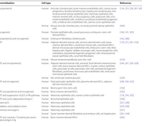
Table 1 Immortalization of primary cells for therapeutics and research
Immortalization Cell type References
Oncoprotein(s) Human Articular chondrocytes, bone marrow endothelial cells, cranial suture progenitors, foreskin keratinocytes, hepatocyte, keratinocytes, liver renal proximal tubular epithelial cells, mammary epithelial cells, marrow stromal cells, nucleus pulposus cells, podocyte cells, sinu-soidal endothelial cells, umbilical cord blood endothelial progenitor cells, umbilical vein endothelial cells, uterine cervix epithelial cells
[154, 233, 238, 287–303]
Animal Mouse articular chondrocytes, rat renal proximal tubular epithelial
cells [304]
Oncogene(s) Human Prostate epithelial cells, neural precursors, embryonic stem
cell-derived MSCs [160, 161, 305] Oncoprotein(s) and oncogene(s) Human Embryonic fibroblasts, keratinocytes [162, 306] TERT Human Adipose-derived stromal cells, amnion-derived stem cells, bone
marrow-derived MSCs, cementum-lining cells, cord blood MSCs, dermal microvascular endothelial cells, embryonic stem cells, fetal hepatocytes, hepatic stellate scavenger cells, neural progenitor cells, osteoblasts, periodontal ligament progenitor cells, renal proximal tubule epithelial cells, vocal fold fibroblasts
[178, 215, 307–318]
Animal Mouse temporomandibular joint disc cells [319] TERT and oncoprotein(s) Human Adipose-derived stromal cells, amniotic fluid-derived mesenchymal
stem cells, bone marrow-derived MSCs, ovarian surface epithelial cells, pancreatic β cells, pancreatic islet cells, periodontal ligament fibroblasts, pulmonary microvascular endothelial cells, renal proxi-mal tubule epithelial cells
[244, 247, 320–328]
Animal Rat ventricular cardiomyocytes [329] TERT and oncogene(s) Human Fetal pancreatic epithelial cells, placenta-derived MSCs,
adipose-derived stromal cells [246, 330–332] Animal Bovine germ line stem cells [163] TERT, oncoprotein(s) and oncogene(s) Human Bone marrow-derived MSCs [333] TERT and suppression of p53 or Rb pathway Human Mammary epithelial cells, ovarian surface epithelial cells [176, 334, 335] TERT and cyclin-dependent kinase 4 Human Bronchial epithelial cells [336] Mutant p53 Human Mammary epithelial cells [337, 338] Irradiation and oxidative stress Human Mammary epithelial cells [339, 340] Chemical carcinogens Human Mammary epithelial cells [341]
Animal Syrian hamster dermal fibroblasts and embryo cells, rat hepatocytes [342–344] TERT and cytotoxic T
human mesothelial cells, but not in primary fibroblasts
[
151
].
Human papillomavirus (HPV) is a small,
double-stranded DNA virus that infects mucosal and cutaneous
epithelial tissue [
152
]. The high-risk strains including
HPV-8, -16, -18 and -31 cause malignant progression of
lesions, whereas the low-risk strains including HPV-6
and -11 cause benign warts and lesions [
153
]. The E6
and E7 proteins encoded by “high-risk” strains including
HPV-16 and -18 are oncoproteins that have been shown
to have transformation properties [
154
]. When used in
immortalization, E6 causes telomerase activation as well
as accelerating the degradation of p53 by the 26S
protea-some, whereas E7 inactivates Rb by preventing the
bind-ing of pRb to the E2F transcription factor [
155
,
156
].
Human T-lymphotropic virus type 1 (HTLV-1) is
the etiologic agent of adult T cell leukemia. Although
HTLV-2 is less pathogenic than HTLV-1, both of the
HTLV-1 and -2 Tax proteins, p40
tax(Tax1) and p37
tax(Tax2), share the capacity to immortalize lymphocytes
in vitro [
157
]. HTLV-2 protein Tax2 demonstrates much
stronger efficacy than that of Tax1 in immortalizing
human T cells [
158
].
The
myc oncogene family consists of several different
members including c-myc, N-myc, L-myc and B-
myc.
c-myc expression is restricted to proliferating cells while
N-myc and L-myc expression is associated with
cellu-lar differentiation [
159
,
160
]. Moreover, the oncogene
myc fulfils many of the expectations for a gene involved
in immortalization of primary cells alone [
160
,
161
] or
cooperates with oncoproteins [
162
] or TERT [
163
].
B cell-specific Moloney murine leukemia virus
integra-tion site 1 (BMI1) which was identified as a
c-myc-coop-erating oncogene, is a critical transcriptional repressor
for maintenance of proper gene expression during
devel-opment [
164
–
166
]. INK4a locus, which encodes p16 and
p19
Arf, is an important target of BMI1 and
overexpres-sion of BMI1 extends replicative lifespan of human
fibro-blasts, probably through suppressing the p16-mediated
senescence pathway [
167
,
168
].
TERT
Telomerase is composed of two core components: the
small nuclear ribonucleic acid (RNA) human telomerase
RNA, which serves as an internal template for the
syn-thesis of telomeric repeats, and the protein TERT (or
hTERT in humans), which serves as a catalytic subunit
that synthesizes the new telomeric DNA from the RNA
template [
22
]. In most human primary cells, telomerase
is either absent or present at an insufficient level for
tel-omere maintenance [
169
]. TERT is the determinant for
the presence of active telomerase [
170
,
171
]. The
intro-duction of ectopic expression of TERT is necessary for
telomere-dependent senescence as it is able to
signifi-cantly extend the lifespan of a variety of cell types, but
it alone is not sufficient to immortalize them [
172
–
174
].
Theoretically, the abrogation of the Rb and p53 pathways
with oncogenes or at a minimum, low p16 expression, is
indispensable for cell immortalization [
175
,
176
].
How-ever, there are still investigations showing that TERT
bypassed Rb and p53 pathway-dependent barriers to
immortalize cells alone [
176
–
180
].
Carcinogenic limitations and strategies
Despite the increasingly sophisticated strategies to
immortalize human cells, there is still some debate over
the risks upon integration of oncogenes into
chromo-somes. The primary safety concern with the use of a cell
line is the transmission of an oncogenic factor to the
host cells. Indeed, cells transduced with these oncogenes
underwent additional changes including full
carcinogen-esis-associated changes (Table
2
). The persistent
infec-tion by a subset of HPVs, especially HPV-16 and HPV-18,
is etiologically linked to cervical cancer in women [
181
].
Deregulated overexpression of HPV E6 and E7 led to
sev-eral alterations in cellular pathways and functions, which
is associated with malignant transformation of cells and
tumorigenesis [
182
]. In addition, HPV E6 oncoprotein
can interact with hTERT to promote carcinogenesis in
keratinocytes [
183
,
184
].
SV40 or SV40 sequences were found in several types of
human cancers located in bone, brain, chest, etc. [
185
–
188
]. Evidence has shown that SV40 can successfully
transform cell lines in vitro and induce tumors in
neo-natal hamsters in vivo [
189
–
192
]. The injection of
SV40-transformed cells into terminally ill human patients
caused subcutaneous tumor nodules [
193
]. Moreover,
SV40-transduced cells contained integrated SV40 DNA,
which was integrated at random positions on the cellular
chromosomes of host cells [
194
,
195
], leading to the
con-troversial question of whether the virus poses a threat for
further in vivo use.
injection with HPV-16 E6/E7 immortalized BMSCs into
Nonobese Diabetic/Severe Combined
Immunodefi-ciency (NOD/SCID) mice for 3 days, no tumor mass was
observed compared to those injected with Hela cells in
which tumor mass was observable [
201
]. Even the
intro-duction of TERT and SV40 or HPV-16 E6/E7 was
suffi-cient to immortalize ovarian surface epithelial cells and
dermal papilla cells but not enough for tumor formation
[
179
,
202
–
205
].
However, other groups argue about the increased
potential for tumor development in TERT-immortalized
cells. On monolayer cultures, human MSCs and
fibro-blasts avoid cell-to-cell contact inhibition, anchor to
culture dishes and tend to proliferate limitlessly [
206
,
207
]. In clinical practice, elevated hTERT expression is
a diagnostic marker for tumor and the overexpression
of hTERT is claimed to be associated with an advanced
invasive stage of tumor progression and poor
progno-sis [
208
–
210
]. Moreover, there are still concerns about
genetic instability after TERT transfection or
transduc-tion. Spontaneous changes in c-myc proto-oncogene
expression and other genetic alterations have been
observed during in vitro culture of hTERT-immortalized
human cells [
211
,
212
].
Despite safety concerns for the immortalized cells, there
are still some cases that successfully applied these cells for
in vivo organ and tissue restoration. For liver impairment,
Guo et al. [
213
] found that SV40-immortalized
marmo-set hepatic progenitor cells (MHPCs) injected into the
injured liver of fumarylacetoacetate hydrolase-deficient
mice repopulated with hepatocyte-like cells and MHPCs
were also implanted as cholangiocytes into bile ducts of
3.5-diethoxycarbonyl-1,4-dihydrocollodine-induced bile
ductular injured mice. Meanwhile, SV40-immortalized
human fetal liver cells differentiated into mature
hepat-ocytes after being transplanted into liver injured mice
[
214
]. For brain damage, hTERT-immortalized cord
blood MSCs were injected into the traumatically injured
brain of a rat model and proliferated efficiently at the
injury site for 2 weeks and showed no tumor formation in
SCID mice after a 6-month observation [
215
].
To avoid persistent oncogene expression, conditional
immortalization technology was developed. Conditional
immortalization includes inserting a reagent mediate,
operator controllable gene to create a cell line that can
be expanded in a consistent fashion when the transgene
is active. When desired clinical quantities of cell
mate-rial are achieved, the transgene can be deactivated by
the operator and the cells will return to a normal,
post-mitotic state. The conditional immortalization
technol-ogy c-MycER
TAMuses a combination of growth factor
and 4-hydroxytamoxifen (4-HT) to activate the c-MycER
transgene. In the absence of 4-HT, c-MycER is
inacti-vated and the cells return to a normal phenotype [
216
].
Inactivation of SV40 LT was achieved using a
tempera-ture-sensitive mutant of the LT (SV40 tsA58) that is
bio-logically active at permissive temperature (33.5 °C) but
inactive at a non-permissive temperature (39 °C) [
217
].
Different vectors can have influence on the expression
of transgenes. Unlike lentivirus, adenovirus does not
integrate transgenes into the host genome and thereby
can only provide a transient expression of the transgenes
[
218
]. However, this kind of expression time is not
controllable.
To acquire more accurate excision of oncogenes,
site-specific recombination systems were developed. Cre/
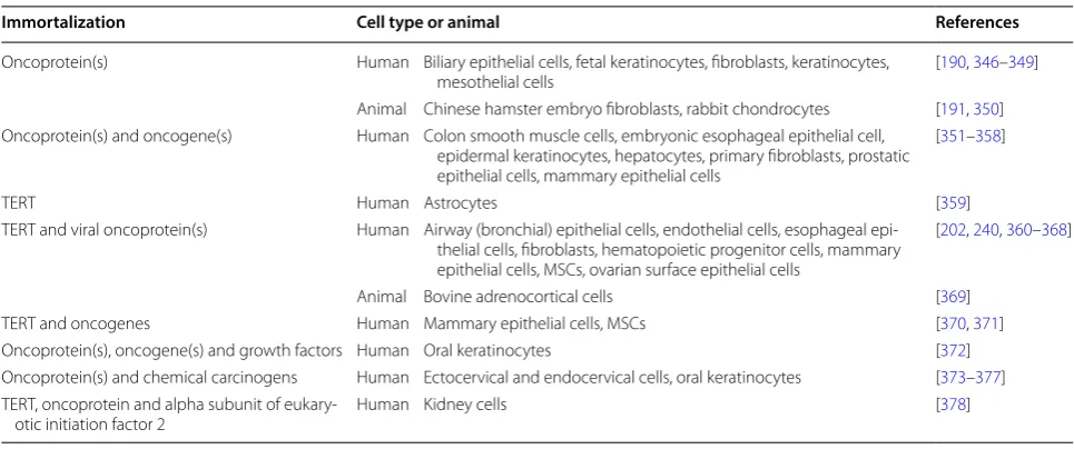
Table 2 Malignant transformation and tumorigenesis during immortalization of primary cells
Immortalization Cell type or animal References
Oncoprotein(s) Human Biliary epithelial cells, fetal keratinocytes, fibroblasts, keratinocytes,
mesothelial cells [190, 346–349] Animal Chinese hamster embryo fibroblasts, rabbit chondrocytes [191, 350] Oncoprotein(s) and oncogene(s) Human Colon smooth muscle cells, embryonic esophageal epithelial cell,
epidermal keratinocytes, hepatocytes, primary fibroblasts, prostatic epithelial cells, mammary epithelial cells
[351–358]
TERT Human Astrocytes [359]
TERT and viral oncoprotein(s) Human Airway (bronchial) epithelial cells, endothelial cells, esophageal epi-thelial cells, fibroblasts, hematopoietic progenitor cells, mammary epithelial cells, MSCs, ovarian surface epithelial cells
[202, 240, 360–368]
Animal Bovine adrenocortical cells [369] TERT and oncogenes Human Mammary epithelial cells, MSCs [370, 371] Oncoprotein(s), oncogene(s) and growth factors Human Oral keratinocytes [372] Oncoprotein(s) and chemical carcinogens Human Ectocervical and endocervical cells, oral keratinocytes [373–377] TERT, oncoprotein and alpha subunit of
LoxP technology involves engineering a transgene
flanked by LoxP sites. The transgene is activated until
Cre recombinase is added. However, the cre-lox
sys-tem is not 100% efficient and cells that have not deleted
the transgene might require elimination [
219
,
220
]. In
addition, a Tet on/off system uses tetracycline
respon-sive elements (TRE) that consist of a Tet operator and
minimal promoter. The activation of the transgene and
the subsequent cell division is related to tetracycline or
doxycycline, which acts as a cue for activation (Tet-On)
or inactivation (Tet-Off) [
221
,
222
]. However, this
tech-nology still has the evident limitation termed “leakiness”,
where the transgene continues to express at a low level
even when the system is off [
223
]. Moreover, the
tran-sient activation of β-catenin was used to efficiently induce
hTERT activation while silencing β-catenin suppresses
the expression of hTERT [
224
]. Meanwhile, therapeutic
strategies concerning transient activation of telomerase
with small molecules, including the administration of 1%
N-acetylcarnosine lubricant eye drops for prevention and
treatment of cataracts, have been proven beneficial for
dogs and other animals [
225
]. Huang et al. have identified
that anthraquinone derivatives might be able to activate
hTERT expression without causing genetic alterations in
cells, whereas these cells fail to possess potent
prolifera-tive ability [
226
]. Interestingly, the introduction of some
adenovirus derived genes, including the early 4 region
(E4) of the adenoviral vector (AdE4), augments survival
of human endothelial cells [
227
,
228
]. However,
investi-gations into AdE4 gene products were largely
overshad-owed by the fact that these proteins not only orchestrated
many viral processes, but also overlapped with oncogenic
transformation of primary cells [
229
,
230
]. Although
creation of conditionally immortalized cell lines has the
potential for therapeutic application, complete silence of
the transgene before introduction into the patient’s body
is still a concern that needs to be addressed.
Potential loss of differentiation capacity
Differentiation capacity of MSCs and progenitor cells
after immortalization is another concern that deserves
more attention. A variety of reports has claimed that
immortalization of progenitor cells will retain
prolif-erative activity without compromising multipotent or
specific differentiation potential of primary cells from
species including human, mouse and porcine [
231
–
237
].
A similar phenomenon has been mentioned in human
MSCs immortalized with SV40 [
238
,
239
]. After serial
transduction with hTERT, SV40 and H-Ras, human
MSCs still retain their multilineage differentiation
poten-tial even during tumorigenesis [
240
,
241
]. Moreover,
Yang et al. [
242
] showed that hTERT-transduced human
BMSCs seeded on porous polylactic glycolic acid (PLGA)
scaffold have better osteogenic differentiation ability than
primary human BMSCs seeded on scaffold. Similarly,
human BMSCs immortalized with hTERT and HPV16
E6/E7 displayed greater differentiation potential far
beyond the primary human BMSCs or even when human
BMSCs expressed HPV-16 E6/E7 alone [
243
].
Interest-ingly, Okamoto et al. [
244
] reported that human BMSCs
immortalized with hTERT and HPV-16 E6/E7
demon-strated significant clonal heterogeneity in differentiation
potential. A similar phenomenon was found in mouse
melanocyte progenitors that displayed distinct
melano-genic differentiation potential [
232
]. More interestingly,
there were opposite results as human primary dental
pulp stem cells (DPSCs) are found to be approximately
60% more effective than hTERT-immortalized DPSCs
in osteogenic differentiation [
245
]. For human
placenta-derived MSCs immortalized with hTERT and BMI1, the
differentiation potential was lost [
246
]. This
discrep-ancy may partially be due to different immortalization
strategies as the lost differentiation potential in ADSCs
due to “SV40
+
hTERT” introduction can be preserved
by “hTERT
+
BMI1” [
247
]. Moreover, cellular
senes-cence counteracts the induction and reprogramming of
induced pluripotent stem cells and senescence related
INK4A/ARF and p53/p21 pathways are considered to be
involved in these processes [
248
–
251
].
Preconditioning of cells through matrix
microenvironment optimization
Although genetic manipulation is a popular strategy for
functional tissue engineering, it has limited clinical
ben-efit due to its inherent risks [
189
,
193
,
194
]. For human
cells that are sensitive to external changes, matrix
micro-environmental alterations may modify intercellular
com-munication, leading to enhanced proliferation ability
without carcinogenic mutation [
252
,
253
].
cells [
265
,
266
] and replicatively senescent porcine
chon-drocytes [
267
]. These dECM-expanded cells were smaller
in size compared to those grown on plastic flasks. These
results are in accordance with the finding from
White-field and coworkers in which, under time-lapse video
microscopy, the smaller cells were observed to continue
proliferation, while the larger cells became senescent and
exited the cell proliferation cycle [
268
].
Furthermore, current data indicate that dECM
depos-ited by MSCs yields human adult SDSCs [
269
,
270
] and
porcine adipose stem cells derived from the infrapatellar
fat pad [
264
] with better chondrogenic potential in vitro
and with better repair capacity for cartilage defects
in vivo [
271
]. Interestingly, BMSCs, a tissue-specific stem
cell for endochondral bone formation, could be greatly
recharged toward chondrogenic differentiation by
expan-sion on dECM deposited by nonchondrogenic human
urine stem cells [
272
] or human BMSCs themselves [
260
].
More interestingly, a recent report showed that, despite
dECMs deposited by BMSCs and ADSCs enhancing the
proliferation ability of MSCs, they failed to yield
expan-sion of cancer cells (HeLa, MCF7 and MDA-MB-231) in
terms of inhibiting the expansion ability of these cancer
cells [
273
]. This finding indicates that normal cell derived
dECM is not favorable for the growth of cancer cells.
Given the undesirable potential of carcinogenesis after
genetic modification from cell immortalization, dECM
tends to be a better alternative.
In addition to rescuing replicative and differentiation
capacities, dECM could reduce intracellular generation of
ROS in aged murine BMSCs [
274
] and in human BMSCs
[
260
] and umbilical cord MSCs [
263
]. Meanwhile, dECM
could enhance the anti-oxidative capacity of human
adult SDSCs [
269
] and protect umbilical cord MSCs
from oxidative stress-induced premature senescence
[
275
] to finally achieve better chondrogenic
differentia-tion. Moreover, dECM could repress osteoclastogenesis
in bone marrow monocytes through the attenuation of
intracellular ROS [
276
]. All the above mentioned studies
confirmed the anti-senescence and anti-oxidative effect
of dECM as a culture substrate.
Mechanical cues, including stiffness and elasticity from
the surrounding matrix microenvironment, are
impor-tant cellular inputs that sustain cell proliferation and
oppose cell senescence. Integrin-based focal adhesions
are the main adhesion complex dominating
mechano-sensing [
277
]. In our previous study, dECM-expanded
human BMSCs demonstrated increased expression of
integrin α2 and β5 [
260
], which are potentially involved
in the process of cell proliferation [
278
,
279
]. As a
power-ful regulator of cell proliferation and survival, YAP/YAZ
act as mechanotransducer that is regulated by F-actin
cytoskeleton [
280
,
281
]. Interestingly, dECM expansion
was found to induce sustained activation of ERK1/2 as
well as phosphorylated cyclin D1 human BMSCs [
260
]
but decreased phosphorylated ERK in human adult
source SDSCs [
261
]. A similar phenomenon was found
in phosphorylated p38 expression in human SDSCs [
261
]
and human umbilical cord MSCs [
263
] after dECM
pre-conditioning. These discrepancies might be explained
by the dual role of ERK [
282
] and p38 [
283
] signals that
play upon cell senescence. The expression of Wnt5a and
Wnt11a were also found to be upregulated following
dECM expansion [
261
].
Conclusions and perspective
Primary cells display a stable and long-term loss of
pro-liferative capacity upon in vitro expansion despite
con-tinued viability and metabolic activity. This inability to
proliferate is due to progressive shortening of telomeres
during each replication which ultimately makes cells
reach their “Hayflick limit”, termed telomere-dependent
or replicative senescence. Meanwhile, there is another
kind of senescence referred to as premature senescence
since it can arrest cell growth long before reaching the
“Hayflick limit”. One type is stress-induced senescence
caused by the failure to simulate the in vivo supportive
environment, which puts pressure on cell proliferation
through the generation of ROS. In addition, both
onco-genes and their counteracting tumor suppressors are
proven to provoke premature senescence. A variety of
signaling pathways are involved in all of these types of
senescence, in which the p53/p21 and p16/Rb pathways
are the two major signals involved. Oncogenes including
SV40 and HPV-16 E6/E7 inhibit the p53/p21 and p16/Rb
pathways, but are not able to immortalize primary cells
unless followed by the introduction of TERT, which
elon-gates telomeres, thereby abrogating the effect of the end
replication problem. However, all of these genetic
modi-fication methods have the risk of virus introduction and
potential oncogenesis, which must be addressed before
its application into tissue engineering.
from degenerative disease and most of their autologous
cells may suffer from senescence, which was identified as
an influential factor in the quality of cells [
286
]. Given the
demand for a younger cell population and the situation
of carcinogenic transformation after genetic modification
for immortalization purposes, it raises the possibility of
combining genetic modification and environmental
opti-mization strategies. In other words, we can immortalize
these senescent cells and utilize their deposited dECM
instead of the cells themselves to achieve a reduced
senescent status and enhanced proliferation potential
of expanded cells. The combination strategy might also
overcome the potential loss of differentiation capacity of
stem cells with the use of immortalization strategy alone.
Further investigation into this matrix
microenvironmen-tal preconditioning-based rejuvenation strategy may
offer important insights into possible means of providing
robust primary cells as therapeutic agents.
Abbreviations
4-HT: 4-hydroxytamoxifen; ADSCs: adipose-derived stromal cells; AKT: protein kinase B; ATM: ataxia-telangiectasia mutated; BMI1: B cell-specific Moloney murine leukemia virus integration site 1; BMP: bone morphogenetic protein; BMSCs: bone marrow-derived MSCs; BTG3: B cell translocation gene 3; CKIs: cyclin-dependent kinase inhibitors; DDR: DNA damage response; dECM: decellularized cell-deposited extracellular matrix; DNA: deoxyribonucleic acid; DPSCs: dental pulp stem cells; ECM: extracellular matrix; ERK1/2: extracellular signal-regulated kinase 1/2; FGF: fibroblast growth factor; H2O2: hydrogen
peroxide; HIF: hypoxia inducible factor; HPV: human papillomavirus; HTLV-1: human T-lymphotropic virus type 1; IGF: insulin-like growth factor; JNK: c-Jun N-terminal kinase; LATS1: large tumor suppressor 1; LRP5: low-density lipoprotein receptor-related protein 5; MAPK: extracellular signal-regulated kinase; MEK: extracellular signal-regulated kinase kinase; MSCs: mesenchymal stem cells; mTOR: mammalian target of rapamycin; NF1: neurofibromatosis 1; OH−: hydroxyl radical; O
2−: superoxide anion; PI3Ks:
phosphatidylinosti-tide 3 kinases; PLGA: polylactic glycolic acid; PTEN: phosphatase and tensin homolog; RNA: nuclear ribonucleic acid; Rb: retinoblastoma protein; ROS: reactive oxygen species; SASP: senescence-associated secretory phenotype; SDSCs: synovium-derived stem cell; SV40: simian virus 40; SV40 LT: SV40 large T; SV40 ST: SV40 small t antigen; TERT: telomerase reverse transcriptase; TGFβ: transforming growth factor β; TRE: tetracycline responsive elements; VHL: von Hippel-Lindau tumor suppressor; Wnt: Wingless/IntMDM2: mouse double minute-2 homolog; YAP: yes-associated protein 1.
Authors’ contributions
YW and MP conceived the original idea; YW and SC drafted the manu-script; YW, SC, ZY and MP revised. All authors read and approved the final manuscript.
Author details
1 Stem Cell and Tissue Engineering Laboratory, Department of Orthopaedics,
West Virginia University, PO Box 9196, 64 Medical Center Drive, Morgantown, WV 26506-9196, USA. 2 Department of Orthopaedics, Zhongshan Hospital
of Fudan University, 180 Fenglin Road, Shanghai 200032, China. 3
Depart-ment of Orthopaedics, Chengdu Military General Hospital, Chengdu 610083, Sichuan, China. 4 WVU Cancer Institute, Robert C. Byrd Health Sciences Center,
West Virginia University, Morgantown, WV 26506, USA.
Acknowledgements
We thank Suzanne Danley for editing the manuscript.
Competing interests
The authors declare that they have no competing interests.
Availability of data and materials
Not applicable.
Ethics approval and consent to participate
Not applicable.
Funding
This work was supported by the National Institute of Arthritis and Musculo-skeletal and Skin Diseases of the National Institutes of Health under Award Number (AR067747-01A1) and the Musculoskeletal Transplant Foundation to Ming Pei, the National Natural Science Foundation of China (81672157) to Zuoqin Yan and Natural Science Foundation of China (81601889) to Song Chen.
Publisher’s Note
Springer Nature remains neutral with regard to jurisdictional claims in pub-lished maps and institutional affiliations.
Received: 18 September 2018 Accepted: 21 December 2018
References
1. Persidis A. Tissue engineering. Nat Biotechnol. 1999;17(5):508–10. 2. Sengupta D, Waldman SD, Li S. From in vitro to in situ tissue
engi-neering. Ann Biomed Eng. 2014;42(7):1537–45.
3. Shay JW, Wright WE. The use of telomerized cells for tissue engineer-ing. Nat Biotechnol. 2000;18(1):22–3.
4. Hayflick L, Moorhead PS. The serial cultivation of human diploid cell strains. Exp Cell Res. 1961;25:585–621.
5. Perez-Mancera PA, Young AR, Narita M. Inside and out: the activities of senescence in cancer. Nat Rev Cancer. 2014;14(8):547–58. 6. Blackburn EH. Telomeres—no end in sight. Cell. 1994;77(5):621–3. 7. Allsopp RC, Chang E, Kashefiaazam M, Rogaev EI, Piatyszek MA, Shay
JW, et al. Telomere shortening is associated with cell-division in-vitro and in-vivo. Exp Cell Res. 1995;220(1):194–200.
8. Harley CB, Futcher AB, Greider CW. Telomeres shorten during ageing of human fibroblasts. Nature. 1990;345(6274):458–60.
9. Allsopp RC, Harley CB. Evidence for a critical telomere length in senescent human fibroblasts. Exp Cell Res. 1995;219(1):130–6. 10. Shay JW, Wright WE. Senescence and immortalization: role of
telom-eres and telomerase. Carcinogenesis. 2005;26(5):867–74. 11. di Fagagna FD. Living on a break: cellular senescence as a
DNA-damage response. Nat Rev Cancer. 2008;8(7):512–22.
12. Li Y, Nichols MA, Shay JW, Xiong Y. Transcriptional repression of the D-type cyclin-dependent kinase inhibitor p16 by the retinoblastoma susceptibility gene product pRb. Cancer Res. 1994;54(23):6078–82. 13. Campisi J, di Fagagna FD. Cellular senescence: when bad things
hap-pen to good cells. Nat Rev Mol Cell Biol. 2007;8(9):729–40. 14. Shay JW, Pereirasmith OM, Wright WE. A role for both Rb and P53
in the regulation of human cellular senescence. Exp Cell Res. 1991;196(1):33–9.
15. Ozer HL, Banga SS, Dasgupta T, Houghton J, Hubbard K, Jha KK, et al. SV40-mediated immortalization of human fibroblasts. Exp Gerontol. 1996;31(1–2):303–10.
16. Ben-Porath I, Weinberg RA. The signals and pathways activating cel-lular senescence. Int J Biochem Cell Biol. 2005;37(5):961–76. 17. Campisi J. Senescent cells, tumor suppression, and organismal aging:
good citizens, bad neighbors. Cell. 2005;120(4):513–22. 18. Brenner AJ, Stampfer MR, Aldaz CM. Increased p16 expression
with first senescence arrest in human mammary epithelial cells and extended growth capacity with p16 inactivation. Oncogene. 1998;17(2):199–205.
20. Bond JA, Wyllie FS, Wynford-Thomas D. Escape from senescence in human diploid fibroblasts induced directly by mutant p53. Oncogene. 1994;9(7):1885–9.
21. Shay JW, Wright WE, Werbin H. Defining the molecular mechanisms of human cell immortalization. Biochim Biophys Acta. 1991;1072(1):1–7. 22. Vaziri H, Benchimol S. Reconstitution of telomerase activity in normal human cells leads to elongation of telomeres and extended replicative life span. Curr Biol. 1998;8(5):279–82.
23. Toussaint O, Medrano EE, von Zglinicki T. Cellular and molecular mecha-nisms of stress-induced premature senescence (SIPS) of human diploid fibroblasts and melanocytes. Exp Gerontol. 2000;35(8):927–45. 24. Scott JE. Oxygen and the connective tissues. Trends Biochem Sci.
1992;17(9):340–3.
25. Moussavi-Harami F, Duwayri Y, Martin JA, Moussavi-Harami F, Buckwal-ter JA. Oxygen effects on senescence in chondrocytes and mesenchy-mal stem cells: consequences for tissue engineering. Iowa Orthop J. 2004;24:15–20.
26. Prasad KN, Wu MX, Bondy SC. Telomere shortening during aging: attenuation by antioxidants and anti-inflammatory agents. Mech Age-ing Dev. 2017;164:61–6.
27. Ramirez RD, Morales CP, Herbert BS, Rohde JM, Passons C, Shay JW, et al. Putative telomere-independent mechanisms of replicative aging reflect inadequate growth conditions. Genes Dev. 2001;15(4):398–403. 28. Ho PJ, Yen ML, Tang BC, Chen CT, Yen BL. H2O2 accumulation mediates
differentiation capacity alteration, but not proliferative decline, in senescent human fetal mesenchymal stem cells. Antioxid Redox Signal. 2013;18(15):1895–905.
29. Giorgio M, Trinei M, Migliaccio E, Pelicci PG. Hydrogen peroxide: a metabolic by-product or a common mediator of ageing signals? Nat Rev Mol Cell Biol. 2007;8(9):722–8.
30. Johnson TM, Yu ZX, Ferrans VJ, Lowenstein RA, Finkel T. Reactive oxygen species are downstream mediators of p53-dependent apoptosis. Proc Natl Acad Sci U S A. 1996;93(21):11848–52.
31. Dohi Y, Ikura T, Hoshikawa Y, Katoh Y, Ota K, Nakanome A, et al. Bach1 inhibits oxidative stress-induced cellular senescence by impeding p53 function on chromatin. Nat Struct Mol Biol. 2008;15(12):1246–54. 32. Yin Y, Solomon G, Deng C, Barrett JC. Differential regulation of p21
by p53 and Rb in cellular response to oxidative stress. Mol Carcinog. 1999;24(1):15–24.
33. Serrano M, Lin AW, McCurrach ME, Beach D, Lowe SW. Oncogenic ras provokes premature cell senescence associated with accumulation of p53 and p16INK4a. Cell. 1997;88(5):593–602.
34. Zhu J, Woods D, McMahon M, Bishop JM. Senescence of human fibro-blasts induced by oncogenic Raf. Genes Dev. 1998;12(19):2997–3007. 35. Dimri GP, Itahana K, Acosta M, Campisi J. Regulation of a senescence checkpoint response by the E2F1 transcription factor and p14(ARF) tumor suppressor. Mol Cell Biol. 2000;20(1):273–85.
36. Michaloglou C, Vredeveld LC, Soengas MS, Denoyelle C, Kuilman T, van der Horst CM, et al. BRAFE600-associated senescence-like cell cycle arrest of human naevi. Nature. 2005;436(7051):720–4.
37. Kuilman T, Michaloglou C, Mooi WJ, Peeper DS. The essence of senes-cence. Genes Dev. 2010;24(22):2463–79.
38. Land H, Parada LF, Weinberg RA. Tumorigenic conversion of primary embryo fibroblasts requires at least 2 cooperating oncogenes. Nature. 1983;304(5927):596–602.
39. Di Micco R, Fumagalli M, Cicalese A, Piccinin S, Gasparini P, Luise C, et al. Oncogene-induced senescence is a DNA damage response triggered by DNA hyper-replication. Nature. 2006;444(7119):638–42.
40. Marshall CJ. MAP kinase kinase kinase, MAP kinase kinase and MAP kinase. Curr Opin Genet Dev. 1994;4(1):82–9.
41. Nelson DM, McBryan T, Jeyapalan JC, Sedivy JM, Adams PD. A com-parison of oncogene-induced senescence and replicative senes-cence: implications for tumor suppression and aging. Age (Dordr). 2014;36(3):9637.
42. Mallette FA, Gaumont-Leclerc MF, Ferbeyre G. The DNA damage signal-ing pathway is a critical mediator of oncogene-induced senescence. Genes Dev. 2007;21(1):43–8.
43. Nikiforov YE. Thyroid carcinoma: molecular pathways and therapeutic targets. Mod Pathol. 2008;21(Suppl 2):S37–43.
44. Ren G, Feng J, Datar I, Yeung AH, Saladi SV, Feng Y, et al. A micro-RNA connection in BRaf(V600E)-mediated premature senescence of human melanocytes. Int J Cell Biol. 2012;2012:913242.
45. Wajapeyee N, Serra RW, Zhu X, Mahalingam M, Green MR. Oncogenic BRAF induces senescence and apoptosis through pathways mediated by the secreted protein IGFBP7. Cell. 2008;132(3):363–74.
46. Raabe EH, Lim KS, Kim JM, Meeker A, Mao XG, Nikkhah G, et al. BRAF activation induces transformation and then senescence in human neural stem cells: a pilocytic astrocytoma model. Clin Cancer Res. 2011;17(11):3590–9.
47. Dimova DK, Stevaux O, Frolov MV, Dyson NJ. Cell cycle-dependent and cell cycle-independent control of transcription by the Drosophila E2F/ RB pathway. Genes Dev. 2003;17(18):2308–20.
48. DeGregori J, Kowalik T, Nevins JR. Cellular targets for activation by the E2F1 transcription factor include DNA synthesis- and G1/S-regulatory genes. Mol Cell Biol. 1995;15(8):4215–24.
49. Xie Q, Peng S, Tao L, Ruan H, Yang Y, Li TM, et al. E2F transcription factor 1 regulates cellular and organismal senescence by inhibiting Forkhead box O transcription factors. J Biol Chem. 2014;289(49):34205–13. 50. Larribere L, Wu H, Novak D, Galach M, Bernhardt M, Orouji E, et al. NF1
loss induces senescence during human melanocyte differentiation in an iPSC-based model. Pigment Cell Melanoma Res. 2015;28(4):407–16. 51. Lin TY, Cheng YC, Yang HC, Lin WC, Wang CC, Lai PL, et al. Loss of the
candidate tumor suppressor BTG3 triggers acute cellular senes-cence via the ERK-JMJD3-p16(INK4a) signaling axis. Oncogene. 2012;31(27):3287–97.
52. Young AP, Kaelin WG Jr. Senescence triggered by the loss of the VHL tumor suppressor. Cell Cycle. 2008;7(12):1709–12.
53. Welford SM, Dorie MJ, Li X, Haase VH, Giaccia AJ. Renal oxygenation suppresses VHL loss-induced senescence that is caused by increased sensitivity to oxidative stress. Mol Cell Biol. 2010;30(19):4595–603. 54. Nagata Y, Lan KH, Zhou X, Tan M, Esteva FJ, Sahin AA, et al. PTEN
activation contributes to tumor inhibition by trastuzumab, and loss of PTEN predicts trastuzumab resistance in patients. Cancer Cell. 2004;6(2):117–27.
55. Tan W, Gu Z, Shen B, Jiang J, Meng Y, Da Z, et al. PTEN/Akt-p27(kip1) signaling promote the BM-MSCs senescence and apoptosis in SLE patients. J Cell Biochem. 2015;116(8):1583–94.
56. Chen Z, Trotman LC, Shaffer D, Lin HK, Dotan ZA, Niki M, et al. Crucial role of p53-dependent cellular senescence in suppression of Pten-deficient tumorigenesis. Nature. 2005;436(7051):725–30. 57. Kim YI, Ryu JS, Yeo JE, Choi YJ, Kim YS, Ko K, et al. Overexpression of
TGF-beta1 enhances chondrogenic differentiation and proliferation of human synovium-derived stem cells. Biochem Biophys Res Commun. 2014;450(4):1593–9.
58. Millena AC, Vo BT, Khan SA. JunD is required for proliferation of prostate cancer cells and plays a role in transforming growth factor-beta (TGF-factor-beta)-induced inhibition of cell proliferation. J Biol Chem. 2016;291(34):17964–76.
59. Kawamura H, Nakatsuka R, Matsuoka Y, Sumide K, Fujioka T, Asano H, et al. TGF-beta signaling accelerates senescence of human bone-derived CD271 and SSEA-4 double-positive mesenchymal stromal cells. Stem Cell Reports. 2018;10(3):920–32.
60. Rapisarda V, Borghesan M, Miguela V, Encheva V, Snijders AP, Lujambio A, et al. Integrin beta 3 regulates cellular senescence by activating the TGF-beta pathway. Cell Rep. 2017;18(10):2480–93.
61. Hu C, Zhang Y, Tang K, Luo Y, Liu Y, Chen W. Downregulation of CITED2 contributes to TGFbeta-mediated senescence of tendon-derived stem cells. Cell Tissue Res. 2017;368(1):93–104.
62. Li H, Xu D, Li J, Berndt MC, Liu JP. Transforming growth factor beta suppresses human telomerase reverse transcriptase (hTERT) by Smad3 interactions with c-Myc and the hTERT gene. J Biol Chem. 2006;281(35):25588–600.
63. Hu B, Tack DC, Liu T, Wu Z, Ullenbruch MR, Phan SH. Role of Smad3 in the regulation of rat telomerase reverse transcriptase by TGFbeta. Oncogene. 2006;25(7):1030–41.
65. Wu J, Niu J, Li X, Wang X, Guo Z, Zhang F. TGF-beta1 induces senes-cence of bone marrow mesenchymal stem cells via increase of mito-chondrial ROS production. BMC Dev Biol. 2014;14:21.
66. Zhang Y, Lee JH, Paull TT, Gehrke S, D’Alessandro A, Dou Q, et al. Mitochondrial redox sensing by the kinase ATM maintains cellular antioxidant capacity. Sci Signal. 2018;11(538):eaaq0702. 67. Cipriano R, Kan CE, Graham J, Danielpour D, Stampfer M, Jackson
MW. TGF-beta signaling engages an ATM-CHK2-p53-independent RAS-induced senescence and prevents malignant transforma-tion in human mammary epithelial cells. Proc Natl Acad Sci U S A. 2011;108(21):8668–73.
68. Lin S, Yang J, Elkahloun AG, Bandyopadhyay A, Wang L, Cornell JE, et al. Attenuation of TGF-beta signaling suppresses premature senescence in a p21-dependent manner and promotes oncogenic Ras-mediated metastatic transformation in human mammary epithelial cells. Mol Biol Cell. 2012;23(8):1569–81.
69. Walsh DW, Godson C, Brazil DP, Martin F. Extracellular BMP-antagonist regulation in development and disease: tied up in knots. Trends Cell Biol. 2010;20(5):244–56.
70. Cassar L, Nicholls C, Pinto AR, Chen R, Wang L, Li H, et al. TGF-beta receptor mediated telomerase inhibition, telomere shortening and breast cancer cell senescence. Protein Cell. 2017;8(1):39–54. 71. Kaneda A, Fujita T, Anai M, Yamamoto S, Nagae G, Morikawa M, et al.
Activation of Bmp2-Smad1 signal and its regulation by coordinated alteration of H3K27 trimethylation in Ras-induced senescence. PLoS Genet. 2011;7(11):e1002359.
72. Clevers H, Nusse R. Wnt/beta-catenin signaling and disease. Cell. 2012;149(6):1192–205.
73. Gu Z, Tan W, Feng G, Meng Y, Shen B, Liu H, et al. Wnt/beta-catenin sign-aling mediates the senescence of bone marrow-mesenchymal stem cells from systemic lupus erythematosus patients through the p53/p21 pathway. Mol Cell Biochem. 2014;387(1–2):27–37.
74. Zhang DY, Wang HJ, Tan YZ. Wnt/beta-catenin signaling induces the aging of mesenchymal stem cells through the DNA damage response and the p53/p21 pathway. PLoS ONE. 2011;6(6):e21397.
75. Zhang DY, Pan Y, Zhang C, Yan BX, Yu SS, Wu DL, et al. Wnt/beta-catenin signaling induces the aging of mesenchymal stem cells through pro-moting the ROS production. Mol Cell Biochem. 2013;374(1–2):13–20. 76. Gajjar M, Candeias MM, Malbert-Colas L, Mazars A, Fujita J,
Olivares-Illana V, et al. The p53 mRNA-Mdm2 interaction controls Mdm2 nuclear trafficking and is required for p53 activation following DNA damage. Cancer Cell. 2012;21(1):25–35.
77. Jeoung JY, Nam HY, Kwak J, Jin HJ, Lee HJ, Lee BW, et al. A decline in Wnt3a signaling is necessary for mesenchymal stem cells to proceed to replicative senescence. Stem Cells Dev. 2015;24(8):973–82.
78. Hiyama A, Sakai D, Risbud MV, Tanaka M, Arai F, Abe K, et al. Enhance-ment of intervertebral disc cell senescence by WNT/beta-catenin signaling-induced matrix metalloproteinase expression. Arthritis Rheum. 2010;62(10):3036–47.
79. Tivey HS, Brook AJ, Rokicki MJ, Kipling D, Davis T. p38 (MAPK) stress signalling in replicative senescence in fibroblasts from progeroid and genomic instability syndromes. Biogerontology. 2013;14(1):47–62. 80. Borodkina A, Shatrova A, Abushik P, Nikolsky N, Burova E. Interaction
between ROS dependent DNA damage, mitochondria and p38 MAPK underlies senescence of human adult stem cells. Aging (Albany NY). 2014;6(6):481–95.
81. Wang W, Chen JX, Liao R, Deng Q, Zhou JJ, Huang S, et al. Sequen-tial activation of the MEK-extracellular signal-regulated kinase and MKK3/6-p38 mitogen-activated protein kinase pathways medi-ates oncogenic ras-induced premature senescence. Mol Cell Biol. 2002;22(10):3389–403.
82. Iwasa H, Han J, Ishikawa F. Mitogen-activated protein kinase p38 defines the common senescence-signalling pathway. Genes Cells. 2003;8(2):131–44.
83. Shin J, Yang J, Lee JC, Baek KH. Depletion of ERK2 but not ERK1 abrogates oncogenic Ras-induced senescence. Cell Signal. 2013;25(12):2540–7.
84. Choi SH, Jung SY, Yoo SY, Yoo SM, Kim DY, Kang S, et al. Regulation of ROS-independent ERK signaling rescues replicative cellular senescence in ex vivo expanded human c-kit-positive cardiac progenitor cells. Int J Cardiol. 2013;169(1):73–82.
85. Asur R, Balasubramaniam M, Marples B, Thomas RA, Tucker JD. Involvement of MAPK proteins in bystander effects induced by chemicals and ionizing radiation. Mutat Res. 2010;686(1–2):15–29. 86. Liu ZG, Baskaran R, Lea-Chou ET, Wood LD, Chen Y, Karin M, et al.
Three distinct signalling responses by murine fibroblasts to geno-toxic stress. Nature. 1996;384(6606):273–6.
87. Derijard B, Hibi M, Wu IH, Barrett T, Su B, Deng TL, et al. Jnk1—a pro-tein-kinase stimulated by Uv-light and Ha-Ras that binds and phos-phorylates the C-Jun activation domain. Cell. 1994;76(6):1025–37. 88. Hibi M, Lin AN, Smeal T, Minden A, Karin M. Identification of an
oncoprotein-responsive and Uv-responsive protein-kinase that binds and potentiates the C-Jun activation domain. Gene Dev. 1993;7(11):2135–48.
89. Dasgupta J, Kar S, Liu R, Joseph J, Kalyanaraman B, Remington SJ, et al. Reactive oxygen species control senescence-associated matrix metalloproteinase-1 through c-Jun-N-terminal kinase. J Cell Physiol. 2010;225(1):52–62.
90. Spallarossa P, Altieri P, Barisione C, Passalacqua M, Aloi C, Fugazza G, et al. p38 MAPK and JNK antagonistically control senescence and cytoplasmic p16INK4A expression in doxorubicin-treated endothelial progenitor cells. PLoS ONE. 2010;5(12):e15583.
91. Das M, Jiang F, Sluss HK, Zhang C, Shokat KM, Flavell RA, et al. Sup-pression of p53-dependent senescence by the JNK signal transduc-tion pathway. Proc Natl Acad Sci U S A. 2007;104(40):15759–64. 92. Jia D, Lu W, Zhang X, Cai G, Teng L, Wang X, et al. Calf Spleen
Extractive Injection (CSEI), a small peptides enriched extraction, induces human hepatocellular carcinoma cell apoptosis via ROS/ MAPKs dependent mitochondrial pathway. J Pharmacol Sci. 2016;132(2):122–30.
93. Khan KH, Yap TA, Yan L, Cunningham D. Targeting the PI3K-AKT-mTOR signaling network in cancer. Chin J Cancer. 2013;32(5):253–65. 94. Stout MC, Asiimwe E, Birkenstamm JR, Kim SY, Campbell PM.
Analyz-ing Ras-associated cell proliferation signalAnalyz-ing. Methods Mol Biol. 2014;1170:393–409.
95. Tan P, Wang YJ, Li S, Wang Y, He JY, Chen YY, et al. The PI3K/Akt/mTOR pathway regulates the replicative senescence of human VSMCs. Mol Cell Biochem. 2016;422(1–2):1–10.
96. Miyauchi H, Minamino T, Tateno K, Kunieda T, Toko H, Komuro I. Akt negatively regulates the in vitro lifespan of human endothelial cells via a p53/p21-dependent pathway. EMBO J. 2004;23(1):212–20. 97. Vredeveld LC, Possik PA, Smit MA, Meissl K, Michaloglou C, Horlings
HM, et al. Abrogation of BRAFV600E-induced senescence by PI3K pathway activation contributes to melanomagenesis. Genes Dev. 2012;26(10):1055–69.
98. Datta SR, Brunet A, Greenberg ME. Cellular survival: a play in three Akts. Genes Dev. 1999;13(22):2905–27.
99. Li BS, Huang JY, Guan J, Chen LH. Camptothecin inhibits the progres-sion of NPC by regulating TGF-beta-induced activation of the PI3K/ AKT signaling pathway. Oncol Lett. 2018;16(1):552–8.
100. Horowitz JC, Lee DY, Waghray M, Keshamouni VG, Thomas PE, Zhang H, et al. Activation of the pro-survival phosphatidylinositol 3-kinase/ AKT pathway by transforming growth factor-beta1 in mesenchymal cells is mediated by p38 MAPK-dependent induction of an autocrine growth factor. J Biol Chem. 2004;279(2):1359–67.
101. Fernandez LA, Kenney AM. The Hippo in the room: a new look at a key pathway in cell growth and transformation. Cell Cycle. 2010;9(12):2292–9.
102. Varelas X. The Hippo pathway effectors TAZ and YAP in development, homeostasis and disease. Development. 2014;141(8):1614–26. 103. Zhao B, Wei X, Li W, Udan RS, Yang Q, Kim J, et al. Inactivation of YAP
oncoprotein by the Hippo pathway is involved in cell contact inhibi-tion and tissue growth control. Genes Dev. 2007;21(21):2747–61. 104. D’Addario I, Abbruzzese C, Lo Iacono M, Teson M, Golisano O, Barone
V. Overexpression of YAP1 induces immortalization of normal human keratinocytes by blocking clonal evolution. Histochem Cell Biol. 2010;134(3):265–76.
106. Vigneron AM, Vousden KH. An indirect role for ASPP1 in limiting p53-dependent p21 expression and cellular senescence. EMBO J. 2012;31(2):471–80.
107. Aylon Y, Yabuta N, Besserglick H, Buganim Y, Rotter V, Nojima H, et al. Silencing of the Lats2 tumor suppressor overrides a p53-dependent oncogenic stress checkpoint and enables mutant H-Ras-driven cell transformation. Oncogene. 2009;28(50):4469–79.
108. Blaumueller CM, Qi H, Zagouras P, Artavanis-Tsakonas S. Intracellular cleavage of Notch leads to a heterodimeric receptor on the plasma membrane. Cell. 1997;90(2):281–91.
109. Yin L, Velazquez OC, Liu ZJ. Notch signaling: emerging molecular targets for cancer therapy. Biochem Pharmacol. 2010;80(5):690–701. 110. Wilson A, Radtke F. Multiple functions of Notch signaling in
self-renewing organs and cancer. FEBS Lett. 2006;580(12):2860–8. 111. Dotto GP. Notch tumor suppressor function. Oncogene.
2008;27(38):5115–23.
112. Liu ZJ, Xiao M, Balint K, Smalley KS, Brafford P, Qiu R, et al. Notch1 signaling promotes primary melanoma progression by activating mitogen-activated protein kinase/phosphatidylinositol 3-kinase-Akt pathways and up-regulating N-cadherin expression. Cancer Res. 2006;66(8):4182–90.
113. Mittal S, Subramanyam D, Dey D, Kumar RV, Rangarajan A. Coopera-tion of Notch and Ras/MAPK signaling pathways in human breast carcinogenesis. Mol Cancer. 2009;8:128.
114. Purow BW, Haque RM, Noel MW, Su Q, Burdick MJ, Lee J, et al. Expression of Notch-1 and its ligands, Delta-like-1 and Jagged-1, is critical for glioma cell survival and proliferation. Cancer Res. 2005;65(6):2353–63.
115. Moriyama H, Moriyama M, Ozawa T, Tsuruta D, Iguchi T, Tamada S, et al. Notch signaling enhances stemness by regulating metabolic pathways through modifying p53, NF-kappaB, and HIF-1alpha. Stem Cells Dev. 2018. https ://doi.org/10.1089/scd.2017.0260.
116. Rampias T, Vgenopoulou P