STRUCTURAL CHANGES IN THE SHOOT APEX OF
CALLISTEMON VIMINALIS AND THEIR
RELATION-SHIP WITH INITIATION AND DEVELOPMENT OF LEAVES AND FLORAL BUDS
A. N. PuROHIT* and K. K. NANDA
Panjab University Botany Department, Chandigarh-14
SUMMARY
The shoot apex of Callistemon viminalis exhibits tunica-corpus pattern and passes through a sequence of morphogenetic events during its annual growth cycle, which consists of three phases: (i) a reproduc-tive phase; (ii) a vegetareproduc-tive phase and (iii) a long dormant phase. The vegetative phase exhibits 4 growthflushes with alternating periods of active growth and rest. The length of branches increases appreci-ably during growth flushes 1 and 4. The growth curves during all the growth flushes are sigmoid in nature. The initial period of slow growth, however, decreases from growth flush l to 4.
The number of leaves produced during each growth flush increases from growth flush l to 4 and their size and nature is depen-dent upon the duration of the active growth period.
The floral buds are initiated during dormant phase but they separate apart after the onset of active growth. The changes in the number of tunica layers at different stages of development and the appearance of a central cone-shaped zone representative of zonate pattern, are observed towards the later stqges of inflorescence development.
These results along with changes in some metabolites during different growth phases have been discussed in the light of recent literature available on these aspects.
INTRODUCTION
Purohit and Nanda ( 1968) have shown that the annual growth cycle of Callistemon viminalis consists of 3 phases, (i) a repro-ductive phase; (ii) a vegetative phase; and (iii) a long dormant phase and the cycle exhibits 4 growth flushes with alternating
*Present address: Plant Physiologist, Section of Plant Physiology, Central Potato
periods of active growth and rest. The duration of successive rest periods increases, leading to the onset of long dormant phase. It was considered of interest to study the growth pattern of branches during different growth flushes and to correlate this with the number of leaves produced during the individual growth flushes. The structural and metabolic changes in the shoot apex during the initiation and development of floral buds, which starts somewhere during the long dormant phase and continues till the onset of vegetative phase, were also studied. The results are communicated in this paper.
MATERIALS AND METHODS
For details of the methods used to study changes in the rate of growth and number of leaves during different growth flushes, a reference may be made to Purohit and Nanda (1968). The linear growth added to 10 tagged branches during each growth flush was recorded separately and was measured from the base of the apical resting bud after each growth flush to the tip each time. The rate of extension growth was calculated from the data oflinear growth by taking the differences in the Naperian Logrithms of successive weekly readings. Counts were also made of the number of leaves produced by 10 branches during each growth flush.
Since it was possible to obtain branches at different stages of development at a given time on the same tree, these were used for studying histological and metabolic changes. For this purpose the sequence of events occurring in the growing apex beginning with the long dormant phase through the completion of reproductive development and onset of first rest phase were arbitrarily divided into following stages on the basis of visible morphological
changes:-Stage 1.-Apex in dormant state.
Stage 2.-Apex covered with numerous scaly leaves and begins to swell.
Stage 3.-Apex begins to elongate, a few lower floral buds become visible.
Stage 4.-Scaly leaves begin to separate, all floral buds become visible.
STRUCTURAL CHANGES IN SHOOT APEX OF CALLISTEMON 109
Stage 6.-All acropetall.y developing floral buds become visible, fall of scaly leaves completed, axis covered with crowded small green leaves.
Stage 7.-Flowers begin to open, vegetative axis above the inflorescence begins to elongate and newly emerging leaves separate and expand.
Stage 8.-All flowers open, lower ones develop into fruits and vegetative axis
2-3
em long. The stages at which samples were taken are shown in Fig. 1.~<
1
1
2
- 3
4
7
8
The apical growing segmen.ts of branches at these stages were sampled and used for studying histological and metabolic changes. These were fixed in aceto-alcohol during growing season 1963-64, sections were cut, stained and studied for changes in configuration as described elsewhere (Purohit and Nanda, 1968). Another group of apices representing the stages described above, were separated into individual parts, viz., apex, stem, leaves, young leaves, floral buds, flowers and fruits and determinations were made of the contents of ascorbic acid (AA) and free amino acids.
The AA content was determined photocolorimetrically using Bausch and Lomb "Spectronic 20" by the method of Evelyn, Malloy and Rosen (1938) modified in this laboratory. Five replicate determinations were made for each plant part and the contents were expressed in terms of mg of AA per g of fresh weight.
The determination of free amino acids was made by multiple ascending chromatography using Whatman No. I chromato• graphic paper (16" X 14"), and n-butanol: acetic acid: water in a ratio of 4 : 1 : 5, as solvent. The dried chromatograms were developed by spraying with 0 · 2 per cent ninhydrin in acetone, and dried at 80oC for 15 minutes. The Rfvalues were compared for identification with those of the known amino acids. The concentration of individual amino acids was arbitrarily determined based on the area and colour intensity of the spot and represented by plus signs (
+ ).
Thus, three plus marks indicate high concentration, two, lower and one still lower concentration. Amino acids present in traces were recorded as 'T' and those absent at a stage as 'M'. The spots obtained were traced on butter paper for permanent records.RESULTS
STRUCTURAL CHANGES IN SHOOT APEX OF CALLISTEMO.V 111
19
2
<>-<> G IIOWTH Fl.USif·l
...., GROWTH FWSH·Z ._.GROWTH FlUSH·)
<>-<>GROWTH FWSH·4
. .
.
.
I 2 3 4 5 :1 ~ E 9 9 10 II 12 IJ 14 t5 16 17 -WEEKS
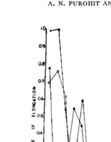
Fw. 2. Length of branches during different gtowth flushes in the annual growth cycle of Callistemon viminalis.
the first 8 weeks, did not increase later and the ultimate length during these growth flushes, therefore, remained much lower than that during growth flush 1. A much greater increase in length occurred during growth flush 4 (triangles). It .is interesting to note that the initial period of slow growth decreased from growth flush 1 to 4 and the period through which branch length continued to increase also decreased with the growth flush. Thus, it continued for 13, II, 7 and 4 weeks in growth flushes
1, 2, 3 and 4, respectively.
Number and size of leaves.-The number and size of leaves produced during different growth flushes are presented in Figs. 4 and 5, respectively. It can be seen that the number of leaves increased with the growth flush, so that it was the least in growth flush 1 and the highest in growth flush 4. The size of leaves produced during different growth flushes also followed similar trends.
z 0 ;:
c
~
0
-' w().
o.z
0·1
3
0---<> GROWTH FlUSH· I -GROWTH FLUSH·2 .._.GROWTH FLUSH·3 .>-<>GROWTH FLUSH-4
FIG. 3. Rate of extension growth of branches during different growth flushes in the annual growth cycle of Callistemon viminalis.
the size of those produced in growth flush 4 decreased with their position towards the apical growing point. Thus, the earlier formed leaves were longer than the later produced ones. It was also found that the leaves produced during the reproductive phase (January-February) were small and scally in nature and fell during the elongation of floral axis. Even some of the leaves produced in the region just above the inflorescence abscised, indicating an inverse relationship between reproductive develop-ment and vegetative growth.
Structural changes in shoot apex during different developmental
stages.-Figs. 6-16 are the photomicrographs showing changes
, ...
...
STRUCTURAL CHANGES IN SHOOT APEX OF CALLISTEMON 113
---.... ·-·-···-···--··· .. '·,
SS• ... :,, ...
::~':·":::.:.:····
... . .. ..,ci·~::-.... :~:-··· ... ~-:-... _ ~-... -....
---·.
25"· ... : ... :, ..
4
-PERIOD OF INFLORESCENCE HONGATION =PERIOD OF ACTIVE GROWTH -PERIOD OF INACTIVE GROWTH
3 ; 4'
GR9WTH FlUSH
6
5
JAPI•MAil' ~'f MA..-·JIJLY JUlY - DlCfMih
TIME- MONTHS
5
- PERIOD OF INFLORESCENCE ELONGATIOI" =PERIOD OF ACTIVE GROWTH
··· ... t-20•: ... :·.,,.
•·. "'4> · .. , i- ·.
'., "-. 15''·.
-PERIOD OF INACTIVE GROWTH
··
... ,_..,.
· ..'·· ... "'>
10'', ... '.: ... ··-.......
1 .... ,=t.~2~.~~3~.====~
..
~4...
~i.ii=Miit.l :~; ~OWT-Fl~-f.,:H-DECEMIEII
TIME-MONTHS
li
FIGs. 4 and 5. Three-dimensional diagrams showing the number and size of leaves
produced in different growth flushes inC, viminalis, Length ofleaves (Fig. 4);
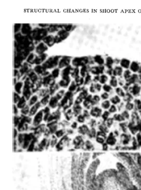
Fws. 6-16. Median longitudinal sections of shoot apices at different developmental stages. Fig. 6: Shoot apex at stage I. Note the flat apical meristem and two layered tunica (x783). Fig. 7: Shoot apex at stage 2, dome shaped apex with three layered tunica and a bud primordium on the right flank
(X 780). Fig. 8: Shoot apex at stage 2 under low magnification showing scaly leaves with one floral bud primordium in the axil of each scaly leaf
STRUCTURAL CHANGES IN SHOOT APEX OF CALLJSTEMON 115
Figs. 12 and 13: Shoot apices at stages 5 and 6, respectively showing large apical domes and cells with more cytoplasmic contents (X 783). Fig. 14: Shoot apex at stage 7 showing large intercellular spaces in the central region and a low apical dome (X 783).
,.
STRUCTURAL CHANGES IN SllOOT APEX OF CALLISTEMON
1J7
Fig. 15. Apex at stage 8 showing a distinct central cone shaped zone (X 783); and Fig. 16: same as in Fig. 15: under low magnification showing undeveloped vegetative buds in the axils of leaf primordia (X 116).
this stage are small and more or less isodiametric. These are deeply stained, non-vacuolated and are characteristically meriste-matic. A few outgrowths in the axils of scaly leaves representing the floral bud primordia are also visible (Fig. 8).
The height of the apical dome increases slightly at stage 3. The central cells of tunica and upper corpus are small with dense cytoplasm and a large nucleus in each. All these are indicative of the high rate of meristematic activity. The intercellular spaces also increase at this stage (Fig. 9). The axillary meristems, initiated earlier during the period of long dormancy, develop into floral buds in acropetal manner with the resumption of growth. This is observed in Fig. 10, which is the photomicrograph of shoot apex at stage 3 at a lower magnification.
The tunica becomes very distinct with anticlinal divisions in some of its cells at stage 4 (Fig. 11). The cells of the outer tunica layer are rectangular. In the central region of tunica and corpus the cells are large with less dense cytoplasm and less prominent nuclei than at stage 3. The intercellular spaces, however, increase considerably. All these indicate that at stage 4 there is a decrease in the rate of cell division.
At stages 5 and 6 (Figs. 12 and 13) the height of the apical dome increases and the content of cells in the centre of tunica and corpus, becomes dense with an appreciable decrease in the intercellular spaces. At stage 6 (Fig. 13), the cells of tunica have large nuclei. The size of the dome reaches its maximum at stage 7. The intercellular spaces in the central region increase and the cells, which are large in size, show divisions in different planes (Fig. 14).
The apical dome becomes more or less flat at stage 8 (Fig. 15). The tunica is single layered. The intercellular spaces increase and the cell content becomes more dense. The develop-ment of axillary meristem at this stage slows down and no young leaf primordia can be observed (Fig. 16). It is interesting to note that a central cone-shaped zone characteristic of "cyto-histo-logical zonate pattern" becomes apparent at this stage of develop-ment (Fig. 15).
Ascorbic acid (AA) content
STRUCTURAL CHANGES IN SHOOT APEX OF CALLISTEMON 119 '-' 0 0
--
'-' ~ I1--fS,oo
1--z
0 0 <( <(Jl
IYil
2
~
jIt
4
STAGE
OF
17
.--. GROWING APE X (>-~ STEM
c·~-~ LEAF
.~---<> SCALY LEAF
~-~ FLORAL BUD
.--..FLOWER
o--o
FRUITf
w
5 6 7 8
DEVELOPMENT
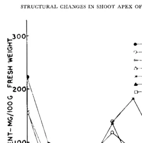
FIG. 17. Ascorbic acid content (mg per 100 g fresh weight) of different plant parts of C. viminalis at different developmental stages.
Apical growing point.-The AA content of the growing apex at stage 1 is high (Fig. 17, blackened circles). The content falls rapidly and reaches a low level at stages 3 and 4, during which the axis elongates (Fig. 1). A slight increase at stage 5 is followed by a fall at stage 6 and subsequently a gradual in-crease during stages 7 and 3.
Stem.-The trends of AA content of stem are similar to those of apical growing point, although it is lower till stage 3 and becomes higher than that of growing apex at subsequent stages.
18
®
@) 0@
®
@ it0
0
(G ·1)
@) €)
®
0
0
0 0
0
\D
\YQ)
0
0
0 0 CZl <2') @) (I!)QJ
898
0
0~
~
\[]
\!1
0
§§
~®
0
<12&
Q'J
\Y'
\D
fB
\!)
Q) 0 0
(!) <D
Q) <Z> Q) <D <2::> <D
CD ('_u
· - - - .
I 2 l4 ~ 6 8
DEVELOPMENTAL STAGES
19
@ @) '7-l)
-...;;, ~[!j
C)
0
0
0
€)e
®
®
G
8
e
<V
<?)
~
(B
0
®
0
0
0
C2J
G
0
~
0
0
C)
0
0
Q]
e
ffi
®
0
~
CD
ffi
8
CJ
8
Q) CD 0Q)
(3
QJ
8
0
Q)
8
0
\!)
G
QJ
\D
CD (!_":
V8 P8 FB
FB F F FR
STRUCTURAL CHANGES 1N SHOOT APEX OF CALLISTEMON 121
Leaves.-The AA content of young leaves emerging above
the inflorescence at stage 5, is very high to start with (circles with dotes) but falls during subsequent stages with a slight increase at stage 8.
Floral buds.-The AA content of floral buds (crosses)
increases with an increase in their size, reaches its maximum at stage 6 and then falls with their opening into flowers (blackened triangles) and their development into fruits (empty squares).
Free amino acids:
The content of free amino acids only of growing apex and vegetative and floral buds was determined at different stages of development.
Apical growing point.-The free amino acids found in the
apical growing point are shown in Fig. 18 and their relative concentrations, as judged from the area and intensity of colour of spots, in Table I. About 9-13 amino acids were found in
Table I. Free amino acids in the shoot apex
of
Callistemon viminalisat different stages in its development
s.
Stage of developmentNo. Amino acid
2 3 4 5 6 7 8
Cystine
++ ++
++
++
++
+
+
+
2 Lysine M M M
++
++
+
+
+
3 Histidine
++ ++ +++ +++ +++ ++ ++ ++
4 Aspartic acid
+ ++
++
++
+
+
+
+
5 Glycine
+ ++ +++
M M M++ ++
6 Proline
++ ++ +++
++
++ ++ ++ ++
7 Unknown M
++ +++
++
+
M+
+
8 Glutamic acid
+ ++ +++ +++
++ ++ ++ ++
9 Theronine
++ ++ +++ +++
++ ++ ++ ++
10 Phenylalanine M M
++
++
M M M++
II Methionine M M
++
++
M M+
+
12 Isoleucine
+
+
+
T M M M Mthe growing apex at various developmental stages. Six of these, namely, histidine, proline, threonine, glutamic acid, cystine and aspartic acid,were present at all stages, while the other 6 appeared at some stages. Thus, methionine, lysine and phenylalanine were more or less completely absent at stages 1 and 2, when the apex had not started active growth, but appeared at stages 3 and 4, when axis started elongating. Isoleucine and leucine, on the other hand, were present only at stages 1 to 3.
Buds.-Only a few free amino acids were present in vegeta-tive buds (Fig. 19), cystine, glutamic acid, isoleucine and leucine being more or less completely absent. In marked contrast to this, aspartic acid, alanine, proline and threonine were present in high concentrations at all stages in the development of floral buds, flowers and fruits. The concentration of histidine, gluta-mic acid and methionine decreased with the opening of floral buds into flowers and their development into furits. Cystine, isoleucine and leucine, which were present till stages 3-4, and were completely absent in flowers and fruits.
Table II. Free amino acids in the vegetative and floral buds of Calliste-mon viminalis at different stages in its development
Stage of development of buds
s.
No. Amino Acid Vege- Floral
tative - - - Flowers
Fru-l 2 3 4 its
Cystine M
+
++
+
+
T T2 Histidine M
+++ +++ +++
++
+
M3 Aspartic acid
++
+
++ +++ +++ +++ ++
4 Alanine
+ +++ +++ +++
++
++ ++
5 Proline
+ +++ +++
++
++
++ ++
6 Unknown M
+++ +++ +++
++
++ ++
7 Glutamic acid T
+++ +++
++
++
+
+
8 Threonine
++ +++ +++ +++
++
++ ++
9 Phenylala nine M M M T
+
+
M10 Methionine T
++
++
+
+
+
+
11 Isoleucine M +
+
+
T M MSTRUCTURAL CHANGES IN SHOOT APEX OF CALUSTEMON 123
Comparing Tables I and II, it is apparent that the behaviour of free amino acids in floral buds was more or less identical to that in the shoot apex at the corresponding develop-mental stage. It is interesting to note that lysine, which ap-peared in the apical growing point after stage 4, was totally absent in buds, whether floral or vegetative. On the other hand alanine, which was absent in the apical growing point, was present in the buds in appreciable concentrations.
DISCUSSION
The morphological changes that the branches of C. viminalis
undergo and their relationship with histological changes in the shoot apex during the annual growth cycle have been reported earlier (Purohit and Nanda, 1968). The growth in this plant takes place in recurrent flushes with alternating periods of growth and rest. During growth flush I cells of the rib meristem show elongation twice during the active period of growth, first during January and again during March. The elongation of cells in rib meristem during this flush corresponds with the two peaks in the rate of growth during this period (Fig. 3). A slow rate of growth during growth flushes 2 and 3 and a high rate for about 4 weeks in growth flush 4, also correspond with the slow and fast rates of elongation of rib meristem during these growth flushes (Purohit and Nanda, 1968). All these demonstrate that the rate and duration of elongation of a branch are a conse-quence of events that occur in the rib meristem during each growth flush.
The number, size and nature of leaves that expand during each growth flush seem to be closely related with the duration of the preceding rest period. Thus, although the rate of elongation is high during growth flushes 1 and 4, and very low during growth flushes 2 and 3, the number and size of leaves increases with the growth flush, being the least during growth flush 1 and the highest during growth flush 4.
of tunica layers, the degree of vacuolation and in the mitotic activity of the cells of the peripheral zone. The divisional acti-vity of the cells in the peripheral zone increases considerably during the initiation of leaf primordia as well as the initiation of floral buds. The apical dome is more flat during leaf initiation but becomes conical during floral bud initiation.
An upsurge in the free AA content of the growing apex with its transformation into the reproductive state has been reported in a number of determinate type of plants (Chinoy, 1962, Chinoy and Mansuri 1965, Tayal, 1965). C. viminalis belongs to a group of plants that are indeterminate in nature. The lack of upsurge in the free AA content in the growing apex observed in this plant even when floral buds appear in the axils of leaves down below it, therefore, is not contrary to the findings in determinate plants. The relationship between free AA content and reproductive development, however, is apparent in the developing floral buds. Thus, the content increases in the buds with an increase in their size, reaches a maximum by stage 6 and decreases subsequently with the opening of flowers reaching the lowest level in fruits. These results are in accord with those of wheat, oat and millets reported earlier (Chinoy, 1962, Tayal, 1965). The massive concentration of AA in the shoot apex at stage 1 when it is still in dormant state, supports the findings reported in a previous paper (Purohit and Nanda, 1966).
The AA content of leaves increases with development but decreases rapidly when approaching maturity. Similar results were obtained in lemon leaves by Earks (1964). While nothing can be said at this stage regarding the mechanism involved and the significance of changes in the level of free endogenous AA, the decrease in content with maturity may be ascribed to either (i) a decrease in the rate of destruction, or (ii) an increase in the rate of its synthesis or (iii) the production of new enzyme(s) or enzyme system(s) that cause its rapid mobilization.
STRUCTURAL CHANGES IN SHOOT APEX OF CALLISTEMON 125
abnormal increase in the number of leaves. These two amino acids are present in the growing apex of this plant during the developmental stages 1-3 and may be concerned in the increased production of leaves and their limited growth, resulting in scaly form. The compact scaly leaves probably restrict the entry of oxygen to the apex and as a consequence of which a rapid synthesis of proteins occurs due to the metabolism of the products of glycolysis as proposed by Ruhland et al. ( 1936, 1938). A high concentration of cystine and glutamic acid along with the high concentration of free AA in the floral buds prior to their opening into flowers is of particular significance, as they are interrelated through the synthesis of reduced glutathione. Further work is needed to prove their importance in this case.
REFERENCES
Biivat, R. (1952). Structure, evolution et fontionnement du meristeme apical d!' quelques dicoty!edones. Ann. Sci. Nat. Bot., XI. 13: 199-300.
Chinoy, J. ]. (1962). Formatior; and utilization of ascorbic acid in the shoot apex of wheat as factors of growth and development. Indian J Plant Physiol.,
5: 172-201.
- - - - a n d Mansuri, A. D. (1965). Further evidencein support of the ascorbic acid-nucleic acid-protein metabolism concept of flowering in Plants. In-Proc. Seminar in Plant Physiol., Panjab Univ. Chandigarh. (ed. R. D. Asana and K. K. Nanda) pp. 68-83.
Earks, I. L. (1964). Ascorbic acid content of citrus during growth and development.
Bot. Gaz., 125: 186-91.
Evelys, K. A., M:a!loy, N. T. and Rosen, C. (1938). J. Biol. Chern., 126: 645 (Seen in-Methods of Bioch. Analysis (eel. Glick, D.). Inter. Pub!. Inc., New York. 1954: 115-40.
Lance, A. (1957). Recherches cytologiques sur !'evolution DE quelques mcristemcs APICAUX ET SUR ses variations provoquees par des traitenments photoperiodiques. Ann. Sci. Nat. Bot., XI. 18: 91-422.
Purohit, A. N. and Nanda, K. K. (1966). Seasonal variations in ascorbic acid content of shoot apex and its relationship with extension growth of Callistemon
viminalis. Plant and Cell Physiol., 7: 499-501.
- - - - and - - - - (1968). Morphophysiological studies of the shoot apex I. Recurrent growth flushes and their relationship with structural changes in the growing apex of Callistemon viminalis. Cand. J. Bot., 46: 1287-95.
Ruhland, W. and Vllirich, H. (1936). Aerobe Garung in Wachsenden Pflanzen-geweben. Ber. Sachs. Akad. Wiss. Leipzgi, M:ath-phys. KI. 88: II-20.
- - - - a n d Ramshorn, V. K. (1938). Aerobe Garung in aktiven Pflan-zlich Meristemen. Planta, 28: 471-514.
Steinbery, R. A. (1947). Growth responses to organic compounds by tobacco seed-lings in aseptic culture. ]. Agr. Res., 75: 81-92.
- - - - (1949). Symptoms of amino acid action or tobacco culture. ]. Agr. Res.,
78: 733-41.