Journal of Nature and Science, Vol.1, No.4, e74, 2015
Biological Chemistry
Control of the cell cycle and mitosis by phosphorylated
activating transcription factor 2 and its homologue 7
Chia-Chen Ku1-3, Hitomi Hasegawa5,+, Chang-Shen Lin1-4, Ming-Ho Tsai1-3, Kenly Wuputra1-3, Richard Eckner6, Naoto Yamaguchi5,*, and Kazunari K. Yokoyama1-3,*
1Center for Stem Cell Research, 2Center for Environmental Medicine, 3Graduate Institute of Medicine, Kaohsiung Medical University Hospital,
Kaohsiung Medical University, Kaohsiung, Taiwan. 4Department of Biological Sciences, National Sun Yat-sen University, Kaohsiung, Taiwan. 5
Department of Molecular Cell Biology, Graduate School of Pharmacological Sciences, Chiba University, Chiba, Japan. 6Department of Biochemistry and Molecular Biology, Rutgers New Jersey Medical School, Rutgers, The State University of New Jersey, Newark, USA
Activating transcription factors (ATFs) comprise a family of sequence-specific DNA-binding proteins that possess a basic region/leucine zipper (bZIP) and they play multiple roles in the mammalian cells. Despite their diverse physiological roles, they all share the ability to respond to environmental stresses and maintain cellular homeostasis. ATF2 and ATF7 are structurally very similar, especially in terms of their bZIP DNA-binding and dimerization domains, where they share >90% identity. In response to stress stimuli, ATFs activate a variety of targets, including cell cycle, DNA damage, antiproliferation, and apoptosis regulators, although most of these targets have been studied only G1/S phase events. Recent research demonstrates
that the cycling-dependent kinase 1 specifically phosphorylates ATF2 and ATF7 in the M phases, and phosphorylated ATF2 and ATF7 are required for G2/M progression, partly by activating Aurora kinase. This review describes the phosphorylation mode of ATF2/ATF7 proteins and their potential functions in cell cycle progression and oncogene addiction. Journal of Nature and Science, 1(4):e74, 2015
ATF2 | ATF7 | DNA damage | G2/M progression | phosphorylation | oncogenesis transcription
Introduction
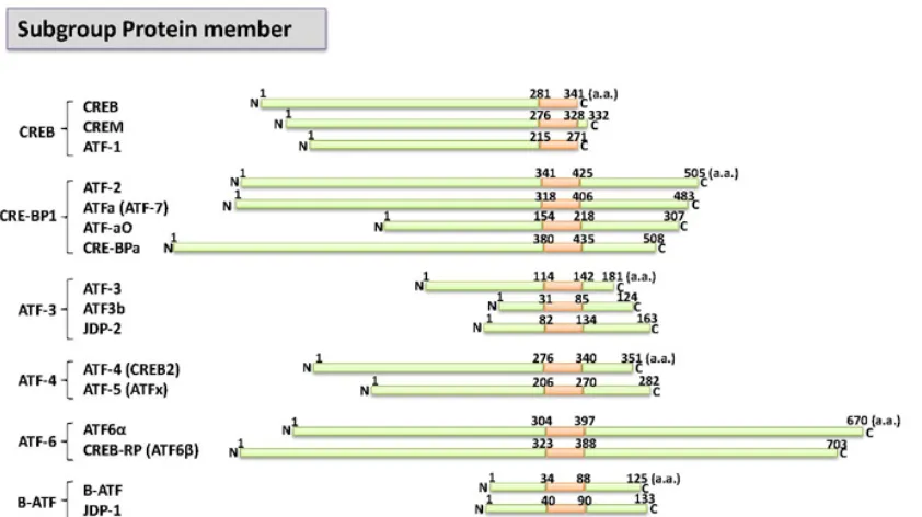
The activation transcription factor (ATF)/cAMP-response element- binding (CREB) family comprises 16 cellular stress-responsive transcription factors, which are divided into six subgroups, according to their sequence similarity (Figure 1) [1-4]. A common feature shared by all of these proteins is the basic zipper (bZIP) element, which allows them to dimerize and bind to specific DNA sequences [5-10]. The bZIP element comprises a leucine zipper subdomain and a basic region subdomain, which are connected by a short fork [11]. The bZIP transcription factors such as the ATF/cAMP-responsive-element (CRE) family and AP-1 (activation protein 1) family proteins homodimerize, but they can also selectively heterodimerize with each other, e.g., c-Fos, Fra2, and c-Jun [12, 13]. However, they share little similarity apart from the bZIP domain, and their binding sequences, such as binding to the ATF/CRE consensus 5ˊ-TGACGTC/AC/A-3ˊ [12, 14] or the AP-1 consensus 5ˊ-TGACTCA-3ˊ. Among the bZIP transcription factors, particularly the ATF/CREB and AP-1 families, ATF2, ATF7 and CREB5 comprise a similar subfamily based on their sequence conservation [5, 8, 12, 15, 16]. The transcriptional activation and DNA-binding domains of ATF2 and ATF7 are highly conserved and their specificity is governed mainly by posttranslational modifications and interactions with specific cofactors [17-20].
The ATF2 gene is located on chromosome 2q32 and it is encoded by a 505-amino-acid protein, which is expressed ubiquitously, with more abundant expression in the brain [21, 22]. ATF2 exhibits diverse, tissue-dependent functions [23, 24]. For example, ATF2 has been implicated in malignant and non-malignant skin tumor development [25, 26]. ATF2 also elicits a suppressor functions in mammary tumors [27]. In addition, ATF2 has transcription-independent functions in the DNA damage response, chromatin remodeling, and mitochondrial membrane organization.
The ATF7 gene is located on chromosome 12q13 and it encodes
a 483-amino-acid protein, which is also expressed ubiquitously. Depending on the cellular context, the composition of the dimeric complex determines the regulation of growth, survival, and apoptosis. ATF7 is known to share similarity with TATA-binding protein associated proteins (TAFs) such as TAF 4 [19]. ATF7 is also critical for neuron networks and social stress-related responses [28]. These multiple functions of ATF2 and ATF7 in oncogenesis and mammalian development are related mainly posttranslational regulation including phosphorylation.
This short review summarizes our current understanding of the phosphorylation status of ATF2 and ATF7 during the cell cycle and the possible functions of phosphorylated ATF2 and ATF7.
Complex isoforms of ATF2 and ATF7
ATF2 has several splice variants, including mouse orthologues (CRE-BP1, CRE-BP2, and CRE-BP-3) and one human isoform (ATFs-sm). The structures of CRE-BP1 and CRE-BP3 are nearly identical, except that eight hydrophobic amino acids in CRE-BP3 replace the first 15 amino acids in CRE-BP1 [29]. By contrast, CRE-BP2 has a 98-amino-acid N-terminal internal deletion. The splice variants of these three murine isoforms occur predominantly in the N- and extreme C-termini, whereas the bZIP domain is conserved, thereby suggesting that the transcription factor function of these variants is conserved, although their regulation might differ due to the loss of various regulatory elements in the N-termini [21, 29]. In agreement with this hypothesis, CRE-BP3 and CRE-BP1 exhibit only weak or no transcriptional activity in murine T cells, respectively, whereas CRE-BP2 exhibits strong transcriptional activity. The human ATF2 splice isoform ATF2-sm lacks the entire bZIP domain and it only retains the first and the last two exons of the full-length ATF2. However, despite the absence of the bZIP domain, ATF2-sm still exhibits its transcriptional activity [30]. ATF2-sm is specifically expressed in endometrial tissue and its protein levels fluctuate dynamically throughout pregnancy [30]. This distinct expression pattern suggests that ATF2 splice variants might elicit tissue- and temporal-specific functions, which may be distinct from those regulated by the full-length ATF2 [31].
In the case of ATF7, three major different transcripts are encoded in humans. i.e., ATF7-1, ATF7-2, and ATF7-4, whereas only one, ATF7-2, is encoded in the mouse [32, 33]. The ATF7-4 protein contains the common ATF7 N-terminal moiety (88 residues), which encompasses the transcriptional activation domain [34], but it lacks the entire C-terminal bZIP domain (residues 89–494), and the DNA binding/nuclear localization signal, and a dimerization domain. The ATF7-4 isoform is potentially encoded in many mammalian genomes, but not in the mouse and rat, and it is localized mainly in the cytoplasm [33]. The human ATF2 and human ATF7 genes generate 13 and six transcripts variants,
__________
Conflict of interest: No conflicts declared.
+Present address; Education & Research Center for Pharmaceutical
Sciences, Faculty of Pharma Sciences, Teikyo University, Tokyo, Japan *Corresponding Authors: Naoto Yamaguchi ([email protected]), and Kazunari K. yokoyama ([email protected]).
Figure 1. Schematic representation of members of the ATF/CREB transcription factor family. ATF/CREB protein members can be categorized into six subgroups according to their sequence similarity. The red boxes indicate the bZIP domain. (GenBank Accession numbers: AAC60616, Q02930, AAC02258, P18848, AAD51372, and AAB49921). The figure is modified as reported previously (Ref. 24, Figure 2).
respectively, including noncoding RNA. In order to prepare specific ATF2 and ATF7 short hairpin (sh) knockdown constructs, the care should be taken when selecting the corresponding sequences, as they might present splice variants or noncoding transcripts.
Specificity of antibodies against ATF2 and ATF7
Hasegawa et al. (2014) reported a problem with reagents such as antibodies against ATF2 and ATF7 [35]. Surprisingly, most of the widely used ATF2 antibodies such as N-96 (sc-63233; Santa Cruz Biotechnology Inc.) and phopsho-ATF2 (pT71) (#922; Cell Signaling Technology) can cross-react with ATF7. Thus, most previous studies that used these antibodies also analyzed the ATF7 protein. It is possible to differentiate ATF2 and ATF7 proteins using antibodies from Sigma-Aldrich Co. ATF2 [ss-16] is specific for ATF2 (#A4086; Sigma-Aldrich) and anti-ATF7 antibodies (#SAB250013 and #HPA003384; Sigma-Aldrich) are specific for ATF7. Thus, we consider that the critical observation might lead to re-investigation of previously obtained results.
Role of ATF2 in human cancer
Analyses of tumor samples in breast cancer and melanoma suggest that ATF2 may possess tumor suppressive and promoting functions, respectively [27, 36]. Furthermore, the enhanced presence of activated (Thr71 phosphorylated) ATF2 has been reported in common types of skin carcinoma, e.g., squamous cell carcinoma, Bowen’s disease, and basal cell carcinoma, thereby suggesting the involvement of ATF2 in this type of tumor [37]. However, activation does not preclude a functional role for ATF2 in the development of this type of tumor, and thus further experimental testing is required. Interestingly, a recent global genomic analysis identified a comprehensive array of gene mutations and the core signaling pathways instrumental in pancreatic cancer development [38]. This study confirmed that mutations in Kras and associated genes are major features of pancreatic tumors. In addition, significant numbers of gene mutations have been identified in MAPK (Jun Nh2-terminal protein kinase; JNK) components as well as in ATF2 protein in cancer specimens. This may suggest that JNK signaling and/or ATF2 are instrumental in the progression of this cancer type, but this remains to be examined experimentally. ATF2 mutations have also been identified in mammary carcinomas
(see Catalogue of Somatic Mutations in Cancer at http://www.sanger.ac.uk/genetics/CGP/cosmic/). Therefore, it is still unclear whether the mutations identified in cancer cell lines have any significant effects on the function of ATF2 s well as on the tumor phenotype.
Loss of function of ATF2 and ATF7 in mouse models
Reimold et al. (1996) reported that the first Atf2 mouse knockout
led to decreased postnatal viability and reduced growth in animals, as well as defects in endochondral ossification and in the central nervous system [39]. However, this knockout was subsequently shown to be hypomorphic because it retained some of the ATF2 activity due to alternative splicing in the targeted allele [40]. Another knockout of ATF2 (Atf20/0) was generated by the deletion
of the DNA-binding domain, which produced, at least transcrip- tionally, a functional null mutation. All of the Atf20/0 mutant mice
died shortly after birth because of severe respiratory distress [40]. Furthermore, Maekawa et al. demonstrated that a knock-in mutant mouse line where the Thr69 and Thr71 (Thr51 and Thr53 in mouse ATF2) phosphorylation sites in the transcription activation domain were mutated into alanine (Atf2AA) yielded to a similar phenotype, and to invariably led to death at birth, thereby confirming the importance of these phosphorylation sites for the activity of ATF2 [41]. Ackermann et al. (2011) demonstrated that the perinatal death caused by the loss of ATF2 functions in mouse embryos is probably caused by defects during the development of the brainstem, where ATF2 is involved in functions that prevent specific sets of facial motor neurons from undergoing neuro- degeneration and apoptosis. Previous studies of the deletion of the DNA-binding domain of ATF7 in the presence of functional ATF2 have not identified exposed any discernible developmental phenotypes in mice [41]. However, ATF7 has an important role in the adult central nervous system, specifically in the regulation of gene expression in dorsal raphe nuclei of the brainstem in response to isolation stress. By contrast, the ATF2/ATF7 double deletion (Atf2−/−; Atf7−/−) results in severe hypoplasia in the embryonic
Journal of Nature and Science, Vol.1, No.4, e74, 2015
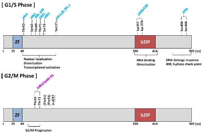
Figure 2. Phosphorylation of ATF2 protein and ATF7 protein. Schematic representation of various kinases that can phosphorylate ATF2 protein. The model
designated as (Cell cycle) was modified (33). Most of the phosphorylation sites are shown during the G1/S phase. Hasegawa et al. (35) reported the cdk1–cycling B complex was phosphorylated in the G2/M phase.
MKK4 (MAPK kinase 4) [43, 44] and it occurs around the same time, but significantly before the liver defect observed in c-Jun-knockout embryos [45, 46], which suggests that ATF2 and ATF7 are essential substrates for embryonic JNK signaling. ATF2/ATF7 mutant liver progenitor cells (hepatoblasts) undergo high rates of apoptosis in embryos as well as in culture. This high rate of apoptosis in cultured hepatoblasts can be rescued by the chemical inhibition of p38 activity, which suggests that these transcription factors may be involved in negative-feedback regulation of their upstream activating kinases. Double mutant livers express reduced levels of specific MAPK phosphatases, including Dusp1, Dusp8 and Dusp10, which are direct targets for ATF2/ATF7- mediated transcription.
Posttranslational regulation of ATF2 and ATF7
ATF-2 is phosphorylated by many upstream kinases including stress-activating kinase or protein kinase C (PKC) in the G1/S phase (Figure 2). JNK, p38, and Erk, which are activated by stress stimuli, phosphorylate ATF2 at Thr-69 and Thr-71 (Thr-69/Thr-71) and lead to its transcriptional activation [47-52]. Moreover, the phosphorylation of ATF2 at Ser-121 by several PKC isoforms (including α, βI, βII, and γ) plays a role in the c-Jun-mediated activation of transcription in response to 12-O-tetradecanoylphorbol-13-acetate [53]. It is known that ATF7 is phosphorylated by p38 at Thr-51 and Thr-53, which correspond to Thr-69 and Thr-71 in ATF2, thereby leading to its transcriptional activation [54, 55]. In contrast to its transcriptional functions, ATF2 has several functions that are independent of transcriptional activation [56]. ATF2 is phosphorylated at Thr-52 by PKCε, which negatively regulates the outer-membrane permeability of mitochondria and inhibits apoptosis during genotoxic stress [57]. During the DNA damage response, ataxia telangiectasia mutated (ATM) phosphorylates ATF2 at Ser-490 and Ser-498, to facilitate DNA repair [58]. In this context, the interaction between ATF2 and the histone acetyltransferase TIP60 comprises a positive feedback loop, which allows ATF2 to promote the activity of ATM. Genotoxic stress attenuates the interaction between TIP60 and ATF2, which stabilizes TIP60 and promotes the subsequent acetylation and activation of ATM [59]. Thus, ATF2 and ATF7 play important roles in the G1 and S phases.
Recently, Hasegawa et al. (2014) identified a new kinase that phosphorylates ATF2 and ATF7 in the G2 and M phases [35]. ATF2 (at Thr-69/Thr-71) and ATF7 (at Thr-51/Thr-53) are phosphorylated by cyclin-dependent kinase 1 (Cdk1) in the M phase. Similar to the knockdown of ATF7, the expression of mitotically nonphosphorylatable ATF7 inhibits the entry of cells into the M phase [35]. These results suggest that the phosphorylation of ATF7 at Thr-51/Thr-53 during the M phase plays an important role in G2/M progression, partly by activating Auora kinases.
ATF-2 is also acetylated on Lys-357 and Lys-374 by p300 or CREB-binding protein (CBP, also known as CREBBP), which contributes to its transcriptional activity [60]. The binding of ATF2 suppresses the acetyltransferase activity of the transcriptional coactivator p300/CBP. The cross-regulation between the acetylation and phosphorylation of ATF2 has yet to be elucidated in the context of its transcriptional activities.
The stability of ATF2 protein is regulated by ubiquitylation and SUMOylation. However, the exact model has not been elucidated in detail. N-terminal phosphorylation and heterodimerization of ATF2 reduce its transcriptional activity by promoting ubiquitylation-dependent degradation [61]. The binding of JNK to ATF2 serves to limit the availability of ATF2 by promoting its degradation, but the E3 ubiquitin ligases involved in the ubiquitylation and degradation of ATF2 have not been identified. The SUMO-conjugating enzyme Ubc9 has been shown to interact with ATF2 and to affect its stability [62], although ATF2 SUMOylation has not been demonstrated formally.
Interplay between mitochondria and nuclear ATF2
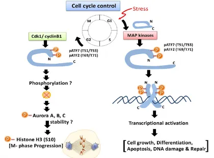
Figure 3. Possible roles of ATF2 and ATF7 phosphorylation in G2/M progression. During the late stages of the G2 and M phases, Thr-51/Thr-53 in ATF7 and
Thr-69/Thr-71 in ATF2 are phosphorylated by cdk1–cyclin B1, which promotes M-phase entry by stabilizing Aurora kinase family.
ATF7 phosphorylation is linked to Aurora signaling
Hasegawa et al. (2014) showed that the mitotic phosphorylation of ATF7 is involved in Aurora signaling [35]. The Aurora family comprises three members; Aurora A, B, and C. Aurora A is an oncogene in a variety of cancers and it plays a role in centrosome maturation during the function the G2/M transition [63]. Disruption of the function of Aurora A delays mitotic entry [64, 65]. Inhibition of Aurora kinases is also known to induce apoptosis [66], so the downregulation of Aurora kinase signaling by ATF7 knockdown may explain the increased apoptosis during the sub-G1 phase after the G2/M-phase arrest following ATF7 knockdown.
Inhibition of the kinase activity of Cdk1 induces Aurora A inactivation, although Cdk1 does not directly phosphorylate Aurora A [67]. Activated Aurora B phosphorylates histone H3 at Ser-10 [68-71]. However, after Cdk1 is activated, the mitotic phosphorylation of ATF7 precedes the phosphorylation of histone H3 at Ser-10, which suggests that ATF7 is located upstream of the Aurora kinases. Thus, it is assumed that the mitotic phosphorylation of ATF7 by the Cdk1–cyclin B1 complex promotes the activation of Aurora kinases to allow mitotic entry via the stabilization of Aurora kinases (see Figure 3).
Potential functions of ATF2/ATF7 function in cell division It is well known that the correct assembly and timely disassembly of the mitotic spindle is crucial for the propagation of the genome during cell division [72]. The main aim of mitosis is ensuring that the replicated sister chromatids are segregated with the highest possible accuracy among the daughter cells. In principal, this is a mechanical problem related to the generation of a force that segregates the two sister chromatids of each chromosome and moves them to opposite ends of the cell division plane. The mitotic spindle provides a platform that facilitates the accurate alignment of the condensed chromosomes and it comprises a molecular machine that segregates the sister chromatids [73]. It is essential that the segregation process is only initiated when each chromosome is aligned in the center of the spindle and bi-oriented, so the sister chromatids in each network are subjected to various
Journal of Nature and Science, Vol.1, No.4, e74, 2015 The proteasome is also known to function at the onset of mitosis
[78, 79]. Thus, Hesegawa et al. (2014) examined the role of ATF7 in M-phase progression in ATF7-wt- or ATF7-TA-inducible cells using the proteasome inhibitor MG132 at the onset of mitosis, which showed that the proteosomal control of inhibitor proteins or associated proteins was induced by phosphorylated ATF2/ATF7 and the M-phase then proceeded smoothly [35]. Thus, it is hypothesized that the mitotic phosphorylation of ATF7 promotes Aurora signaling at the onset of M phase in a manner that is dependent on the activity of the proteasome. Although no physical associations have been detected between ATF7, phosphorylated Aurora kinases, and its related proteins, it is of interest to determine precisely how the mitotic phosphorylation of ATF7 is involved in Aurora signaling, specifically the stability of Aurora kinase, which allows to the active dividing machinery, such as histone H3 phosphorylation, to promote cell division.
Future studies will reveal the roles of this novel mechanism of ATF2/ATF7 during mitosis progression in cell growth control, transformation, and cancer development.
Future perspectives
ATF2 and ATF7 are known to be important as transcription factors, DNA-damage response proteins and during G2/M progression, but
they also implicated in the regulation of cellular growth and cell division control. ATF2 and ATF7 have functions in checkpoint control during the intra-S phase and probably in mitosis, as well as in transcriptional regulation via distinct upstream regulatory kinases. It is still necessary to clarify whether different pools of ATF2 and ATF7 are utilized for each of these functions, and whether their roles in the DNA repair axis affects the transcriptional activities of ATF2 and ATF7. Novel functions of ATF2 and ATF7 during G2/M progression might control Aurora signaling in cell division. Our growing understanding of the complex regulatory roles of ATF2 and ATF7 indicates that they may have the capacity to elicit oncogenes or anti-oncogenic function, as well as being involved in mitotic regulation.
Acknowledgments
We thank Drs. O Lee, K Kato, K Nagata, S Lin, YC, G Gachelin and M. Horikoshi for their advice and discussions. This study was supported by grants from the Taiwan government:
NSC-101-2320-B-037-047-My3; NSC-103-2314-B-037-063; NHRI-Ex102-10109BI; NHRI-EX104-10416SI; KMU-DT103001; KMU-TP103G00, KMU-TP103G03, KMU-TP103G04,
KMU-TP103G05, KMU-TP103A04.
1. Pescini R, Kaszubska W, Whelan J, DeLamarter JF,& Hooft van Huijsduijnen R. ATF-a0, a novel variant of the ATF/CREB transcription factor family, forms a dominant transcription inhibitor in ATF-a heterodimers. J. Biol. Chem. 1994; 269: 1159-1165.
2. Hai T & Hartman MG. The molecular biology and nomenclature of the activating transcription factor/cAMP responsive element binding family of transcription factors: activating transcription factor proteins and homeostasis. Gene 2001; 273: 1-11.
3. Haze K, Okada T, Yoshida H, Yanagi H, Yura T, Negishi M, & Mori K. Identification of the G13 (cAMP-response-element-binding protein-related protein) gene product related to activating transcription factor 6 as a transcriptional activator of the mammalian unfolded protein response. Biochem. J. 2001; 355: 19-28.
4. Wang J, Cao Y, & Steiner DF. Regulation of proglucagon transcription by activated transcription factor (ATF) 3 and a novel isoform, ATF3b, through the cAMP-response element/ATF site of the proglucagon gene promoter. J. Biol. Chem. 2003; 278: 32899-32904.
5. Gaire M, Chatton B, & Kedinger C. Isolation and characterization of two novel, closely related ATF cDNA clones from HeLa cells. Nuc. Acids Res. 1990; 18: 3467-3473.
6. Liu F, Thompson MA, Wagner S, Greenberg ME, & Green MR.Activating transcription factor-1 can mediate Ca(2+)- and
cAMP-inducible transcriptional activation. J. Biol. Chem.1993; 268: 6714-6720.
7. Masquilier D, Foulkes NS, Mattei MG, & Sassone-Corsi P. Human CREM gene: evolutionary conservation, chromosomal localization, and inducibility of the transcript. Cell Growth Differentiation 1993; 4: 931-937.
8. Nomura N, Zu YL, Maekawa T, Tabata S, Akiyama T, & Ishii S. Isolation and characterization of a novel member of the gene family encoding the cAMP response element-binding protein CRE-BP1. J. Biol. Chem. 1993; 268: 4259-4266.
9. Dorsey MJ, Tae HJ, Sollenberger KG, Mascarenhas NT, & Johansen LM, B-ATF: a novel human bZIP protein that associates with members of the AP-1 transcription factor family. Oncogene 1995; 11: 2255-2265. 10.Forgacs E, Gupta SK, Kerry JA, & Semmes OJ. The bZIP transcription
factor ATFx binds human T-cell leukemia virus type 1 (HTLV-1) Tax and represses HTLV-1 long terminal repeat-mediated transcription. J. Virol. 2005; 79: 6932-6939.
11.Landschulz WHHH, Johnson PF, & McKnight SL. The leucine zipper: a
hypothetical structure common to a new class of DNA binding proteins. Science 1988; 240: 1759-1764.
12.Hai TW, Liu F, Coukos WJ, & Green MR. Transcription factor ATF cDNA clones: an extensive family of leucine zipper proteins able to selectively form DNA-binding heterodimers. Genes Dev. 1989; 12: 2083-2090.
13.Vinson CR, Hai T, & Boyd SM. Dimerization specificity of the leucine zipper-containing bZIP motif on DNA binding: prediction and rational design. Genes Dev. 1993; 7: 1047-1058.
14.Lin YS, & Green MR. Interaction of a common cellular transcription factor, ATF, with regulatory elements in both E1a- and cyclic AMP-inducible promoters. Proc. Natl. Acad. Sci. USA 1988; 85: 3396-3400.
15.Lee KA, Fink JS, Goodman RH, & Green MR. Distinguishable promoter elements are involved in transcriptional activation by E1a and cyclic AMP. Mol. Cell. Biol. 1989: 9: 4390-4397.
16.Maekawa T, Sakura H, Kanei-Ishii C, Sudo T, Yoshimura T, Fujisawa J, Yoshida M, & Ishii S. Leucine zipper structure of the protein CRE-BP1 binding to the cyclic AMP response element in brain. EMBO J. 1989; 8: 2023-2038.
17.De Graeve F, Bahr A, Chatton B, & Kedinger C. A murine ATFa-associated factor with transcriptional repressing activity. Oncogene 2000; 19: 1807-1819.
18.Duyndam MC1, van Dam H, Smits PH, Verlaan M, van der Eb AJ, &
Zantema A. The N-terminal transactivation domain of ATF2 is a target for the co-operative activation of the c-jun promoter by p300 and 12S E1A. Oncogene 1999; 18: 2311-2321.
19.Hamard PJ, Dalbies-Tran R, Hauss C, Davidson I, Kedinger C, & Chatton B. A functional interaction between ATF7 and TAF12 that is modulated by TAF4. Oncogene 2005; 24: 3472-3483.
20.Hong S, Choi HM, Park MJ, Kim YH, Choi YH, Kim HH, Choi YH, & Cheong J. Activation and interaction of ATF2 with the coactivator ASC-2 are responsive for granulocytic differentiation by retinoic acid. J Biol. Chem. 2004; 279: 16996-167003.
21.Kara CJ, Liou HC, Ivashkiv LB, & Glimcher LH. A cDNA for a human cyclic AMP response element-binding protein which is distinct from CREB and expressed preferentially in brain. Mol. Cell. Biol. 1990; 10: 1347-1357.
22.Takeda J, Maekawa T, Sudo T, Seino Y, Imura H, Saito N, Tanaka C, & Ishii S. Expression of the CRE-BP1 transcriptional regulator binding to the cyclic AMP response element in central nervous system, regenerating liver, and human tumors. Oncogene 1991; 6: 1009-1014. 23.Bhoumik A, Lopez-Bergami P, Ronai Z. ATF2 on the double-activating transcription factor and DNA damage response protein. Pigment Cell Res. 2007; 20498-20506.
24.Vlahopoulos SA, Logotheti S, Mikas D, Giarika A, Gorgoulis V, & Zoumpourlis V. The role of ATF-2 in oncogenesis. Bioessays 2008; 30: 314-327.
25.Bhoumik A, Gangi L, & Ronai Z. Inhibition of melanoma growth and metastasis by ATF2-derived peptides. Cancer Res. 2004; 64: 8222-8230. 26.Bhoumik A, Fichtman B, Derossi C, Breitwieser W, Kluger HM, Davis S, Subtil A, Meltzer P, Krajewski S, Jones N, & Ronai Z. Suppressor role of activating transcription factor 2 (ATF2) in skin cancer. Proc. Natl. Acad. Sci. USA 2008; 105: 1674-1679.
27.Maekawa T, Shinagawa T, Sano Y, Sakuma T, Nomura S, Nagasaki K, Miki Y, Saito-Ohara F, Inazawa J, Kohno T, Yokota J, & Ishii S. Reduced levels of ATF-2 predispose mice to mammary tumors. Molecular Cellular Biology 2007; 27:1730-1744.
28.Maekawa T, Kim S, Nakai D, Makino C, Takagi T, Ogura H, Yamada K, Chatton B, & Ishii S. Social isolation stress induces ATF-7 phosphorylation and impairs silencing of the 5-HT 5B receptor gene. EMBO J. 2010; 29: 196-208.
29.Georgopoulos K, Morgan BA, & Moore DD. Functionally distinct isoforms of the CRE-BP DNA-binding protein mediate activity of a T-cell-specific enhancer. Mol. Cell. Biol. 1992; 12:747-757.
Europe-Finner GN. Characterization and functional analysis of cAMP response element modulator protein and activating transcription factor 2 (ATF2) isoforms in the human myometrium during pregnancy and labor: identification of a novel ATF2 species with potent transactivation properties. J. Clin.Endocrinol Metab. 2002; 87:1717-1728.
31.Bailey J, & Europe-Finner GN. Identification of human myometrial target genes of the c-Jun NH2-terminal kinase (JNK) pathway: the role of activating transcription factor 2 (ATF2) and a novel spliced isoform ATF2-small. J. Mol. Endocrinol. 2005; 34:19-35.
32.Goetz J, Chatton B, Mattei MG, & Kedinger C. Structure and expression of the ATFa gene. J. Biol. Chem. 1996; 271: 29589-29598. 33.Diring J, Camuzeaux B, Donzeau M, Vigneron M, Rosa-Calatrava M,
Kedinger C, & Chatton B. A cytoplasmic negative regulator isoform of ATF7 impairs ATF7 and ATF2 phosphorylation and transcriptional activity. PLoS One. 2011; 6: e23351.
34.Chatton B, Bocco JL, Goetz J, Gaire M, Lutz Y, & Kedinger C. Jun and Fos heterodimerize with ATFa, a member of the ATF/CREB family and modulate its transcriptional activity. Oncogene. 1994; 9: 375-385. 35.Hasegawa H, Ishibashi K, Kubota S, Yamaguchi C, Yuki R, Nakajo H,
Eckner R, Yamaguchi N, Yokoyama KK, & Yamaguchi N. Cdk1-Mediated Phosphorylation of Human ATF7 at Thr-51 and Thr-53 Promotes Cell-Cycle Progression into M Phase. PLoS One. 2014; 9: e116048.
36.Berger AJ, Kluger HM, Li N, Kielhorn E, Halaban R, Ronai Z, & Rimm DL. Subcellular localization of activating transcription factor 2 in melanoma specimens predicts patient survival. Cancer Res. 2003; 63: 8103-8107.
37.Chen SY, Takeuchi S, Urabe K, Hayashida S, Kido M, Tomoeda H, Uchi H, Dainichi T, Takahara M, Shibata S, Tu YT, Furue M, & Moroi Y. Overexpression of phosphorylated-ATF2 and STAT3 in cutaneous angiosarcoma and pyogenic granuloma. J Cutan Pathol. 2008; 35:722-730.
38.Jones S, Zhang X, Parsons DW, Lin JC, Leary RJ, Angenendt P, Mankoo P, Carter H, Kamiyama H, Jimeno A, Hong SM, Fu B, Lin MT, Calhoun ES, Kamiyama M, Walter K, Nikolskaya T, Nikolsky Y, Hartigan J, Smith DR, Hidalgo M, Leach SD, Klein AP, Jaffee EM, Goggins M, Maitra A, Iacobuzio-Donahue C, Eshleman JR, Kern SE, Hruban RH, Karchin R, Papadopoulos N, Parmigiani G, Vogelstein B, Velculescu VE, & Kinzler KW. Core signaling pathways in human pancreatic cancers revealed by global genomic analyses. Science. 2008; 321: 1801-1806.
39.Reimold AM, Grusby MJ, Kosaras B, Fries JW, Mori R, Maniwa S, Clauss IM, Collins T, Sidman RL, Glimcher MJ, & Glimcher LHHH.
Chondrodysplasia and neurological abnormalities in ATF-2-deficient mice. Nature. 1996; 279: 262-265.
40.Maekawa T, Bernier F, Sato M, Nomura S, Singh M, Inoue Y, Tokunaga T, Imai H, Yokoyama M, Reimold A, Glimcher LH, & Ishii S. Mouse ATF-2 null mutants display features of a severe type of meconium aspiration syndrome. J Biol Chem. 1999; 274: 17813-9. 41.Breitwieser W, Lyons S, Flenniken AM, Ashton G, Bruder G,
Willington M, Lacaud G, Kouskoff V, & Jones N. Feedback regulation of p38 activity via ATF2 is essential for survival of embryonic liver cells. Genes Dev. 2007; 21: 2069-82.
42.Ackermann J, Ashton G, Lyons S, James D, Hornung JP, Jones N, & Breitwieser W. Loss of ATF2 function leads to cranial motoneuron degeneration during embryonic mouse development. PLoS One 2011; 6: e19090.
43.Ganiatsas S, Kwee L, Fujiwara Y, Perkins A, Ikeda T, Labow MA, & Zon LI. SEK1 deficiency reveals mitogen-activated protein kinase cascade crossregulation and leads to abnormal hepatogenesis. Proc. Natl. Acad. Sci. U S A 1998; 95:6881-6886.
44.Nishina H, Vaz C, Billia P, Nghiem M, Sasaki T, De la Pompa JL, Furlonger K, Paige C, Hui C, Fischer KD, Kishimoto H, Iwatsubo T, Katada T, Woodgett JR, & Penninger JM. Defective liver formation and liver cell apoptosis in mice lacking the stress signaling kinase SEK1/MKK4. Development 1999; 126: 505-516.
45.Hilberg F, Aguzzi A, Howells N, & Wagner EF. c-jun is essential for normal mouse development and hepatogenesis. Nature 1993; 365: 179-181.
46.Johnson RS, van Lingen B, Papaioannou VE, & Spiegelman BM. A null mutation at the c-jun locus causes embryonic lethality and retarded cell growth in culture. Genes Dev. 1993; 7: 1309-1317.
47.Gupta S, Campbell D, Dérijard B, & Davis RJ. Transcription factor ATF2 regulation by the JNK signal transduction pathway. Science 1995; 267: 389-393.
48.van Dam H, Wilhelm D, Herr I, Steffen A, Herrlich P, & Angel P. ATF-2 is preferentially activated by stress-activated protein kinases to mediate c-jun induction in response to genotoxic agents. EMBO J. 1995; 14: 1798-1811.
49.Livingstone C, Patel G, & Jones N. ATF-2 contains a
phosphorylation-dependent transcriptional activation domain. EMBO J. 1995; 14: 1785-1797.
50.Ouwens DM, de Ruiter ND, van der Zon GC, Carter AP, Schouten J, van der Burgt C, Kooistra K, Bos JL, Maassen JA, & van Dam H. Growth factors can activate ATF2 via a two-step mechanism: phosphorylation of Thr71 through the Ras-MEK-ERK pathway and of Thr69 through RalGDS-Src-p38. EMBO J. 2002; 21: 3782-3793. 51.Morton S, Davis RJ, & Cohen P. Signalling pathways involved in
multisite phosphorylation of the transcription factor ATF-2. FEBS Lett. 2004; 572: 177-83.
52.Lopez-Bergami P, Lau E, & Ronai Z. Emerging roles of ATF2 and the dynamic AP1 network in cancer. Nat. Rev. Cancer. 2010; 10: 65-76. 53.Yamasaki T, Takahashi A, Pan J, Yamaguchi N, & Yokoyama KK.
Phosphorylation of Activation Transcription Factor-2 at Serine 121 by Protein Kinase C Controls c-Jun-mediated Activation of Transcription. J. Biol. Chem. 2009; 284: 8567-8581.
54.Camuzeaux B, Diring J, Hamard PJ, Oulad-Abdelghani M, Donzeau M, Vigneron M, Kedinger C, & Chatton B. p38beta2-mediated phosphorylation and sumoylation of ATF7 are mutually exclusive. J. Mol. Biol. 2008; 384: 980-991.
55.Gozdecka M, & Breitwieser W. The roles of ATF2 (activating transcription factor 2) in tumorigenesis. Biochem Soc Trans. 2012; 40: 230-234.
56.Lau E, & Ronai ZA. ATF2-at the crossroad of nuclear and cytosolic functions. J. Cell Sci. 2012; 125: 2815-2824.
57.Lau E, Kluger H, Varsano T, Lee K, Scheffler I, Rimm DL, Ideker T, & Ronai ZA. PKCε promotes oncogenic functions of ATF2 in the nucleus while blocking its apoptotic function at mitochondria. Cell 2012; 148: 543-555.
58.Bhoumik A, Lopez-Bergami P, & Ronai Z. ATF2 on the
double-activating transcription factor and DNA damage response protein. Pigment Cell Res. 2007; 20: 498-506.
59.Bhoumik A, Singha N, O'Connell MJ, & Ronai ZA. Regulation of TIP60 by ATF2 modulates ATM activation. J. Biol. Chem. 2008; 283: 17605-17614.
60.Karanam B, Wang L, Wang D, Liu X, Marmorstein R, Cotter R, & Cole PA. Multiple roles for acetylation in the interaction of p300 HAT with ATF-2. Biochemistry 2007; 46: 8207-8216.
61.Fuchs SY, & Ronai Z. Ubiquitination and degradation of ATF2 are dimerization dependent. Mol. Cell. Biol. 1999; 19: 3289-3298.
62.Firestein R, & Feuerstein N. Association of activating transcription factor 2 (ATF2) with the ubiquitin-conjugating enzyme hUBC9. Implication of the ubiquitin/proteasome pathway in regulation of ATF2 in T cells. J. Biol. Chem. 1998; 273: 5892-5902.
63.Barr AR, & Gergely F. Aurora-A: the maker and breaker of spindle poles. J. Cell Sci. 2007; 120: 2987-2996.
64.Marumoto T, Honda S, Hara T, Nitta M, Hirota T, Kohmura E, & Saya HHH. Aurora-A kinase maintains the fidelity of early and late mitotic
events in HeLa cells. J. Biol. Chem. 2003; 278: 51786-51795.
65.Hirota T, Kunitoku N, Sasayama T, Marumoto T, Zhang D, Nitta M, Hatakeyama K, & Saya H. Aurora-A and an interacting activator, the LIM protein Ajuba, are required for mitotic commitment in human cells. Cell 2003; 114: 585-598.
66.Pérez de Castro I, de Cárcer G, Montoya G, & Malumbres M. Emerging cancer therapeutic opportunities by inhibiting mitotic kinases. Curr. Opin. Pharnmacol. 2008; 8: 375-383.
67.Marumoto T, Hirota T, Morisaki T, Kunitoku N, Zhang D, Ichikawa Y, Sasayama T, Kuninaka S, Mimori T, Tamaki N, Kimura M, Okano Y, & Saya H. Roles of aurora-A kinase in mitotic entry and G2 checkpoint in mammalian cells. Genes Cells 2002; 7:1173-1182.
68.Hsu JY, Sun ZW, Li X, Reuben M, Tatchell K, Bishop DK, Grushcow JM, Brame CJ, Caldwell JA, Hunt DF, Lin R, Smith MM, & Allis CD. Mitotic phosphorylation of histone H3 is governed by Ipl1/aurora kinase and Glc7/PP1 phosphatase in budding yeast and nematodes. Cell 2000; 102: 279-291.
69.Crosio C, Fimia GM, Loury R, Kimura M, Okano Y, Zhou H, Sen S, Allis CD, & Sassone-Corsi P. Mitotic phosphorylation of histone H3: spatio-temporal regulation by mammalian Aurora kinases. Mol. Cell. Biol. 2002; 22: 874-885.
70.Ruchaud S, Carmena M, & Earnshaw WC. Chromosomal passengers: conducting cell division. Nat. Rev. Mol. Cell. Biol. 2007; 8: 798-812. 71.Giet R, & Glover DM. Drosophila aurora B kinase is required for
histone H3 phosphorylation and condensin recruitment during chromosome condensation and to organize the central spindle during cytokinesis. J. Cell. Biol. 2001; 152: 669-682.
72.Kops GJ, Weaver BA, & Cleveland DW. On the road to cancer: aneuploidy and the mitotic checkpoint. Nat Rev Cancer. 2005; 5: 773-785.
Journal of Nature and Science, Vol.1, No.4, e74, 2015 74.Sullivan M, & Morgan DO. Finishing mitosis, one step at a time. Nat.
Rev. Mol. Cell. Biol. 2007; 8: 894-903.
75.Kapoor TM, Lampson MA, Hergert P, Cameron L, Cimini D, Salmon ED, McEwen BF, & Khodjakov A. Chromosomes can congress to the metaphase plate before biorientation. Science. 2006; 311: 388-391. 76.Tanenbaum ME, Macurek L, van der Vaart B, Galli M, Akhmanova A,
& Medema RH. A complex of Kif18b and MCAK promotes microtubule depolymerization and is negatively regulated by Aurora kinases. Curr. Biol. 2011; 21: 1356-1365.
77.Hégarat N, Smith E, Nayak G, Takeda S, Eyers PA, & Hochegger H. Aurora A and Aurora B jointly coordinate chromosome segregation and anaphase microtubule dynamics. J. Cell Biol. 2011; 195:1103-1113. 78.Acquaviva C, & Pines J. The anaphase-promoting complex/cyclosome:
APC/C. J. Cell Sci. 2006; 119:2401-2404.