R E S E A R C H A R T I C L E
Open Access
Evaluating performance of dental caries detection
methods among third-year dental students
Heini Parviainen
1, Hannu Vähänikkilä
1, Marja-Liisa Laitala
1, Leo Tjäderhane
1,2,3and Vuokko Anttonen
1,2*Abstract
Background:Reliable caries detection is a cornerstone in the modern caries treatment schema. This study aimed to evaluate adopting traditional and new caries detection methods by third-year dental students.
Methods:Fifty-seven students were given lectures on caries detection, after which they evaluated 27 extracted carious teeth using traditional clinical assessment (CE), Nyvad’s, and ICDAS methods. On three teeth they also performed DIAGNOdent pen® (LF) scanning. Histological scores of the sectioned teeth (ICDAS, LF) and activity estimations of the lesions by the supervisors were used as golden standards (Nyvad, CE). For the ICDAS method , sensitivity and specificity were calculated using dentine caries (D3) as a cut-off point. Mean ICC and kappa values were calculated to evaluate interexaminer agreement for all lesions and methods. Spearman’s correlation coefficient evaluated LF scanning.
Results:ICDAS method presented good sensitivity(0.78) and specificity (0.87). The inter-examiner agreement for different methods was fair or good (CE ICC = 0.69,κ= 0.53; Nyvad’s method ICC = 0.68,κ= 0.48, ICDAS ICC = 0.66, κ= 0.47). Variation in LF values was the greatest with lesions extending to middle third of dentin. In that case, the Spearman’s correlation coefficient was also the weakest.
Conclusions:To follow the guidelines by the European Core Curriculum on Cariology, the third year dental students are introduced to methods for detecting lesion depth and assessing lesion activity as well as using new caries detection methods. Their performance in estimating lesion depth is good, and fair to good in estimating lesion activity even after basic training only.
Keywords:Caries detection methods, Dental student, ICDAS
Background
The base in the new schema of non-invasive and invasive caries treatments lies on treating dental caries or control-ling the course of the disease by choosing the right me-thod of treatment at the right time. The new schema is based on detecting the early signs of the disease. However, it has been shown that for clinical practitioners, detection of caries lesions reliably, and particularly detecting early signs of the disease, is challenging [1,2]. Teaching caries detection, assessment and synthesis remains as a core topic in the dental curriculum. This was also emphasized in the recent European Core Curriculum on Cariology, which was published with an aim to provide a framework
of content and goals for education in cariology to under-graduate dental students [3].
With an ideal caries diagnostic method, all caries lesions from the earliest signs to the cavitation stage could be reli-ably detected and the activity of the lesions assessed. An ideal detection method would also determine the fully sound teeth intact, and the outcome of the detection should be able to be repeated independently of the exam-iner or the environment. The detection method should be easy to learn, simple to use and applicable to all surfaces of the teeth. Currently, none of the traditional methods completely fulfills these requirements.
Ekstrand et al. [4] were the first ones to introduce a classification of caries lesions connecting the specific clinical signs with histological findings (Table 1) [4]. The ICDAS system (the International Caries Detection and Assessment System) is the most recent effort to create
* Correspondence:[email protected]
1Institute of Dentistry, Department of Cariology, Endodontology and
Paedodontics, University of Oulu, POB 5281 Oulu, Finland 2University Hospital of Oulu, Oulu, Finland
Full list of author information is available at the end of the article
an accurate and reproducible diagnostic tool on the basis of the best available evidence on caries detection me-thods (Table 2) [5]. The European Core Curriculum in Cariology recommends teaching ICDAS to dental stu-dents [6]. The ICDAS method has been shown to be easily adopted by dental students [7,8], with no differ-ences between the faculty and dental students in classify-ing occlusal caries lesions in vitro [9]. Gimenez et al. [10] in their in vivo study also reported no difference in capability of examiners with different work experience, in detecting caries lesions or assessing their acitivity [10]. Yet, in a recent work by Foley [11] intra- and inter-and intraexaminer reliability in using ICDAS method by dental students was fair [11].
To assess caries lesion activity in addition to lesion depth, e.g. Nyvad’s method as well ICDAS-LAA-method have been introduced (Table 3). In both of these methods the activity of the lesion is based on the colour and texture of the tooth surface considering plaque stag-nation. Nyvad’s method can be called descriptive, whereas ICDAS-LAA is prescriptive i.e. cilinical signs of activity are given numeric values and the sum score indicates le-sion activity [12]. In the health centers in Finland, the ac-tivity of initial lesions has been considered and registered for decades (CE), yet in the method, the decision is made
without specific criteria (descriptive). This protocol has been used in earlier studies and changes in clinical stauts have been reported being well associated with i.e. change in laser fluorescence values over time [13].
In the recent two decades, numerous new quantitative caries detection methods have been introduced to im-prove especially early detection of caries lesions. Teach-ing these methods is not part of the main core of the curriculum for dental students, but the students should be aware of them [3]. One of the most popular, and also clinically used, additional method is laser fluorescence or LF (DIAGNOdent®, KaVo Dental GmbH, Biberach, Germany) introduced two decades ago [14]. The laser fluorescence method has been found to possess good sensitivity and reproducibility. The method has been recommended to be used as an adjunct to visual in-spection [15].
Because caries detection is a corner stone in control-ling caries and core I in dental curriculum, the aim in this study was to investigate how easily caries detection and activity assessment are adopted by dental students previously unfamiliar with caries diagnostics. Another aim was to investigate how accurate their performance is using different methods after basic training. The methods used here, were ICDAS, Nyvad’s method, clin-ical estimation protocol used widely in Finland (CE) and Table 1 Criteria for the scores of the histological caries lesion depth [4]
Score Visual criteria Histological criteria
0 No or slight change in enamel after prolonged air drying Sound
1 Opacity or discoloration hardly visible on wet surface but distinct after air drying
Caries/Radiolucency extending to the outer half of the enamel
2 Opacity distinctly visible without air drying Caries/Radiolucency extending to the inner half of the enamel or the outer third of dentine
3 Localized enamel break- down in opaque or discolored enamel and/or grayish discoloration from the under lying dentine
Caries/Radiolucency extending to the middle third of the dentine
4 Cavitation in opaque or discolored enamel exposing the dentine Caries/Radiolucency extending to the inner third of the dentine
These criteria were used as golden standard values of the dissected teeth.
Table 2 Criteria for the ICDAS scores (international caries detection and assessment system coordinating
committee 2005b)
Score ICDAS criteria
0 Sound
1 First visual change in enamel (seen only after prolonged air drying or restricted to within the confines of a pit or fissure)
2 Distinct visual change in enamel
3 Localized enamel breakdown (without clinical visual signs of dentinal involvement)
4 Underlying dark shadow from dentine
5 Distinct cavity with visible dentine
6 Extensive distinct cavity with visible dentine
Table 3 Criteria for a modified version of the scores by Nyvad et al. [12]; scores 7–9, which relate to restored surfaces are omitted
Score Modified Nyvad
0 Sound
1 Active caries (intact surface)
2 Inactive caries (intact surface)
3 Active caries (surface discontinuity)
4 Inactive caries (surface discontinuity)
5 Active caries (cavity)
laser fluorescence (LF). Teaching was done by combin-ing introductory lectures and anin vitrohands-on work-shop. It was hypothesized that the accuracy of dentine caries lesion detection by third year dental students using the ICDAS method would be good. It was also hy-pothesized that reproducibility i. e. inter-examiner agree-ment between the students would be fair to good for using different caries detection and lesion activity assess-ment methods.
Methods Subjects
To teach caries detection, the third-year dental students (n = 57) in the Institute of Dentistry, University of Oulu, Finland, had three hours of lectures followed by a hands-on course in a simulation laboratory. The lectures comprised an introduction to caries disease and its clin-ical manifestations, progression and arrest of the lesions as well as clinical and histological findings at different stages. Lesion depth and respective histological changes as well as lesion activity were illustrated by clinical pho-tographs of teeth (whole and sectioned). The students were also introduced to lesion depth according to clin-ical signs and different caries detection systems and the criteria there of (Tables 1, 2 and 3). The students had no prior clinical experience and most of them had never be-fore seen a carious tooth.
Study material
After the theoretical part, caries detection was practiced on extracted teeth in the hands-on session. A sample of 27 permanent molars was collected of the pool of ex-tracted teeth donated for research purposes to the Insti-tute of Dentistry, University of Oulu, Finland. All the teeth had been stored in denatured alcohol after the ex-traction and were only removed for the time of the evaluation. The teeth were selected by two supervisors (HP and VA). For the assessment session, the teeth were placed in numbered plastic containers (1–27).
Ethics
No donors of the study material could be identified. Ac-cording to The Medical Research Act (No. 488/1999), no ethical evaluation is required for non-interventional studies. Therefore the present study did not require a statement from the Ethics Committee.
Examination protocol
The students were working in teams of two, but each student was advised to record his/her own assessment manually on an evaluation sheet. During the workshop, the students were given a written document on the cri-teria of the detection methods (Tables 1, 2 and 3). For assessing each tooth, the pairs were given three minutes.
The teeth were examined visual-tactilely by a ballpoint probe and a 3-in-1 air syringe under the light of the den-tal unit in a simulation laboratory. They used DIAGNO-dent pen® on only three teeth. The device was calibrated by each student according to the manufacturer’s direc-tions on a ceramic standard block and individually on a caries free surface before scanning each tooth.
Scoring
The most severe site of the occlusal caries lesion of 24 teeth was registered. The students gave scores using ICDAS score (0–6) (Table 2), activity assessment by modified Nyvad’s method (0–6) (Table 3), and. trad-itionalclinical assessment, CE (0 = sound, 1 = inactive initial caries, 2 = active initial caries, 3 = dentine car-ies), The students assessed additionally three teeth using CE (0–3) and DIAGNOdent pen® scanning, scoring the peak value (0–99) of the occlusal surface.
No instructions were given about the order of scoring, but the students were allowed to ask about the criteria and the use of the DIAGNOdent pen® device.
Validation
Clinical validation for Nyvad’s method and (CE)
Before the workshop digital photographs had been taken of all the teeth. Also, all 27 teeth were assessed visual-tactilely using the traditional clinical (CE) and lesion activ-ity assessment (Nyvad) by the supervisors (HP and VA). HP and VA used in their assessment a ballpoint probe, light of the unit, a magnifying glass, and digital photo-graphs. The obtained scores were used as a clinical golden standard for the traditional clinical (CE) and Nyvad scores by the students.
Histological validation for ICDAS and LF
Statistical analyses
For the analyses, the ICDAS scores were re-coded into four categories as follows; 0➔0, 1➔1, 2➔2, 3&4➔3, 5&6➔4, to make them comparable with the histological golden standard scores (Tables 1 and 2). The LF scores were categorized according to the manufacturer’s guide-lines (0–13➔1, sound; 14–20➔2, enamel caries; 21–29 ➔3, deep enamel caries; >30 ➔4, dentine caries). The categorized scores were compared with the histological golden standard (Table 1). To evaluate the correlation between the LF values and histological lesion depths, Spearman’s correlation coefficient (ƥ) was calculated for all three teeth.
To evaluate the validity of the ICDAS method, mean sensitivity and specificity values (SD) were calculalted for each tooth (dentine caries D3 vs. sound tooth). Sen-sitivity was calculated using the cut-off point ICDAS values 3–6 and histological score 3–4 (lesion depth to middle dentine). To evaluate inter-examiner agreement, weighted κ (SD) values and intra-class correlation coefficient (ICC, 95% CI) values were calculated considering all lesions. The data were analyzed using SPSS (version 16.0, SPSS, Inc., Chicago, Il, USA).
Results and discussion Results
Sectioning of the teeth revealed that two out of the 27 teeth were caries free, 5 had caries extending histologi-cally into the inner half of the enamel or outer third of the dentine, 9 into the middle third of the dentine, and 10 into the inner third of the dentine.
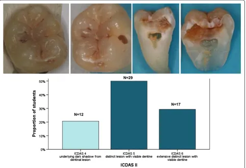
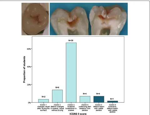
The sensitivity and specificity values for the ICDAS scores at level D3 were very good, 0.78 (0.12) and 0.87 (0.13) respectively. Theκand ICC values considering all lesions of different lesion depth, suggested fair to good accuracy of the performance among the students for all of the assessed methods (Table 4). Evaluating lesions correctly was the easiest for the students concerning teeth with the most severe lesions (Figure 1), whereas in the distribution of scores of initial lesions the range was wide (Figure 2).
DIAGNOdent pen® values of the sound tooth agreed 100% with the histological appearance (median 3; min 0, max 7). In the case where caries extended into the inner
third of dentine, the agreement was 78% (median 99; min 73, max 99). When caries histologically extended into the middle third of dentine, the DIAGNOdent pen® values had a wide range (median 24; min 5, max 88); only 25.5% of the values fell within the category >30, dentinal caries according to the manufacturer. These findings were sup-ported by the Spearman’s correlation coefficient; ƥ was 1.000 when the tooth was distinctly decayed, 0.302 when the tooth was sound (p = 0.021), and 0.240 when the tooth was moderately decayed (p = 0.240).
Discussion
Caries risk assessment, diagnosis and synthesis is consid-ered as one of the five main domains in cariology, and the base of clinical decision-making [3]. Particularly the assessment of activity of lesions is essential in selecting between non-operative and operative treatment. Reliable caries detection and lesion activity assessment are cor-nerstones in turning the philosophy from the operative treatment schema in today’s clinical practice to non-operative caries control schema in the future [3]. It is essential for all dentists to know the new schema to be applied in their clinical practice. This schema as well as caries detection ad lesion activity assessment must be emphasized in dental curriculum. Combining lectures and hands-on workshops as a teaching method of caries detection is one of the possibilities recommended by the European Core Curriculum [16] but until this study, it had not been used in Finland. This work investigated how easily caries detection is adopted by dental students, and how accurate their performance is using different methods after basic training. According to the present results, third-year dental students are very well capable of embracing different caries detection and lesion activ-ity assessement methods when introduced by lectures and a hands-on workshop. Our findings are in concord-ance with other studies supporting the capability of effi-cient learning of caries detection methods even by beginners [1,8,10,17].
It was the easiest for the students to evaluate lesions correctly in the most severe cases. The lesions that were difficult for the students to score (ICDAS) could have surprised even an experienced examiner by their depth. The accuracy of their decisions using ICDAS method (dentine caries D3, as the cut off point) was very good. Inter-examiner agreement concerning lesion depth and activity of all lesions was fair to good using all methods. In calculating sensitivity and specificity (accuracy) D3 was used as cut off point, whereas all scores were con-sidered in evaluating inter-examiner agreement. The findings are in line with other studies reporting good ac-curacy and inter-examiner agreement both by Nyvad’s and ICDAS methods [18-21], even if some of those stud-ies were carried out on primary teeth. Methods using Table 4 Intra-examiner agreement with golden standard
using different caries detection methods by the third-year dental students expressed as kappa (κ) and intra-class correlation coefficient (ICC) values
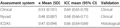
Assessment system κMean (SD) ICC mean (95% CI) Validation
Clinical 0.53 (0.101) 0.69 (0.65-0.70) Clinical
Nyvad 0.48 (0.087) 0.68 (0.66-0.71) Clinical
classifications are easily introduced and adapted, are ac-curate and reproducible, which will hopefully make them popular among clinicians. To our knowledge they are mainly used in research, so far.
Validation i.e. “golden standard” is a major issue in evaluating methods for caries detection and lesion activity assessment. Validating lesion depth detection in in vitro studies can be accomplished reliably by sectioning the ob-served teeth and estimating lesion depth using magnifica-tion (microscope, magnifying glasses, computer screen). Using PC screen has been shown to be a valid and applic-able method comprapble with the findings through a microscope [22]. Validation can also be radiographical considering its limitations especially in detecting occlusal lesions and secondary caries lesions. Inin vivostudies and studies estimating lesion activity, validation is subjective, done by a reference examiner i.e.“golden standard”. The histological validation was carried out in this study in evaluating the outcome by the ICDAS and LF methods whereas lesion activity assessment was validated clinically by reference scores (Nyvad’s method and CE). It was em-phasized to the students that estimating and evaluating
lesion depth reliably on extracted teeth is difficult if possible at all. However, every dentist must be aware of signs indicating lesion depth and activity of the lesions. Gimenez et al. [10] reported that use of prescriptive methods (like ICDAS, or Nyvad’s method) is easier for undergraduate students whereas adopting such new approaches (classification of lesions) is challenging for experienced colleagues, who rely on their experience i.e. their approach is heuristic, prone to bias.
Nyvad’s system is based on the dynamic nature of caries, when environmental changes may affect de-remineralization balance, causing detectable consequences [12] even in weeks, either as progress or arrest of the lesions. In this study, a modified version of Nyvad’s method was used, i.e. the lesions were categorized according to the continuity of the surface to three categories, and activity was assessed in each category. Estimating signs of activity on extracted teth is not fully comparable with a clinical situation. This may explain confusion of the students when estimating lesion activity. The results showed, however, good agree-ment on estimation of activity of the lesions by the stu-dents according to clinical validation by the supervisors,
which again supports the validity of the method [12]. In the respective works on dental students and staff by Zandona et al. [9] and Gimenez et al. [10], all groups were equally capable of evaluating lesion activity either using descrip-tive or prescripdescrip-tive methods. However, activity evalu-ation using clinical signs was quicker to perform than the one with scoring (LAA) [10]. Authentic clinical situ-ation is always easier for evaluating lesion activity which was emphasized to the students of the present study.
To describe reliability for detecting and assessing differ-ent kinds of lesions by all methods and by all examiners, ICC and kappa values were calclulated. The proportion of fully sound teeth and teeth with initial lesions in our sam-ple was smaller compared to the situation in the clinical practice. This most likely influences the outcome in the present study, as it is easier for the dentist to diagnose a tooth sound than to find caries lesions. In the present study, on the other hand, the teeth with most severe le-sions were correctly diagnosed.
Working as pairs may be considered a factor causing some bias in the present study; pairs often had exactly same scores (5 pairs of the total 28 pairs) even though they had been told to determine the scores individually. Another factor distorting our results may have been the use of probes by the students. On the photographs taken before and after the workshop, we could notice some micro cavities having turned into true ones during the workshop. The reason for that might have been the difficulty in determining lesion activity on extracted tooth surfaces and the fact that the soft surface wears away easily after multiple probing. This change of sur-face continuity might have caused some variation in the scores on lesion depth. Today, probing of suspected le-sions is recommended to be carried out by feeling tac-tilely the surface structure. For students, it should be emphasized that probing does not promote sensitivity of the caries detection, but may cause irreversible tooth damage [23].
The students were allowed to question about the cri-teria and use of DIAGNOdent Pen, this might have in-fluenced the results. This was done, however, to support the students in a situation with the time limit and facing many new criteria simultaneously. Again, it can be spec-ulated, if sequential decisions on the same tooth was a source of bias. Even if the students were just learning the criteria it is possible that some quick learners could adopt the respective scores by different methods which may have influenced the results.
To eliminate the cross infection, the storing solution for the extracted teeth had been denatured alcohol. In
vitro, of course, makes estimating lesion activity more difficult that in vivo. Additionally, compared with, for example, freezing, storing in alcohol may have had some effect on the tooth surface and therefore complicate ac-curate diagnostics [24]. The inter-examiner agreement was not affected by this. However, it might have affected laser fluorescence measurements because fluorescence is believed to be caused by bacterial metabolic porphyrins in caries lesions [25].
Sectioning of the teeth in two halves was done to re-veal the most severe-looking site on the occlusal surface. In magnified digital photographs, the evaluation of the depth of the lesions was unproblematic [20]. On the other hand, it can be discussed if the most representative site of the lesion was reached by the free-hand section-ing of the lesion, considersection-ing also that cuttsection-ing destroys a small part of the lesion. This could have caused some bias in the histological scores or golden standard. The optimal way would have been chemical fixation and cut-ting the teeth in several very thin slices and examining them with more accurate histopathological techniques [26]. For the learning/teaching process, teeth should be sectioned immediately after the hands-on session and should be available at the feedback lecture to demon-strate lesion depth and activity. However, this was not possible here.
Histological agreement with the DIAGNOdent pen® scores by the students was excellent on the fully intact teeth and on the teeth with an extensive caries lesion. When caries extended into the middle third of dentine, the range of the scores was wide, reported also elsewhere [27]. The performance of the students using DIAGNO-dent pen® scanning in the present study is in concordance with the literature [28]. However, the results on LF scan-ning shall be considered with great precaution due to the limited number of the teeth (n = 3) evaluated.
Conclusions
Accuracy and reproducibility of different caries detection and lesion activity assessment methods seems to be good among the third-year dental students without previous diagnostic experience even after basic training only.
Combining lectures with a hands-on practice can be con-sidered as a good method for introducing dental students to caries detection using classification of the lesions as well as lassessing lesion activity. With new caries treat-ment schema, teaching new caries detection methods is essential for future dentists. Education must be continu-ously up-dated and evaluated.
Competing interests
The authors declare that they have no competing interests.
Authors’contributions
VA is the designer, supervisor and conductor of this project, HP, a fourth year dental student at the time of the field phase of the study, participated in the project in all its phases (design, implementing, writing). HV’s biggest contribution is statistical, he also participated in the writing process. LT gave his scientific expertise to the project. M-LL has actively participated in writing the paper. All authors read and approved the final manuscript.
Acknowledgements
The help of the charge nurse, oral hygienist Päivi Haataja and laboratory technician Jaana Laitinen working in the simulation laboratory is highly appreciated. They helped to provide the examined teeth and helped with photography and organizing the teaching sessions.
Author details
1Institute of Dentistry, Department of Cariology, Endodontology and
Paedodontics, University of Oulu, POB 5281 Oulu, Finland.2University Hospital of Oulu, Oulu, Finland.3Institute of Dentistry, Department of Cariology, University of Turku, Turku, Finland.
Received: 23 August 2013 Accepted: 28 November 2013 Published: 6 December 2013
References
1. Brocklehurst P, Ashley J, Walsh T, Tickle M:Relative performance of different dental professional groups in screening for occlusal caries. Community Dent Oral Epidemiol2012,40:239–246.
2. Lussi A:Comparison of different methods for the diagnosis of fissure caries without cavitation.Caries Res1993,27:409–416.
3. Schulte AG, Pitts NB, Huysmans MC, Splieth C, Buchalla W:European core curriculum in cariology for undergraduate dental students.Eur J Dent Educ2011,15(Suppl 1):9–17.
4. Ekstrand KR, Ricketts DN, Kidd EA:Reproducibility and accuracy of three methods for assessment of demineralization depth of the occlusal surface: an in vitro examination.Caries Res1997,31:224–231. 5. International Caries Detection and Assessment System.[http://www.icdas.org/];
adapted 18.10.2013.
6. Pitts N, Melo P, Martignon S, Ekstrand K, Ismail A:Caries risk assessment, diagnosis and synthesis in the context of a European core curriculum in cariology.Eur J Dent Educ2011,15(Suppl 1):23–31.
7. Diniz M, Lima L, Santos-Pinto L, Eckert G, Zandoná A, de Cássia Loiola Cordeiro R:Influence of the ICDAS e-learning program for occlusal caries detection on dental students.J Dent Educ2010,74:862–868.
8. Jablonski-Momeni A, Busche JF, Struwe C, Lange J, Heinzel-Gutenbrunner M, Frankenberger R, Pieper K:Use of the international caries detection and assessment system two-digit coding method by predoctoral dental students at philipps university of Marburg, Germany.J Dent Educ2012, 76:1657–1668.
9. Zandona AG, Al-Shiha S, Eggertsson H, Eckert G:Student versus faculty performance using a new visual criterion for the detection of caries on occlusal surfaces: an in vitro examination with histological validation. Oper Dent2009,34:598–604.
10. Gimenez T, Bittar D, Piovesan C, Guglielmi C, Fujimoto K, Matos R, Novaes T, Braga M, Mendes F:Influence of the examiners’experience on linical performance of visual inspection in detecting and assessing the activity status of caries lesions.Oper Dent2013,38:583–590.
12. Nyvad B, Machiulskiene V, Baelum V:Reliability of a new caries diagnostic system differentiating between active and inactive caries lesions.Caries Res1999,33:252–260.
13. Anttonen V, Seppä L, Hausen H:A follow-up study of the use of DIAGNOdent for monitoring fissure caries in children.Community Dent Oral Epidemiol
2004,32:312–318.
14. Lussi A, Imwinkelried S, Pitts N, Longbottom C, Reich E:Performance and reproducibility of a laser fluorescence system for detection of occlusal caries in vitro.Caries Res1999,33:261–266.
15. Bader JD, Shugars DA:A systematic review of the performance of a laser fluorescence device for detecting caries.J Am Dent Assoc2004, 135:1413–1426.
16. Splieth CH, Innes N, Söhnel A:Evidence-based cariology in clinical and public health practice as part of the European core curriculum in cariology.Eur J Dent Educ2011,15(Suppl 1):45–51.
17. Fung L, Smales R, Ngo H, Moun G:Diagnostic comparison of three groups of examiners using visual and laser fluorescence methods to detect occlusal caries in vitro.Aust Dent J2004,49:67–71.
18. Braga MM, Mendes FM, Martignon S, Ricketts DN, Ekstrand KR:In vitro comparison of Nyvad’s system and ICDAS-II with lesion activity assessment for evaluation of severity and activity of occlusal caries lesions in primary teeth.Caries Res2009,43:405–412.
19. Jablonski-Momeni A, Stachniss V, Ricketts DN, Heinzel-Gutenbrunner M, Pieper K:Reproducibility and accuracy of the ICDAS-II for detection of occlusal caries in vitro.Caries Res2008,42:79–87.
20. Rodrigues JA, Hug I, Diniz MB, Lussi A:Performance of fluorescence methods, radiographic examination and ICDAS II on occlusal surfaces in vitro.Caries Res2008,42:297–304.
21. Shoaib L, Deery C, Ricketts DN, Nugent ZJ:Validity and reproducibility of ICDAS II in primary teeth.Caries Res2009,43:442–448.
22. Jablonski-Momeni A, Ricketts DN, Stachniss V, Maschka R, Heinzel-Gutenbrunner M, Pieper K:Occlusal caries: evaluation of direct microscopy versus digital imaging used for two histological classification systems.J Dent2009, 37:204–211.
23. Neuhaus K, Ellwood R, Lussi A, Pitts N:Traditional lesion detection aids. Monogr Oral Sci2009,21:42–51.
24. Francescut P, Zimmerli B, Lussi A:Influence of different storage methods on laser fluorescence values: a two-year study.Caries Res2006, 40:181–185.
25. Lussi A, Hibst R, Paulus R:DIAGNOdent: an optical method for caries detection.J Dent Res2004,83:Spes No C80–C83.
26. Rodrigues JA, Neuhaus KW, Diniz MB, Hug I, Stich H, Karlsson L, Lussi A: Comparison among gold standard techniques used for the validation of methods for occlusal caries detection.Microsc Res Tech2012,75:605–608. 27. Anttonen V, Seppä L, Hausen H:Clinical study of the use of the laser
fluorescence device diagnodent for detection of occlusal caries in children.Caries Res2003,37:17–23.
28. Kühnisch J, Bücher K, Hickel R:The intra/inter-examiner reproducibility of the new DIAGNOdent Pen on occlusal sites.J Dent2007,35:509–512.
doi:10.1186/1472-6831-13-70
Cite this article as:Parviainenet al.:Evaluating performance of dental
caries detection methods among third-year dental students.BMC Oral
Health201313:70.
Submit your next manuscript to BioMed Central and take full advantage of:
• Convenient online submission
• Thorough peer review
• No space constraints or color figure charges
• Immediate publication on acceptance
• Inclusion in PubMed, CAS, Scopus and Google Scholar
• Research which is freely available for redistribution
![Table 1 Criteria for the scores of the histological caries lesion depth [4]](https://thumb-us.123doks.com/thumbv2/123dok_us/357000.1528264/2.595.55.282.599.732/table-criteria-scores-histological-caries-lesion-depth.webp)