A SMART ARRHYTHMIA CLASSIFICATION SYSTEM BASED ON WAVELET TRANSFORM AND SUPPORT VECTOR MACHINE TECHNIQUES
CHIA NYOKE GOON
A SMART ARRHYTHMIA CLASSIFICATION SYSTEM BASED ON WAVELET TRANSFORM AND SUPPORT VECTOR MACHINE TECHNIQUES
CHIA NYOKE GOON
A thesis submitted in fulfilment of the requirements for the award of the degree of
Master of Engineering (Biomedical)
Faculty of Biosciences and Medical Engineering Universiti Teknologi Malaysia
iii
iv
ACKNOWLEDGEMENT
Firstly, I would like to express my gratitude to my supervisor, Dr. Hau Yuan Wen for her support and motivation in completing my research. Her suggestion, support and motivation have guidances me throughout my days in Universiti Teknologi Malaysia. Besides, I would also like to sincerely thank my co-supervisor, Dr. Najeb for his support and asisstant. I also would like to acknowledge Faculty of Biosciences and Medical Engineering (FBME) for providing me the good environment and lab facilities to do my work.
A special thanks to my family especially my parent for their tremendous support and encouragement throughout the time in completing my research.
Last but not least, I would like to thank Kui Lin, Huey Woan, Siti Nurhayati and Amin who are helpful for their guidance, assistant and support throughout this research. Thank you to all that have involved and helped me in completing this research. Thank you very much.
v
ABSTRACT
Heart disease is still the most common cause of death and contributes large number of death in this modern world. According to the survey conducted by World Health Organization (WHO), cardiovascular disease (CVD) is the number one cause of death globally in 2016. CVD claimed 801,000 lifes and heart disease killed more than 370,000 asian people which is 23.2% according to David S. Siscovick who is the chair of AHA’s Council on Epidemiology and Prevention. Arrhythmia is defined as an irregular heartbeat which will cause abnormal rhythms of the heart that further lead to serious heart disease like stroke and heart attack.Thus, arrhythmia detection and classification is crucial in clinical cardiology to analyze the extracted features from non-invasive electrocardiogram (ECG) testing. However, the arrhythmia classification accuracy based on the commercial classification software is still a remaining issue and it is an extremely time consuming process for manual visual inspection. This research proposed an arrhythmia classification software based on support vector machine (SVM) algorithm due to its advantage of higher accuracy and solve overfitting problem. The proposed system consists of three stages, namely pre-processing, feature extraction using wavelet coefficient and arrhythmia classification using SVM. All the processing stage and intermediate outputs are displayed in user friendly Graphic User Interface (GUI). The system verification is based on offline MIT-BIH database for classification accuracy, benchmarking with the other related works. The classification result shows that the proposed system is able to detect arrhythmia classification up to accuracy of 91.11%. This research output is used as PC-based ECG classification software which able to run at workstation to perform long duration of ECG data, such as 24 hour holter data. For the future work, it is suggested to add an automated R-peak detection algorithm to the system in order to solve the problem of dependency on the R-peak annotation file.
vi
ABSTRAK
Penyakit jantung adalah punca kematian yang sering berlaku dan menyumbang sejumlah besar kematian di dunia moden ini. Menurut kaji selidik yang dijalankan oleh Pertubuhan Kesihatan Sedunia (WHO), penyakit kardiovaskular ialah punca utama kematian global pada tahun 2016. Aritmia ditakrifkan sebagai degupan jantung yang tidak teratur yang akan menyebabkan irama yang tidak normal jantung yang lebih utama kepada penyakit jantung yang serius seperti strok dan serangan jantung.Oleh itu, pengesanan aritmia dan klasifikasi adalah penting dalam kardiologi klinikal untuk menganalisis ciri-ciri yang diekstrak daripada ujian elektrokardiogram tidak invasif (ECG). Walau bagaimanapun, ketepatan klasifikasi aritmia berdasarkan perisian klasifikasi komersial masih menjadi isu yang tunggal dan ia adalah proses yang amat memakan masa untuk pemeriksaan visual secara manual. Kajian ini mencadangkan satu perisian klasifikasi aritmia Matlab berdasarkan algoritma Support Vector Machine (SVM) kerana kelebihan ketepatan yang lebih tinggi dan menyelesaikan penyesuaian masalah. Sistem yang dicadangkan terdiri daripada tiga peringkat, iaitu pra-pemprosesan, pengekstrakan ciri menggunakan pekali ombak dan klasifikasi aritmia menggunakan SVM. Semua peringkat pemprosesan dan output perantaraan dipaparkan pengantaraan muka yang mesra pengguna. Pengesahan sistem adalah berdasarkan offline pangkalan data MIT-BIH untuk ketepatan pengelasan bertanda araskan kepada kerja-kerja yang berkaitan. Hasil pengelasan menunjukkan bahawa sistem yang dicadangkan dapat mengesan klasifikasi aritmia sehingga ketepatan 91.11%. Hasil penyelidikan ini digunakan sebagai perisian berasaskan PC klasifikasi ECG yang dapat beroperasi di stesen kerja untuk melaksanakan data ECG dengan tempoh panjang, seperti 24 jam holter data. Ini adalah dicadangkan menambah R-puncak algoritma pengesanan automatik kepada sistem bagi menyelesaikan masalah pergantungan kepada fail anotasi R-puncak untuk kerja masa depan.
vii
TABLE OF CONTENTS
CHAPTER TITLE PAGE
DECLARATION ii
DEDICATION iii
ACKNOWLEDGEMENT iv
ABSTRACT v
ABSTRAK vi
TABLE OF CONTENTS vii
LIST OF TABLES x
LIST OF FIGURES xi
LIST OF ABBREVIATIONS xiii
1 INTRODUCTION 1 1.1 Problem Background 1 1.2 Problem Statement 3 1.3 Objectives 5 1.4 Scope 5 1.5 Research Contribution 6 1.6 Thesis Organisation 6 2 LITERATURE REVIEW 7
2.1 Human Heart Anatomy and Electrocardiogram
(ECG) 7
2.2 Arrhythmia 9
2.2.1 Premature Ventricular Contraction (PVC) 10 2.2.2 Left Bundle Branch Block (LBBB) 11 2.2.3 Right Bundle Branch Block Beat (RBBB) 12 2.2.4 Atrial Premature Beat (APB) 13 2.2.5 Aberrated Atrial Premature Beat (AAPB) 13 2.2.6 Nodal (Junctional) Premature Beat (NPB) 14 2.2.7 Nodal (Junctional) Escape Beat (NEB) 15
viii
2.2.8 Ventricular Escape Beat (VEB) 15
2.2.9 Paced Beat (PB) 16
2.2.10 Fusion of Ventricular and Normal Beat
(FVN) 17
2.2.11 Fusion of Paced and Normal Beat (FPN) 17
2.3 Arrhythmia Detection Algorithm 18
2.4 Pre-processing 19
2.4.1 Pan and Tompkins Algorithm (PT) 19
2.4.2 Derivative Based Method 20
2.4.3 Heartbeat Segmentation Method 20
2.5 Feature Extraction 22
2.5.1 Heart Rate Variability (HRV) Analysis 22
2.5.2 Wavelet Transform (WT) 25
2.5.3 Hermite Polynomial (HP) 27
2.6 Classification 29
2.6.1 Artificial Neural Network (ANN) 30
2.6.2 Fuzzy Logic 31
2.6.3 Rule Based Algorithm 32
2.6.4 Support Vector Machine (SVM) 32
2.7 Previous Work 35
2.8 Tools 37
2.8.1 MIT-BIH Offline Database 37
2.8.2 MATLAB 37
2.8.3 Support Vector Machine Library
(LIB-SVM) 38
2.9 Chapter Summary 39
3 ALGORITHM MODELLING AND SOFTWARE
DE-VELOPMENT 40
3.1 Research Methodology 40
3.2 Overview of Proposed Arrhythmia Detection
Algorithm 42
3.3 ECG Pre-processing 42
3.4 Feature Extraction based on Wavelet Transform,
Statistical Features and Time Interval Features 46 3.5 Arrhythmia Classifier based on SVM 51 3.6 Arrhythmia Classification Software Development 54 3.6.1 Software Welcoming Screen 54
ix
3.6.3 SVM Testing 57
3.7 Chapter Summary 59
4 RESULT AND DISCUSSION 60
4.1 ECG Signal Pre-processing Validation 60
4.2 Feature Extraction Validation 64
4.3 Arrhythmia Classification 69
4.4 Related Work Brenchmarking 76
4.5 Computation Timing Performance 78
4.6 Graphical User Interface (GUI) Development 78
4.7 Chapter Summary 80
5 CONCLUSION 81
5.1 Conclusion 81
5.2 Recommendation of Future Works 82
REFERENCES 83
x
LIST OF TABLES
TABLE NO. TITLE PAGE
2.1 Types of arrhythmia 10
2.2 Comparison of pre-processing techniques 21
2.3 Comparison of three domain 24
2.4 Comparison of feature extraction techniques 29
2.5 Comparison of the classifier 35
2.6 Previous work of arrhythmia detection 36
3.1 Statistical features 50
3.2 Timing interval features 50
4.1 The mean values of absolute value in each sub-band 68
4.2 The accuracy of arrhythmia detection 71
4.3 Results of arrhythmia detection 74
4.4 The exploration accuracy in term of exclusion timing interval
features 75
4.5 Previous work of arrhythmia detection 77
xi
LIST OF FIGURES
FIGURE NO. TITLE PAGE
1.1 The statistic of United States 2
1.2 ECG signal 3
1.3 Research scope 6
2.1 Human heart anatomy 8
2.2 An ECG signal 9
2.3 ECG waveform sample of MIT-BIH offline database 10
2.4 ECG characteristic of PVC 11
2.5 ECG characteristic of LBBB 12
2.6 ECG characteristic of RBBB 12
2.7 ECG characteristic of APB 13
2.8 ECG characteristic of AAPB 14
2.9 ECG characteristic of nodal (junctional) premature beat 14 2.10 ECG characteristic of nodal (junctional) escape beat 15 2.11 ECG Characteristic of Ventricular Escape Beat 16
2.12 ECG characteristic of paced beat 16
2.13 ECG characteristic of fusion of ventricular and normal beat 17 2.14 ECG characteristic of fusion of paced and normal beat 18 2.15 Overview of arrhythmia detection algorithm 18
2.16 Pan and Tompkins algorithm 19
2.17 Derivative based method 20
2.18 Heartbeat segmentation method 20
2.19 Discrete wavelet transform for feature extraction 27
2.20 ANN layer [6] 31
2.21 Fuzzy logic system [13] 32
2.22 SVM 33
2.23 MATLAB 38
3.1 Work flow 41
3.2 Overview of the proposed arrhythmia classifier 42
3.3 The flow of the ECG pre-processing 43
xii
3.5 ECG pre-processing behavioural flowchart 45
3.6 Six times decomposition of ECG signal 48
3.7 Timing interval features 49
3.8 Feature extraction behavioural flowchart 51
3.9 The concept of SVM 52
3.10 The SVM behavioral flowchart 54
3.11 The welcoming page 55
3.12 SVM training 56
3.13 SVM training behaviour flowchart 57
3.14 SVM testing 58
3.15 SVM testing behaviour flowchart 59
4.1 The raw ECG signal 61
4.2 The filtered ECG signal 61
4.3 The first four heart beat ECG segmentation 62
4.4 The first four heart beat offset removal 63
4.5 The first four heart beat normalisation 63
4.6 The first ECG heart beat in decomposition result of details
output,Dk,1≤k ≤6 64
4.7 The first ECG heart beat in decomposition result of
approximation feature,Ak,1≤k≤6 65 4.8 The second heart beat in decomposition result of details
output,Dk,1≤k ≤6 65
4.9 The second heart beat in decomposition result of
approxima-tion feature,Ak,1≤k ≤6 66
4.10 The third heart beat in decomposition result of details output,
Dk,1≤k ≤6 66
4.11 The third heart beat in decomposition result of approximation
feature,Ak,1≤k≤6 67
4.12 The forth heart beat in decomposition result of details output,
Dk,1≤k ≤6 67
4.13 The forth heart beat in decomposition result of approximation
feature,Ak,1≤k≤6 68
4.14 Confusion Matrix 72
4.15 SVM Training 79
xiii
LIST OF ABBREVIATIONS
AHA - American Heart Association
ANN - Artificial Neuron Network
AF - Atrial Fibrillation
APB - Atrial Premature Beat
AV - Atrial Ventricular
BPM - Beat Per Minute
CVD - Cardiovascular Disease
CVT - Cardio Vascular Technologist
ECG - Electrocardiogram
FN - False Negative
FP - False Positive
GUI - Graphical User Interface
HRV - Heart Rate Variability
LBBBB - Left Bundle Branch Block Beat
N - Normal
PT - Pan and Tompkin
PVC - Premature Ventricular Contraction RBBBB - Right Bundle Branch Block Beat
SA - Sino Atrial
SVM - Support Vector Machine
TN - True Negative
TP - True Positive
CHAPTER 1
INTRODUCTION
This chapter presents the research background, problem statement, research objectives and associated project scope, research significance and the organization of the thesis.
1.1 Problem Background
In this modern world, heart disease is still the most common cause of the death and contributes large number of death even in developed countries. According to the survey conducted by World Health Organization (WHO), cardiovascular disease (CVD) is the number one cause of death globally in 2016. 17.5 millions of people which about 31% of global population died from CVD in 2012 due to coronary heart disease and stroke from WHO [1]. According to a study conducted in 2014, approximately 36% of Malaysians lose their life due to cardiac diseases as shown in Figure1.1which contribute the death rate among Malaysians due to heart disease rank at 33 in worldwide [2]. For the country USA in 2016, CVD claimed 801,000 lifes and heart disease killed more than 370,000 Asian people which is 23.2% according to David S. Siscovick who is the chair of AHA’s Council on Epidemiology and Prevention and senior vice president for research at the New York Academy of Medicine in New York City [3].
2
Figure 1.1: The statistic of United States
An irregular heartbeat is known as arrhythmia which causes abnormal heart rhythms [4].This can be treated as an indicator of potential heart problem which may lead to stroke or sudden cardiac death. For instance, Premature Ventricular Contraction (PVC) is one of the most common arrhythmia which contribute approximately 50% of death among the various heart diseases [5].Thus, arrhythmia classification with high accuracy is crucial in clinical cardiology to analyse the extracted features from non-invasive electrocardiogram (ECG) testing.
Electrocardiogram (ECG) is an essential human vital signal which can be used to detect heart rate. It can also be defined as an electrical activation of heart and is a non-stationary signal. A typical ECG heartbeat consists of P wave, QRS complex and T wave as shown in Figure1.2. This signal is useful for arrhythmia detection and may consist of indicator or alert about various heart diseases [6].
3
Figure 1.2: ECG signal
The ECG analysis has been proven to be a conventional non-invasive approach for heart disease detection in clinical setting [7]. The characteristic of ECG signal gives important information about the amplitudes, shapes and the duration of PQRST waves. These information are very essential to extract the key features for arrhythmia detection. Moreover, it provides effective information about the heart deficiency [8]. As a result, ECG analysis have been greatly researched and remain as an active research area to detect heart abnormality for early cardiac disease detection to increase survival rate [9,10,11].
On the other hand, Matlab is universally accepted as one of the most powerful software modelling platform and algorithm verification tool. This software have connected with many advanced programming language such as C, Java and Visual Basic and equipped with rich set of toolbox. As a result, this research propose a Matlab-based arrhythmia classification software based on wavelet transform and timing interval as feature extraction and SVM classifier as clinical decision support tool.
1.2 Problem Statement
ECG data obtained from ECG medical devices are normally printed and need to be manually analysed by specialists to detect any heart abnormalities of a patient. For example, the ECG data duration recorded using Holter monitor are normally 24 to 48 hours, hence the human interpretation of the ECG data through visual inspection is a very time consuming procedure during attachment at Institut Jantung Negara (IJN). Therefore, an automated computerized ECG signal analysis and classification software
4
is desired to reduce the time required by cardiologists or cardiovascular technologists (CVTs) to improve their work effectiveness and efficiency.
ECG biosignal processing, feature extraction and arrhythmia classification are computed intensively and time consuming computations [12,13]. The current commercial arrhythmia classification software take a relatively long time to analyse the patient ECG data, especially 24-hour ECG data retrieved from holter. In addition to that, the arrhythmia classification accuracy of the existing commercial clinical software solution is still a remaining questioning issue. They may not apply the latest state-of-the-art research algorithm to classify the arrhythmia. For example, some of algorithm provide fast computation but low accuracy or high accuracy but great algorithm complexity. Hence, manual visual inspection of the computerized classification result is still required by the CVTs for further verification and confirmation. For example, a CVT at Institut Jantung Negara or the National Heart Institute of Malaysia takes average 15-30 minutes to manually analyse single 24 hour ECG record. This will definitely increase the CVT’s workload when there are many ECG data need to be analyse during routine clinical examination. For these reasons, it is important to develop an automated ECG analysis and arrhythmia classififcation software with high accuracy and fast processing performance as clinical decision support tool to help specialists improve their work effectiveness and efficiency.
In order to provide key features for feature extraction, there are many techniques have been carried out such as Heart Rate Variability (HRV) analysis, Wavelet Transform, Hermite Polynomial and many more. Time and frequency domain analysis are the conventional techniques applied in Heart Rate Variability analysis. Due to non-stationationary nature of signal analysis, this technique unable to discriminate some types of arrhythmia clearly and cannot provide time-frequency analysis. In addition, timing interval features are also important to extract arrhythmia features. For example, Premature Ventricular Contraction (PVC) can be identified clearly by using timing interval features. Therefore, wavelet transform and timing interval features are proposed for feature extraction technique.
In terms of arrhythmia classification, there are many methods have been proposed such as Artificial Neuron Network (ANN), fuzzy logic, rule based algorithm and Support Vector Machine (SVM). However, each technique has their own drawback and limitation. For example, ANN generally provides a high accurate classification result but it does not extrapolate the result and has over fitting problem. Ruled based algorithm is a simple algorithm but it provides low accuracy meanwhile fuzzy logic
5
required prior knowledge for the computation. Therefore, SVM has also been proposed as a solution to overcome these limitations. SVM is used due to its generalisation ability. Thus, computer based monitoring system is needed and proposed to the clinical applications based on support vector machine.
1.3 Objectives
Based on the aforementioned problems, this research derives the objectives as described below:
1. To propose a smart arrhythmia detection algorithm based on combination of ECG pre-processing, wavelet transfrom feature extraction and arrhythmia classification based on support vector machine technique.
2. To develop a MATLAB based arrhythmia classification software based on proposed algorithm in (1) as clinical decision support system.
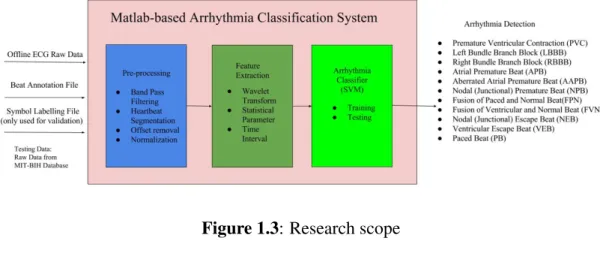
1.4 Scope
Figure 1.3 shows the overall research scope of the proposed work. The arrhythmia classifier is developed using Matlab modelling tool with user-friendly Graphical User Interface (GUI). The system will take three types of files obtain from MIT-BIH ECG offline database measured by lead II as the input data which are the offline ECG raw data, beat annotation file and arrhythmia symbol labelling file to compute the SVM training and testing process.
The system will perform ECG pre-processing, feature extraction at time-frequency domain using wavelet transform method, as well as some statistical parameters and time-interval information and arrhythmia classification based on SVM. The targeted 11 arrhythmia types are Premature Ventricular Contraction (PVC), Left Bundle Branch Block (LBBB), Right Bundle Branch Block (RBBB), Atrial Premature Beat (APB), Aberrated Atrial Premature Beat (AAPB), Nodal (Junctional) Premature Beat, Fusion of Paced and Normal Beat, Fusion of Ventricular and Normal Beat, Nodal (Junctional) Escape Beat, Ventricular Escape Beat and Paced Beat (PB).
6
Figure 1.3: Research scope
1.5 Research Contribution
At the end of this research, a high accuracy arrhythmia classification algorithm which able to detect 11 types of arrhythmia with an acceptable processing time is proposed. A Matlab-based arrhythmia classification software tool with user friendly GUI is developed based on the proposed algorithm to execute ECG pre-processing, feature extraction using wavelet transform and other statistical and time interval parameters, as well as classification using SVM technique. The classification software is designed in a novel and easy way to use GUI which can train SVM classifier, to test ECG input data and can investigate each arrhythmia heart beat in details to assist the cardiologists, physician, CVT as a clinical support tool.
1.6 Thesis Organisation
There is a total of five chapters in this thesis. Chapter 1 explains the overview of this research in term of research background, problem statement, research objectives and the scope of the research. Chapter 2 will cover the literature review for ECG foundation, types of arrhythmia, ECG pre-processing techniques, feature extraction and classification as well as their related research work. The research methodology will also be discussed in this chapter. Chapter 3 will focus on the modelling of the proposed arrhythmia classification algorithm and development of clinical decision support tool. Chapter 4 will discuss the result analysis of the research. Finally, the last chapter will conclude the work and provide the recommendation for future work.
REFERENCES
1. Global Status report on Non-communicable Disease (NCD) Country Profile, 2016. Retrieved from the World Health Organization (WHO) http://www.who.int/nmh/publications/ncd-status-report-2014/en/ 2. Retrieved from the America Heart Association (AHA) 2014
http://newsroom.heart.org/news/new-statistics-show-one-of-every-three-u-s-deaths-caused-by-cardiovascular-disease
3. Retrieved from the Medline Plus: heart disease
http://www.nlm.nih.gov/medlineplus/heartdiseases.html
4. U. Rajendra Acharya, K. Paul Joseph, N. Kannathal, Choo Min Lim, and Jasjit S. Suri. Heart rate variability: A review. Medical and Biological Engineering and Computing, 44(12):1031–1051, 2006. 5. Hussain AL-Ziarjawey. Heart Rate Monitoring and PQRST Detection
Based on Graphical User Interface with Matlab. International Journal of Information and Electronics Engineering, 5(4):311–316, 2015. 6. Babak Mohammadzadeh Asl, Seyed Kamaledin Setarehdan, and
Maryam Mohebbi. Support vector machine-based arrhythmia classification using reduced features of heart rate variability signal. Artificial Intelligence in Medicine, 44(1):51–64, 2008.
7. C.Caner and M.Engin. The programmable ECG simulator. J Med Syst 2008; 32:355-9
8. M.Engin, M.Fedekar, E.Z. Engin, M. Koruyurek. Feature measurements of ECG beats based on statistical classifiers. Measurement 2007; 40:904-12.
9. C.M. Jaylaxmi and Raveendra, Matlab Based ECG Signal Classification, International Journal of Science, Engineering and Technology Research, Vol 3, Issue 7, July 2014.
10. S.H. Chung, Forward-backward non-linear filtering technique for extracting small biological signals from noise, J.Neurosci. Methods 40(1991) 71-86.
11. I.K. Daskaloy, Panda, H.Wong and Christov. Electrocardiogram signal preprocessing for automatic detection of QRS boundaries. Med Eng Phys 1999; 21(1):37-44A.
84 12. Knezevic, Stojanovic, Karadaglic and B. Asanin. Single Chip System
for ECG Feature Extraction. IEEE 2nd Mediterranean Conference in Embedded Computing,88-92
13. H.G. Goovarts, H.H. Ros, T.J. Vander Akker, H.Schneider, A digital QRS detector based on the principle of contour limiting, IEEE Trans Biomed. Eng, vol BME-23, pp154,1997
14. P.D. Chazal and R.B. Reilly. Automatic classification of ECG beats using wavelet shape and heart beat interval features. pp 269-272, 2003.
15. W.R. Schwartz E.J.S.Luz and D.Menotti. ECG-based heartbeat classification for arrhythmia detection: A survey. Computer Methods and Programs in Biomedicine, 7(3):144–164, 2015.
16. L Sornmo,¨ P O Borjesson,¨ M E Nygards,˚ and O Pahlm. A method for evaluation of QRS shape features using a mathematical model for the ECG.IEEE Transactions on Bio-Medical Engineering, 28(10):713–717, 1981.
17. M Bahoura, M Hassani, and M Hubin. DSP implementation of wavelet transform for real time ECG waveforms detection and heart rate analysis.Comput. Meth. Progr. Biomed, 52(2):35–44, 1997.
18. Retrieved from the America Heart Association (AHA) : Arrhythmia http://www.heart.org/HEARTORG/Conditions/Arrhythmia/Arrhythmia_ UCM_002013_SubHomePage.jsp
19. George B Moody and Roger G Mark. The Impact of the MIT-BIH Arrhythmia Database. Number June. 2001.
20. Life in the Fast Lane ECG Library. Premature Ventricular Contraction beat - ECG example, 2016.
21. Life in the Fast Lane ECG Library. Left Bundle Branch Block - ECG example, 2016.
22. Life in the Fast Lane ECG Library. Right Bundle Branch Block - ECG example, 2016.
23. Life in the Fast Lane ECG Library. Atrial Premature beat - ECG example, 2016.
24. Life in the Fast Lane ECG Library. Aberrated Atrial Premature beat -ECG example, 2016.
25. Life in the Fast Lane ECG Library. Nodal (Junctional) Premature beat - ECG example, 2016.
85 26. Life in the Fast Lane ECG Library. Nodal (Junctional) Escape beat
-ECG example, 2016.
27. Marc Gertsch. Atrial Premature Beats. In The ECG Manual, pages 197–200. 2009.
28. Life in the Fast Lane ECG Library. Ventricular Escape Rhythm – ECG example, 2016.
29. Life in the Fast Lane ECG Library. Paced beat - ECG example, 2016.
30. Life in the Fast Lane ECG Library. Fusion of Ventricular and Normal beat – ECG example, 2016.
31. Life in the Fast Lane ECG Library. Fusion of Paced and Normal Beat – ECG example, 2016.
32. Abdelhamid Daamouche, Latifa Hamami, Naif Alajlan, and Farid Melgani. A wavelet optimization approach for ECG signals classification.
Biomedical Signal Processing and Control, 7(4):342–349, 2012
33. Philip De Chazal, Maria O Dwyer, Richard B Reilly, and Senior Member. Automatic Classification of Heartbeats Using ECG Morphology and Heartbeat Interval Features. IEEE Transaction on Biomedical Engineering, 51(7):1196– 1206, 2004.
34. Jiapu Pan and J Willis. A Real-Time QRS Detection Algorithm.IEEE Transaction on Biomedical Engineering, 1(3):230–236, 1985.
35. C.S. Poon Natalia M. Arzeno, Z.D. Deng. Analysis of First-Derivative Based QRS Detection Algorithms. Biomedical Engineering, IEEE Transactions on Biomedical Engineering, Vol. 55, No. 2, Pp. 478 -484, 55(2):478–-484, 2008.
36. R. Jane, S. Olmos, P. Laguna, and P. Caminal. Adaptive Hermite models for ECG data compression: performance and evaluation with automatic wave detection. Proceedings of Computers in Cardiology Conference, pages 389– 392, 1993.
37. Chia Ping Shen, Wen Chung Kao, Yueh Yiing Yang, Ming Chai Hsu, Yuan Ting Wu, and Feipei Lai. Detection of cardiac arrhythmia in electrocardiograms using adaptive feature extraction and modified support vector machines. Expert Systems with Applications, 39(9):7845–7852, 2012.
38. B. S. Raghavendra, Deep Bera, Ajit S. Bopardikar, and Rangavittal Narayanan. Cardiac arrhythmia detection using dynamic time warping of ECG beats in e-healthcare systems. 2011 IEEE International Symposium on a World of Wireless, Mobile and
87 48. Benitez, Gaydeck, Zaidi and P. Filzpatrick. The use of Hilbert Transform
in ECG signals analysis. Computers in Biology & Medicine, 31(5):399-406(2001).
49. Rahime Ceylan. Comparison of FCM, PCA and WT techniques for classification ECG arrhythmias using artificial neural network.Expert Systems with Applications, 33:286–295, 2007.
50. Vishnu P. Nambiar, Mohamed Khalil-Hani, and M. N. Marsono. Evolvable Block-based Neural Networks for real-time classification of heart arrhythmia from ECG signals. 2012 IEEE-EMBS Conference on Biomedical Engineering and Sciences, IECBES 2012, (December):866– 871, 2012.
51. Mehmet Engin. ECG beat classification using neuro-fuzzy network. Pattern Recognition Letters, 25:1715–1722, 2004.
52. Muhammad Amin B Hashim. Real-time smart arrhythmia classification embedded system design using Hermite polynomial and multilayer perceptron neural network. Master thesis,2016
53. Rodrigo V Andreao, Bernadette Dorizzi, and Jerome Boudy. ECG Signal Analysis through Hidden Markov Models.IEEE Transaction on
Biomedical Engineering, 53(8):1541–1549, 2006.
54. Themis P. Exarchos, Markos G. Tsipouras, Costas P. Exarchos, Costas Papaloukas, Dimitrios I. Fotiadis, and Lampros K. Michalis. A methodology for the automated creation of fuzzy expert systems for ischaemic and arrhythmic beat classification based on a set of rules obtained by a decision tree.Artificial Intelligence in Medicine, 40(3):187-200, 2007.
55. Roberta Colloca, Alistair E W Johnson, Luca Mainardi, and Gari D Clifford. A Support Vector Machine Approach for Reliable Detection of Atrial Fibrillation Events. Computing in Cardiology, (2):1047–1050, 2013.
56. Chih-chung Chang and Chih-jen Lin. LIBSVM: A Library for Support Vector Machines. ACM Transactions on Intelligent Systems and Technology (TIST), 2:1–39, 2011.
57. Saibal Dutta, Amitava Chatterjee, and Sugata Munshi. Correlation technique and least square support vector machine combine for frequency domain based ECG beat classification. Medical Engineering and Physics, 32(10):1161– 1169, 2010.
58. M. R. Homaeinezhad, S. A. Atyabi, E. Tavakkoli, H. N. Toosi, A. Ghaffari, and R. Ebrahimpour. ECG arrhythmia recognition via a
neuro-88 SVM-KNN hybrid classifier with virtual QRS image-based geometrical features. Expert Systems with Applications, 39(2):2047–2058, 2012.
59. Majid Moavenian and Hamid Khorrami. Expert Systems with Applications A qualitative comparison of Artificial Neural Networks and Support Vector Machines in ECG arrhythmias classification. Expert Systems with Applications, 37(4):3088–3093, 2010.
60. Dusit Thanapatay, Chaiwat Suwansaroj, and Chusak Thanawattano. ECG beat classification method for ECG printout with Principle Components Analysis and Support Vector Machines. ICEIE 2010 -2010 International Conference on Electronics and Information Engineering, Proceedings, 1(Iceie):72–75, 2010.