Rectal cancer: ESMO Clinical Practice Guidelines for
diagnosis, treatment and follow-up
B. Glimelius
1, L. Pa˚hlman
2& A. Cervantes
3On behalf of the ESMO Guidelines Working Group*
1Department of Oncology, Radiology and Clinical Immunology;2
Department of Surgical Science, Akademiska Sjukhuset, University of Uppsala, Uppsala, Sweden;
3
Department of Hematology and Medical Oncology, INCLIVA, University of Valencia, Valencia, Spain
incidence
The crude incidence of rectal cancer in the European Union is
35% of the total colorectal cancer incidence, i.e. 15–25/100 000 per year. The mortality is 4–10/100 000 per year with lower figures valid for females, the higher for males.
diagnosis
Diagnosis is based on a digital rectal examination including rigid sigmoidoscopy with biopsy for histopathological examination. Tumours with distal extension to£15 cm (as measured by rigid sigmoidoscopy) from the anal margin are classified as rectal, more proximal tumours as colonic.
staging and risk assessment
Complete history and physical examination, complete blood count, liver and renal function tests, carcinoembryonic antigen (CEA), chest X-ray (alternatively CT scan) and CT or MRI or ultrasound of liver and abdomen should be performed.
Endoscopic ultrasound for the earliest tumours (cT1–T2) or rectal MRI for all tumours, including the earliest ones, is recommended in order to select patients for preoperative treatment and extent of surgery [III, B]. Complete colonoscopy pre- or postoperatively is required.
Histopathological examination should include surgical specimen with proximal, distal and circumferential margins and regional lymph nodes (at least 12 nodes are recommended to be examined). The circumferential resection margin (crm) status is very important. There are uncertainties in the interpretation of this and the residual (R) tumour classification, and an expanded classification has been suggested. Moreover, vascular and nerve invasion should be evaluated [III, A].
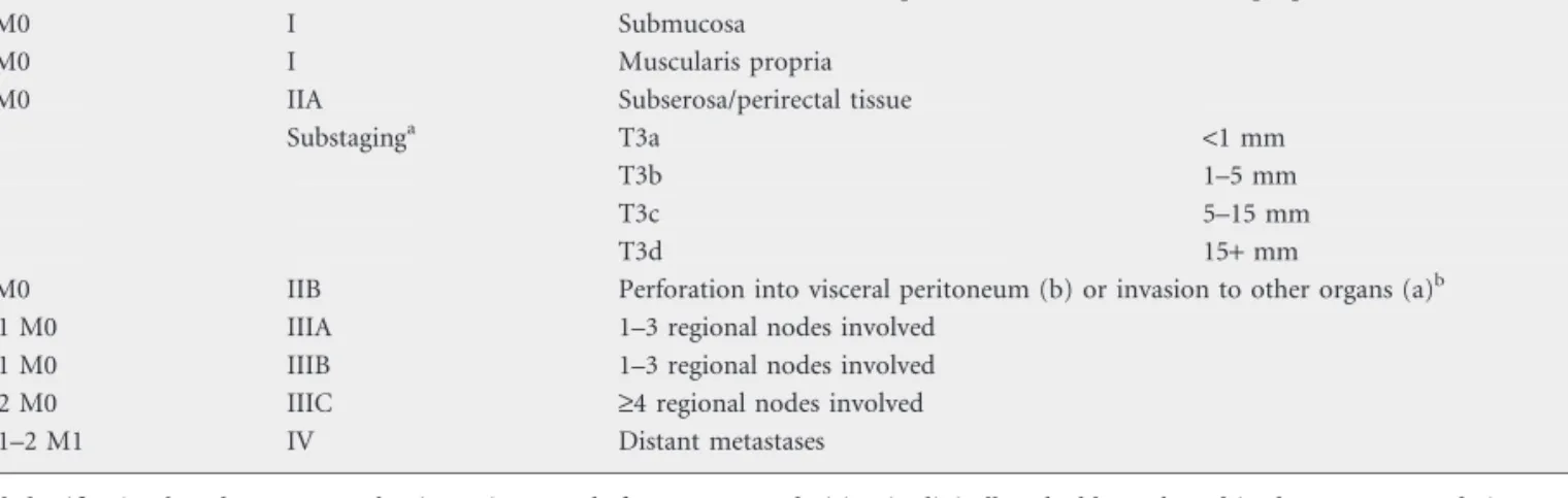
The TNM staging system should be used. There is major controversy about which version to use. In these
recommendations, version 5 from 1997 is preferred over TNM version 6 (2002) and 7 (2010) as the latter show marked interobserver variation in defining stage II and stage III. At the same time, there is a need for further subclassification particularly of cT3, as indicated in Table 1.
T1 tumours could also be classified into the Haggitt’s subclassification if the cancer is in a stalked adenoma and according to the sm-system if in a sessile adenoma. The two systems are overlapping. The level of infiltration into the submucosa (sm) predicts the risk of lymph node metastases and thus the type of surgery [III, B].
treatment
localized disease
overall strategy. An important aim is to treat so that the risk of residual disease in the pelvis, frequently causing a disabling local recurrence, is very low (preferably less than5% in the population in whom curative treatment is intended) and, at the same time, with as little acute and late morbidity as possible. This should be possible in all but the few (£10%) cases presenting with a fixed tumour growing into a non-readily resectable organ (cT4a).
Another aim is to treat with preserved good sphincter function in as many patients as possible.
From a practical point of view influencing treatment, the rectal cancers can be divided into four groups: very early (some cT1), early (cT1–2, some cT3), more advanced (cT3, some cT4) and locally advanced (cT4). Factors other than clinical T-stage, such as tumour height, closeness to the crm, cN-stage, and vascular and nerve invasion are also relevant. It is presently not possible to give a precise definition of which T and N substages belong to these groups.
The terms ‘favourable or early or good’, ‘intermediate or bad’ and ‘locally advanced or ugly’ can be used for categorizing the rectal cancers into these clinical subgroups. In clinical practice and in many recent studies, the term ‘locally advanced’ has been commonly used for the ‘intermediate/bad’ group, but is best reserved for the truly ‘locally advanced/ugly’ tumours. *Correspondence to:ESMO Guidelines Working Group, ESMO Head Office, Via L.
Taddei 4, CH-6962 Viganello-Lugano, Switzerland; E-mail: [email protected]
Approved by the ESMO Guidelines Working Group: August 2002, last update February 2010. This publication supercedes the previously published version—Ann Oncol 2009; 20 (Suppl 4): iv54–iv56.
Conflict of interest: The authors have reported no conflicts of interest.
need for quality assurance and control. Treatment of rectal cancer is demanding and requires great skill in the entire multidisciplinary team (MDT). Good surgery and good pathology as well as good radiation techniques and optimally given chemotherapy together with long-term complete follow-up, also including functional aspects, are important for quality control. Many countries have recently launched quality control programmes in rectal cancer surgery, which has been very beneficial for the outcome.
risk-adapted treatment. In the earliest, most favourable cases, chiefly the malignant polyps [Haggitt 1–3, T1 sm 1 (–2?) N0], a local procedure, e.g. using the transanal endoscopic
microsurgery (TEM) technique, is appropriate [III, A]. The
resection should be radical (R0) and no signs of vessel invasion or poor differentiation should be present. If this is not the case or if the tumour infiltrates deeper into the submucosa [Haggit 4, T1 sm 2(–3?)] or a T2 tumour, the risk of recurrence because of remaining tumour cells or because of lymph node metastases is too high (‡10%) and the patient should have postoperative chemoradiotherapy [III, B] or, more safely, be recommended major [total mesorectal excision (TME)] surgery [II, A]. If the cancer diagnosis is verified in a biopsy, presurgical
chemoradiotherapy is preferred if the intent is to perform a local procedure [III, B].
As an alternative to local surgery, alone or with (preoperative) chemoradiotherapy, local radiotherapy
[brachytherapy or contact therapy (Papillon technique)] can be used [III, C]. Experiences of these treatments is limited outside specialized centres.
In the early, favourable cases {cT1–2, some early cT3, N0 [cT3a(–b) and clear crm (crm–) according to MRI], ‘good’ group} above the levators, surgery alone, meaning a sharp radical dissection using the TME technique [II, A] is
appropriate, since the risk of local failure is very low. The role of TME in tumours situated in the upper third of the rectum has been much discussed and there is no strong evidence supporting TME in those cases. However, to avoid spillage of distal tumour cells a margin of at least 5 cm distally to the tumour on an unfixed specimen must be achieved.
In more locally advanced cases {most cT3 [cT3(b)c+without threatened and involved crm (crm–) according to MRI], some cT4 (e.g. vaginal or peritoneal involvement only), N+, ‘intermediate or bad’ group}, preoperative radiotherapy is recommended followed by TME, since this reduces local recurrence rates [I, A]. Even in the absence of signs of extramural growth on ultrasound or MRI (cT2) in very low tumours, preoperative radiotherapy may be indicated because the distance to the mesorectal fascia is very small. 25 gray, 5 Gy/fraction during 1 week followed by immediate surgery (<10 days from the first radiation fraction) is a convenient, simple and low-toxic treatment [I, A]. More demanding, and
Table 1. TNM classification (version 5, 1997) with subclassifications
TNM Stage Extension to
Tis N0 M0 0 Carcinomain situ: intraepithelial or invasion of lamina propria
T1 N0 M0 I Submucosa
T2 N0 M0 I Muscularis propria
T3 N0 M0 IIA Subserosa/perirectal tissue
Substaginga T3a <1 mm
T3b 1–5 mm
T3c 5–15 mm
T3d 15+mm
T4 N0 M0 IIB Perforation into visceral peritoneum (b) or invasion to other organs (a)b
T1–2 N1 M0 IIIA 1–3 regional nodes involved
T3–4 N1 M0 IIIB 1–3 regional nodes involved
T1–4 N2 M0 IIIC ‡4 regional nodes involved
T1–4 N1–2 M1 IV Distant metastases
aThis subclassification based upon an evaluation using MRI before treatment decision is clinically valuable, and used in these recommendations. It can be
used also in the histopathological classification but is not validated and not incorporated in any of the TNM versions (5–7).
bThis is the subclassification in TNM 5. It has been reversed in TNM 6 and 7.
Table 2. Haggitt’s subclassification of polypoid T1 cancers based upon the extent of invasion of the stalk
Level
0 Absence of invasive carcinoma
1 Invasion into the head of the
polyp
2 Invasion into the neck
3 Invasion into the stalk
4 Invasion into the base
Table 3. Subclassification of T1 cancers based upon depth of invasion into the submucosal layer
sm
1 Upper third
2 Middle third
3 Lower third
not proved more effective alternatives [II, A] are 46–50.4 Gy, 1.8–2 Gy/fraction without or preferably with 5-fluorouracil (5FU) (bolus, continuous infusion or peroral) [III, A]. Whenever possible, preoperative treatment is preferred since it is more effective and less toxic than postoperative treatment [I, A].
In the most locally advanced, frequently non-resectable cases [cT3 crm+, cT4 with overgrowth to organs not readily resectable (cT4a)], preoperative radiochemotherapy, 50.4 Gy, 1.8 Gy/fraction with concomitant 5FU-based therapy should be used [II, A], followed by radical surgery 6–8 weeks later. In very old patients (‡80–85 years) and in patients not fit for radiochemotherapy (CRT), 5·5 Gy with a delay of8 weeks before surgery can be an option, presently under clinical validation [IV, A].
Standard preoperative CRT means a dose of 46–50.4 Gy together with 5FU given either as bolus injections with leucovorin at 6–10 times during the radiation (as in the trials proving that CRT provides better local control than the same RT alone [I, A]), prolonged continuous infusion (likely better than bolus [II, A]) or oral capecitabine or uracil–tegafur (UFT). Extrapolations from other clinical situations and convenience report that oral 5FU is a valid treatment. Combinations of 5FU or other antifolates with other cytostatics like oxaliplatin or irinotecan or targeted biologic drugs have been extensively explored in phase I–II trials, with claimed more favourable results (more downsizing, higher pCR rates), but also more acute toxicity. Several comparative randomized trials are ongoing. The initial results are not favourable (chiefly ASCO 2009), and these combinations are still experimental.
total mesorectal excision.The standard of care today in rectal cancer surgery is TME indicating that the whole mesorectal fat, including all lymph nodes, should be excised [III, A]. In rare situations a local excision can be an option in patients with a T1 tumour or in fragile patients with more advanced tumours. If this is the case, TEM is the procedure of choice.
If an abdominal procedure is performed, there are strong data indicating that a proper TME without damaging the rectal fascia surrounding the mesorectal fat and rectum is
a prognostic indicator. If the fascia has been torn or
damaged outcome is deteriorated and the local recurrence rate will increase. There is also good evidence indicating that surgeons can train and learn this technique and once this technique has been adopted the local recurrence rate will be decreased. In the low-lying rectal cancer there is almost no mesorectal fat and the surgical technique must be changed if an abdominoperineal excision is planned in order to avoid a crm+or an R1/2 resection. The dissection must stop at the levator plane from above and continue from below avoiding a cone effect towards the tumour. Due to the anatomy of the pelvic floor, the dissection from above will end up with a coned specimen with a waste at the entrance of the anal canal, where the tumour is situated. If the procedure is stopped from above and started earlier from below, the surgeon will follow the pelvic floor laterally to the pelvic side-wall and by doing so a cylindrical excision of the tumour-containing distal rectum and anal canal will avoid a positive crm. This
strategy has not yet been studied extensively, but the dissection
plane is likely the most important factor for the high R1 resection rates and local recurrence rates after an
abdominoperineal resection (APR) in low-lying rectal cancers [IV, B].
organ preservation?Besides the earliest tumours that can be treated with a local procedure or local radiation therapy, and described above, it has become increasingly popular to first give radiochemotherapy, wait and restage the tumour with multiple biopsies/excision biopsy of the previous tumour area. If no viable tumour cells are found, i.e. a pathological complete response (pCR) is achieved, no further therapy is provided (organ preservation) and the patient is monitored closely for at least 5 years. It is then assumed that potential lymph node metastases have been eradicated parallel with the excellent response of the tumour. Although this undoubtedly may occur in some patients, this strategy has not been subject to properly controlled prospective studies [IV, D].
evaluation of response after preoperative radio(chemo)therapy.
Since the response to preoperative therapy (5·5 Gy with a delay or prolonged CRT to 46–50.4 Gy) may influence prognosis and thus subsequent therapy, both the extent of surgery and postoperative chemotherapy, attempts to clinically and pathologically restage the tumours have been made.
There is still limited experience in evaluating tumour
response by repeat MRI or PET–CT. Using MRI, decreases in size can be seen as well as increase in fibrosis and mucous degeneration, indicating response. Using PET, decreases in uptake can be seen. At present, the knowledge about the relevance of these changes is too uncertain to modify the extent of surgery [IV, C].
Several systems for pathological tumour regression grading
have been used. The best (reproducibility, prognostic information, etc.) is not known. The tumours should at least be graded into three groups, complete response (pCR), some (potentially in the future good, moderate and poor) response and no response [IV, B].
The proportion of pCRs, meaning absence of tumour cells
after a given treatment for a certain substage is influenced by intensity of dissection. A standardization of the dissection is required if pCR rates are to be used as a valid endpoint.
postoperative therapy.Postoperative CRT (e.g.50 Gy, 1.8–2.0 Gy/fraction) with concomitant 5FU-based chemotherapy is no longer recommended but could be used in patients with positive circumferential margins, perforation in the tumour area or in other cases with high risk of local recurrence if preoperative radiotherapy has not been given [I, A]. Traditionally, all patients with pT3–4 or N+tumours were recommended postoperative CRT, but the routine use of this has been questioned for all pT3 N0 tumours.
Similar to the situation in colon cancer stages III (and ‘high-risk’ stage II), adjuvant chemotherapy can be provided, even if the scientific support for sufficient effect is less than in colon cancer [II, A]. It is possible that the efficacy of adjuvant chemotherapy is less if the tumour has not responded to the (chemo)radiotherapy [IV, C].
radiation therapy volumes and doses.Whenever radiotherapy is indicated to lower the risk of local failure in the ‘intermediate/
bad’ group or to cause downsizing to allow radical surgery in cT4a tumours (‘locally advanced/ugly’ group), the primary tumour with the mesorectum and lymph nodes outside the mesorectum, at risk of containing tumour cells more than exceptionally should be irradiated. A boost of4–6 Gy in two to four fractions to the primary tumour is often given, limiting the radiation dose to the entire volume when long-course CRT is given. The appropriate dose to subclinical dose is not precisely known, but with 5FU chemotherapy should be at least 46 Gy in 1.8- to 2-Gy fractions [III, A].
The entire mesorectum is at great risk of having tumour
deposits, often in the mesorectal lymph nodes, in all tumours except the very earliest [T1 sm1 (–2?)] and should be included in the clinical target volume (CTV). An exception is the high tumours where it is sufficient to include the 4–5 cm distal to the tumour. This means that in these tumours the lower border of the beams can be5–6 cm distal to the tumour.
Besides the mesorectal nodes, the presacral nodes along aa
rectalis superior up to the level of S1–2 (if presacral nodes are radiologically involved, the upper border of CTV should be even higher) and nodes along the internal iliac arteries up to below the bifurcation or to the level of about S1–2 should always be included.
The lateral nodes along aa rectalis inferior and aa obturatorii
and the internal iliac nodes up to the bifurcation from aa iliac communis should be included in tumours below the peritoneal reflection, i.e. in tumours up to9–12 cm from the anal verge. The risk of lateral node involvement in the Western world is not properly known, but studies from Asia show that these lymph nodes are seldomly involved in low– mid-rectal pT1–2 tumours and in high tumours irrespective of T-stage.
External iliac nodes should only be included if an anterior
organ like the urinary bladder, prostate or female sexual organs are involved to such an extent that there is a risk of involvement of these lymph node stations.
Fossae ischiorectalis should only be included when the levator
muscles and the internal and external sphincters are involved.
The medial inguinal nodes need only be included
prophylactically when the tumour grows at or below the dentate line.
When lymph nodes are involved by metastatic disease so that
this can be seen on imaging, there is always a risk of aberrant spread, and the CTV can be enlarged to include other nodal stations than those described above.
local recurrences
Patients with recurrence (if radiotherapy was not given in the
primary situation) should receive preoperative radiotherapy (50 Gy for 5–6 weeks) with concomitant chemotherapy [III, A].
In patients previously irradiated, attempts at providing
additional radiotherapy, externally, using intraoperative
radiotherapy (IORT), or different brachytherapy techniques could be tried [IV, D].
Attempts at radical surgery should take place 6–10 weeks after
radiotherapy [IV, A].
In patients with prior radiotherapy for whom salvage surgery
is not an option, systemic chemotherapy should be considered [I, A].
disseminated disease
Whether patients with primarily disseminated disease (synchronous metastases) should first receive locoregional treatment and then systemic treatment, or the reverse, may be apparent in certain cases, but is otherwise poorly known [IV, D]. Age, co-morbidity, patient preferences, extent of primary and metastatic disease must be considered. Particularly if the number of metastases is limited (oligometastatic) and situated at sites that can be resected or irradiated stereotactically, it is important to consider the sequence and what constitutes the greatest threat for the patient [IV, C].
In selected cases treatment may include surgery of resectable liver or lung metastases [III, A]. Other surgical or stenting procedures [III, A] or radiotherapy should be considered as palliative procedures [II, A].
First-line palliative chemotherapy should be considered early and consists of 5FU/leucovorin in various combinations and schedules with oxaliplatin or irinotecan, with or without an antibody [I, A]. Inhibition of the EGFR receptor with cetuximab or panitumumab is indicated only in w/t k-ras tumours, whereas bevacizumab against VEGF can be used irrespective of k-ras mutation status [II, A].
Second-line chemotherapy should be considered for patients with maintained good performance status [I, A] and third-line therapy for selected patients, also in good performance status [II, A].
follow-up
Follow-up serves to identify patients in need of salvage surgery or other curative treatment modalities or palliative care and to prevent second colorectal cancers. There is no strong proof that regular follow-up after successful treatment improves the outcome of patients with rectal cancer.
A minimum provisional recommendation is:
history and rectosigmoidoscopy (if possible) every 6 months
for 2 years [V, D]. A completion colonoscopy if not done at the time of diagnostic work-up (e.g. if obstruction was present) should be performed within the first year;
history and colonoscopy with resection of colonic polyps
every 5 years [I, B];
the value of regular clinical, laboratory and radiological
examinations are not known. In patients treated with curative intent at least postoperative imaging of the liver and lungs has to be done 1 and 3 years after surgery.
note
Levels of Evidence [I–V] and Grades of Recommendation [A–D] as used by the American Society of Clinical Oncology
are given in square brackets. Statements without grading were considered justified standard clinical practice by the expert authors and the ESMO faculty.
Coordinating authors for the ESMO Guidelines Task Force: B. Glimelius, Department of Oncology, Radiology and Clinical Immunology, Akademiska Sjukhuset, Uppsala University, SE-751 85 Uppsala and Department of Oncology and Pathology, Karolinska Institutet and Hospital, SE-171 76 Stockholm and L. Pa˚hlman, Department of Surgical Sciences, Akademiska sjukhuset, Uppsala University, SE-751 85 Uppsala, Sweden.
literature
1. Bipat S, Glas AS, Slors FJ et al. Rectal cancer: local staging and assessment of lymph node involvement with endoluminal US, CT, and MR imaging–a meta-analysis. Radiology 2004; 232: 773–783.
2. Smith N, Brown G. Preoperative staging in rectal cancer. Acta Oncol 2008; 47: 20–31.
3. Haggitt RC, Glotzbach RE, Soffer EE, Wruble LD. Prognostic factors in colorectal carcinomas arising in adenomas: implications for lesions removed by endoscopic polypectomy. Gastroenterology 1985; 89: 328–336.
4. Kikuchi R, Takano M, Takagi K et al. Management of early invasive colorectal cancer. Risk of recurrence and clinical guidelines. Dis Colon Rectum 1995; 38: 1286–1295.
5. Baatrup G, Endreseth BH, Isaksen V et al. Preoperative staging and treatment options in T1 rectal adenocarcinoma. Acta Oncol 2009; 48: 328–342. 6. Valentini V, Aristei C, Glimelius B et al. Multidisciplinary Rectal Cancer
Management: 2nd European Rectal Cancer Consensus Conference (EURECA-CC2). Radiother Oncol 2009; 92: 148–163.
7. Glimelius B, Holm T, Blomqvist L. Chemotherapy in addition to preoperative radiotherapy in locally advanced rectal cancer – a systematic overview. Rev Recent Clin Trials 2008; 3: 204–211.
8. Pahlman L, Bohe M, Cedermark B et al. The Swedish rectal cancer registry. Br J Surg 2007; 94: 1285–1292.
9. Wibe A, Moller B, Norstein J, Carlsen E et al. A national strategic change in treatment policy for rectal cancer–implementation of total mesorectal excision as routine treatment in Norway. A national audit. Dis Colon Rectum 2002; 45: 857–866. 10. Doornebosch PG, Tollenaar RA, De Graaf EJ. Is the increasing role of transanal
endoscopic microsurgery in curation for T1 rectal cancer justified? A systematic review. Acta Oncol 2009; 48: 343–353.
11. Gerard JP, Ortholan C, Benezery K et al. Contact X-ray therapy for rectal cancer: experience in Centre Antoine-Lacassagne, Nice, 2002–2006. Int J Radiat Oncol Biol Phys 2008; 72: 665–670.
12. Sebag-Montefiore D, Stephens RJ, Steele R et al. Preoperative radiotherapy versus selective postoperative chemoradiotherapy in patients with rectal cancer (MRC CR07 and NCIC-CTG C016): a multicentre, randomised trial. Lancet 2009; 373: 811–820.
13. Folkesson J, Birgisson H, Pa˚hlman L et al. Swedish Rectal Cancer Trial: long lasting benefits from radiotherapy on survival and local recurrence rate. J Clin Oncol 2005; 23: 5644–5650.
14. Kapiteijn E, Marijnen CAM, Nagtegaal ID et al. Preoperative radiotherapy in combination with total mesorectal excision improves local control in resectable rectal cancer. Report from a multicenter randomized trial. New Engl J Med 2001; 345: 638–646.
15. Bujko K, Nowacki MP, Nasierowska-Guttmejer A et al. Long-term results of a randomised trial comparing preoperative short-course radiotherapy vs preoperative conventionally fractionated chemoradiation for rectal cancer. Br J Surg 2006; 93: 1215–1223.
16. Gerard JP, Conroy T, Bonnetain F et al. Preoperative radiotherapy with or without concurrent fluorouracil and leucovorin in T3-4 rectal cancers: results of FFCD 9203. J Clin Oncol 2006; 24: 4620–4625.
17. Bosset JF, Collette L, Calais G et al. Chemotherapy with preoperative radiotherapy in rectal cancer. N Engl J Med 2006; 355: 1114–1123. 18. Sauer R, Becker H, Hohenberger W et al. Preoperative versus
postoperative chemoradiotherapy for rectal cancer. N Engl J Med 2004; 351: 1731–1740.
19. Braendengen M, Tveit KM, Berglund A˚ et al. A randomized phase III study (LARCS) comparing preoperative radiotherapy alone versus chemoradiotherapy in non-resectable rectal cancer. J Clin Oncol 2008; 26: 3687–3694.
20. Radu C, Berglund A˚, Pa˚hlman L, Glimelius B. Short course preoperative radiotherapy with delayed surgery in rectal cancer – a retrospective study. Radiother Oncol 2008; 87: 343–349.
21. Nagtegaal ID, Quirke P. What is the role for the circumferential margin in the modern treatment of rectal cancer? J Clin Oncol 2008; 26: 303–312. 22. Dahlberg M, Glimelius B, Pa˚hlman L. Changing strategy for rectal cancer is
associated with improved outcome. Br J Surg 1999; 86: 379–384. 23. Lehander Martling A, Holm T, Rutqvist L-E et al. Effect of a surgical training
programme on outcome of rectal cancer in the County of Stockholm. Lancet 2000; 356: 93–96.
24. Holm T, Ljung A, Haggmark T, Jurell G, Lagergren J. Extended abdominoperineal resection with gluteus maximus flap reconstruction of the pelvic floor for rectal cancer. Br J Surg 2007; 94: 232–238.
25. Quasar Collaborative Group. Gray R, Barnwell J, McConkey C et al. Adjuvant chemotherapy versus observation in patients with colorectal cancer: a randomised study. Lancet 2007; 370: 2020–2029.
26. Sakamoto J, Hamada C, Yoshida S et al. An individual patient data meta-analysis of adjuvant therapy with uracil-tegafur (UFT) in patients with curatively resected rectal cancer. Br J Cancer 2007; 96: 1170–1177.
27. Collette L, Bosset JF, den Dulk M et al. Patients with curative resection of cT3-4 rectal cancer after preoperative radiotherapy or radiochemotherapy: does anybody benefit from adjuvant fluorouracil-based chemotherapy? J Clin Oncol 2007; 25: 4379–4386.
28. Jeffrey G, Hickey BE, Hider P. Follow-up strategies for patients treated for non-metastatic colorectal cancer (Cochrane review). Cochrane Library, Issue 2. Oxford: Update Software, 2002.