Essentials
for students
PLASTIC SURGERY EDUCATIONAL FOUNDATION ®p l a s t i c s u r g e r y
Essentials
The American Society of Plastic Surgeons®(ASPS®) is the largest organization of board-certified plastic surgeons in the world. With over 6,000 members, the society is recognized as a leading authority and information source on cosmetic and reconstructive plastic surgery. ASPS comprises 94 percent of all board-certified plastic surgeons in the United States. Founded in 1931, the society represents physicians certified by The American Board of Plastic Surgery, Inc.®or The Royal College of Physicians and Surgeons of Canada.
ASPS is recognized as the voice of plastic surgery by the public, organized medicine, industry, and government and works to position its members for success in a highly competitive environment through educational forums and the development of guidelines and products to enhance the profession.
f o r s t u d e n t s
I N T R O D U C T I O N
This book has been written primarily for medical students, with constant attention to the thought, “ Is this something a student should know when he or she finishes medical school?” It is not designed to be a comprehensive text, but rather an outline that can be read in the limited time available in a burgeoning curriculum. It is designed to be read from beginning to end.
Plastic surgery had its beginning thousands of years ago, when clever surgeons in India reconstructed the nose by transferring a flap of cheek and then forehead skin. It is a modern field, stimulated by the challenging reconstructive problems of the unfortunate victims of the World Wars. The advent of the operating microscope has thrust the plastic surgeon of today into the forefront of advances in small vessel and nerve repair, culminating in the successful replantation of amputated parts as small as distal fingers. Further, these techniques have been utilized to perform the first composite tissue transplantations of both hands and partial faces. The field is broad and varied and this book covers the many areas of
involvement and training of today’s plastic surgeons. The American Society of Plastic Surgeons is proud to provide complimentary copies of the Plastic Surgery Essentials for Students
handbook to all third year medical students in the United States and Canada.
Continually updated information about various procedures in plastic surgery and other medical information of use to medical students and other physicians can be found at the ASPS/PSEF website at
www.plasticsurgery.org. Y O U N G P L A S T I C S U R G E O N S C O M M I T T E E
Adam Lowenstein, Chair David H. Song, MD, Vice Chair
Seventh Edition 2007
Essentials for Students Workgroup David H. Song, MD Ginard Henry, MD Russell R. Reid, MD, PhD Liza C. Wu, MD Garrett Wirth, MD Amir H. Dorafshar, MBChB U N D E R G R A D U AT E E D U C AT I O N C O M M I T T E E O F T H E P L A S T I C S U R G E RY E D U C AT I O N A L F O U N D AT I O N First Edition 1979
Ruedi P. Gingrass, MD, Chairman Martin C. Robson, MD Lewis W. Thompson, MD
John E. Woods, MD Elvin G. Zook, MD
Copyright © 2007 by the Plastic Surgery Educational Foundation
444 East Algonquin Road Arlington Heights, IL 60005
14th Printing 2007 All rights reserved.
TA B L E O F C O N T E N T S
Preface:
A Career in Plastic Surgery . . . i
Chapter 1: Wounds . . . 1
Chapter 2: Grafts and Flaps . . . 10
Chapter 3: Skin and Subcutaneous Lesions. . . 18
Chapter 4: Head and Neck . . . 32
Chapter 5: Breast, Trunk and External Genitalia . . . 53
Chapter 6: Upper Extremity . . . 68
Chapter 7: Lower Extremity . . . 81
Chapter 8: Thermal Injuries. . . 89
Chapter 9: Aesthetic Surgery . . . 107
Chapter 10: Body Contouring . . . 113
i P R E FA C E
A C A R E E R I N P L A S T I C S U R G E RY
Originally derived from the Greek “plastikos” meaning to mold and reshape, plastic surgery is a specialty which adapts surgical principles and thought processes to the unique needs of each individual patient by remolding, reshaping and manipulating bone, cartilage and all soft tissues. Not concerned with a given organ system, region of the body, or age group, it is best described as a specialty devoted to the solution of difficult wound healing and surgical problems, having as its ultimate goal the restoration or creation of the best function, form and structure of the body with a superior aesthetic appearance ultimately enhancing a patients quality of life.
Plastic surgeons emphasize the importance of treating the patient as a whole. Whether reconstructing patients with injuries,
disfigurements or scarring, or performing cosmetic procedures to recontour facial and body features not pleasing to the patient, plastic surgeons are concerned with the effect of the outcome on the entire patient. Not necessarily concerned with a set and limited repertoire of surgical procedures, plastic surgery is more a point of view with the ultimate goal of solving problems and thus, exposure to a wide variety of surgical problems and disciplines enhance the ability of the plastic surgeon to care for all patients.
The challenge of plastic surgery then is the wedding of the surgeon’s judgment and problem solving abilities to surgical technique at any given moment. Because of this approach, the plastic surgeon often acts as a “last resort” surgical consultant to surgeons and physicians in the treatment of many wound problems and is often called “the surgeon’s surgeon.”
Plastic surgery not only restores body function, but helps to renew or improve a patient’s body image and sense of self-esteem. Along with psychiatrists, plastic surgeons are especially equipped to handle the patient’s problem of body image and to help the patient deal with either real or perceived problems.
Consistent with these far reaching goals, the scope of the operations performed by plastic surgeons is extremely broad. As outlined by The American Board of Plastic Surgery, “the specialty of plastic surgery deals with the repair, replacement, and reconstruction of
The results of the plastic surgeon’s expertise and ability are highly visible, leading to a high degree of professional and personal satisfaction. The discipline requires meticulous attention to detail, sound judgment and technical expertise in performing the intricate and complex procedures associated with plastic surgery. In addition, plastic surgeons must possess a flexible approach that will enable them to work on a daily basis with a tremendous variety of surgical problems. Most importantly, the plastic surgeon must have creativity, curiosity, insight, and an understanding of human psychology. Because of the breadth of the specialty and its ever changing content, opportunities for individuals with varied backgrounds is particularly important. Individuals with undergraduate majors ranging from art to engineering find their skills useful in various areas of plastic surgery. This need for a broad education continues into medical school.
Students should use elective time to acquire the broadest base of medical knowledge. Experience in surgery and psychiatry are of particular value. Clinical rotations in surgical specialties, such as neurosurgery, orthopaedics, otolaryngology, pediatric surgery, transplantation, or urology may prove more valuable than general surgery since most of the early residency experience will be in general surgery.
While there are several approved types of prerequisite surgical education, most candidates for the traditional plastic surgery residency programs have had from three to five years of training in general surgery after graduating from medical school. Applicants may also apply for a plastic surgery residency after completing a
residency in otolaryngology, orthopaedics, neurosurgery, or urology or oral and maxillofacial surgery (the latter requiring two years of general surgery training in addition to an MD/DDS). Plastic surgery residency in the traditional format is generally for two or three years. Another residency model in plastic surgery is the Integrated Residency. Applicants apply to start immediately following
graduation from medical school and will have either five or six years of training under the leadership of the program director of plastic surgery. Following residency training, many physicians spend an additional six to twelve months of fellowship training in a particular area of plastic surgery such as craniofacial surgery, aesthetic surgery, hand surgery, or microsurgery.
iii
physical defects of form or function involving the skin, musculoskeletal system, craniomaxillofacial structures, hand, extremities, breast and trunk, and external genitalia. It uses aesthetic surgical principles not only to improve undesirable qualities of normal structures, but in all reconstructive procedures as well.” Among the problems managed by plastic surgeons are congenital anomalies of the head and neck. Clefts of the lip and palate are the most common, but many other head and neck congenital
deformities exist. In addition, the plastic surgeon treats injuries to the face, including fractures of the bone of the jaw and face. Craniofacial surgery is a discipline developed to reposition and reshape the bones of the face and skull through inconspicuous incisions. Severe deformities of the cranium and face, which previously were uncorrectable or corrected with great difficulty, can now be better reconstructed employing these new techniques. Such deformities may result from a tumor resection, congenital defect, previous surgery, or previous injury. Treatment of tumors of the head and neck and reconstruction of these regions after the removal of these tumors is also within the scope of plastic surgery.
Another area of expertise for the plastic surgeon is hand surgery, including the management of acute hand injuries, the correction of hand deformities and reconstruction of the hand. Microvascular surgery, a technique that allows the surgeon to connect blood vessels of one millimeter or less in diameter, is a necessary skill in hand surgery for re-implanting amputated parts or in moving large pieces of tissue from one part of the body to another.
Defects of the body surface resulting from burns or from injuries, previous surgical treatment, or congenital deformities may also be treated by the plastic surgeon. One of the most common of such procedures is reconstruction of the breast following mastectomy. Breasts may also be reduced in size, increased in size, or changed in shape to improve the final aesthetic appearance. Operations of this type are sometimes cosmetic in purpose, but in cases where the patient has a significant asymmetry or surgical defect, the procedure serves important therapeutic purposes.
The most highly visible area of plastic surgery is aesthetic or cosmetic surgery. Cosmetic surgery includes facelifts, breast enlargements, nasal surgery, body sculpturing, and other similar operations to enhance one’s appearance.
A D D I T I O N A L R E S O U R C E S O N T H E S P E C I A LT Y O F P L A S T I C S U R G E RY
I. American Society of Plastic Surgeons 444 East Algonquin Road
Arlington Heights, IL 60005-4664 Phone: 847-228-9900
Fax: 847-228-9131 www.plasticsurgery.org
II. Residency Review Committee for Plastic Surgery 515 North State Street, Suite 2000
Chicago, IL 60610 Phone: 312-755-5000 Fax: 312-464-4098
v
The American Board of Plastic Surgery (ABPS) issues a Booklet of Information each year which outlines the training and requirements for eligibility to take the examinations offered by the board. You may request information from ABPS at:
The American Board of Plastic Surgery, Inc. Seven Penn Center, Suite 400
1635 Market Street Philadelphia, PA 19103-2204 Phone: 215-587-9322 Fax: 215-587-9622 Email: [email protected] Web: www.abplsurg.org
Traditionally, plastic surgeons have established their practices in large urban settings. However, there is an increasing need for more plastic surgeons in the smaller communities and rural areas of this country — many metropolitan areas with populations of 65,000 to 268,000 have no plastic surgeons, leaving a large number of areas needing plastic surgery expertise. There are approximately 6,000 board certified plastic surgeons in the United States; many of those currently certified by The American Board of Plastic Surgery received certification in the past ten years. Despite this recent rapid growth, there are opportunities for plastic surgeons in community and academic practice.
Plastic surgery is an old specialty with references that date back thousands of years. It has survived and flourished because it is a changing specialty built by imaginative, creative and innovative surgeons with a broad background and education.
The limit of the specialty is bound only by the imagination and expertise of those in its practice. The opportunities for the future are open to those who wish to be challenged.
C H A P T E R 1 W O U N D S
A wound can be defined as a disruption of the normal anatomical relationships of tissues as a result of injury. The injury may be intentional such as a surgical incision or accidental following trauma. Immediately following wounding, the healing process begins.
I. STAGES OR PHASES OF WOUND HEALING
Regardless of type of wound healing, stages or phases are the same except that the time required for each stage depends on the type of healing.
A. Substratephase (inflammatory, lag or exudative stage or
phase — days 1-4)
1. Symptoms and signs of inflammation
a. Redness (rubor), heat (calor), swelling (tumor), pain (tumor), and loss of function
2. Physiology of inflammation
a. Leukocyte margination, sticking, emigration through vessel walls
b. Venule dilation and lymphatic blockade c. Neutrophil chemotaxis and phagocytosis 3. Removal of clot, debris, bacteria, and other
impediments of wound healing
4. Lasts finite length of time (approximately four days) in primary intention healing
5. Continues until wound is closed (unspecified time) in secondary and tertiary intention healing
B. Proliferativephase (collagen and fibroblastic stage or
phase — approximately days 4-42)
1. Synthesis of collagen tissue from fibroblasts 2. Increased rate of collagen synthesis for 42-60 days 3. Rapid gain of tensile strength in the wound (Fig. 1-1)
C. Remodelingphase (maturation stage or phase — from
approximately three weeks onward)
1. Maturation by intermolecular cross-linking of collagen leads to flattening of scar
2. Requires approximately 9 months in an adult — longer in children
3. Dynamic, ongoing
Fig. 1-1
2
II. WOUND CLOSURE
A. Primaryhealing (by primary intention) — wound closure
by direct approximation, pedicle flap or skin graft 1. Debridement and irrigation minimize inflammation 2. Dermis should be accurately approximated with
sutures (see chart at end of chapter) or skin glue (i.e., Dermabond)
3. Scar red, raised, pruritic, and angry-looking at peak of collagen synthesis
4. Thinning, flattening and blanching of scar occurs over approximately 9 months in adults, as collagen maturation occurs (may take longer in children) 5. Final result of scar depends largely on how the
dermis was approximated
B. Spontaneoushealing (by secondary intention) — wound
left open to heal spontaneously — maintained in inflammatory phase until wound closed
1. Spontaneous wound closure depends on contraction and epithelialization
2. Contraction results from centripetal force in wound margin probably provided by myofibroblasts 3. Epithelialization proceeds from wound margins
towards center at 1 mm/day
3
4. Although contraction (the process of contracting) is normal in wound healing, one must beware of contracture (an end result — may be caused by contraction of scar and is a pathological deformity) 5. Secondary healing beneficial in some wounds,
e.g. perineum, heavily contaminated wounds, scalp
C. Tertiaryhealing (by tertiary intention) — delayed wound
closure after several days
1. Distinguishing feature of this type of healing is the intentional interruption of healing begun as secondary intention
2. Can occur any time after granulation tissue has formed in wound
3. Delayed closure should be performed when wound is not infected (usually 105or fewer bacteria/gram of tissue on quantitative culture except with beta-STREP)
III. FACTORS INFLUENCING WOUND HEALING
A. Local factors most important because we can control them
1. Tissue trauma — must be kept at a minimum 2. Hematoma — associated with higher infection rate 3. Blood supply
4. Temperature 5. Infection
6. Technique and suture materials — only important when factors 1-5 have been controlled
B. General factors — cannot be readily controlled by surgeon; systemic effects of steroids, nutrition,
chemotherapy, chronic illness, etc., contribute to wound healing
IV. MANAGEMENT OF THE CLEAN WOUND
A. Goal — obtain a closed wound as soon as possible to prevent infection, fibrosis and secondary deformity B. General principles
1. Immunization — use American College of Surgeons Committee on Trauma recommendation for tetanus immunization
2. If necessary, use pre-anesthetic medication to reduce anxiety
D. Wounds of face
1. Important to use careful technique
a. Urgency should not override judgement b. There is a longer “period of grace” during which
the wound may be closed since blood supply to face is excellent
c. Do notforget about other possible injuries (chest, abdomen, extremities). Very rarefor patient to die from facial lacerations alone 2. Facial lacerations of secondary importance to airway
problems, hemorrhage or intracranial injury 3. Beware of overaggressive debridement of
questionably viable tissue
4. Isolate cavities from each other by suturing linings, such as oral and nasal mucosa
5. Use anatomic landmarks to advantage, e.g. alignment of vermilion border, nostril sill, eyebrow, helical rim E. Wounds of the upper extremity (See Chapter 6)
F. Special Wounds
1. Amputation of parts
a. Attempt replacement if within six hours of injury
b. Place amputated part in saline soaked gauze in a plastic bag and the bagin ice
2. Cheek injury — examine for parotid duct and/or facial nerve injury
3. Intraoral injuries — tongue, cheek, palate, and lip wounds require suturing
4. Eyelids — align grey line and close in layers — consider temporary tarrsoraphy
5. Ear injuries
a. Hematoma — incision and drainage of
hematoma and well-molded dressing to prevent cauliflower ear deformity
b. Through-and-through laceration requires 3 layer closure including cartilage
6. Animal bites — debridement, irrigation, antibiotics, and possible wound closure. Be particularly careful of cat bites which can infect with a very small puncture wound
5
3. Local anesthesia — use Lidocaine with epinephrine unless contraindicated, e.g. tip of penis
4. Tourniquet to provide bloodless field in extremities 5. Cleansing of surrounding skin — do NOTuse strong
antiseptic in the wound itself 6. Debridement
a. Remove clot and debris, necrotic tissue b. Copious irrigation good adjunct to sharp
debridement
7. Closure — use atraumatic technique to approximate dermis. Consider undermining of wound edges to relieve tension
8. Dressing — must provide absorption, protection, immobilization, even compression, and be aesthetically acceptable
C. Types of wounds and their treatment
1. Abrasion — cleanse to remove foreign material a. Consider scrub brush or dermabrasion to
remove dirt buried in dermis to prevent traumatic tattoos (permanent discoloration due to buried dirt beneath new skin surface) — needs to be accomplished within 24 hours of injury
2. Contusion — consider need to evacuate hematoma if collection is present
a. Early — minimize by cooling with ice (24-48 hours)
b. Later — warmth to speed absorption of blood 3. Laceration — trim wound edges if necessary (ragged,
contused) and suture 4. Avulsion
a. Partial (creates a flap) — revise and suture if viable
b. Total — do not replace totally avulsed tissue except as a skin graft after fat is removed 5. Puncture wound — evaluate underlying damage,
possibly explore wound for foreign body, etc. Animal bites — debride and close primarily or leave open, depending upon anatomic location, time since bite, etc. Use antibiotics
3. Systemic antibiotics of little use
4. Topical antibacterial creams — silver sulfadiazine (Silvadene®) and mafenide acetate (Sulfamylon®) a. Continual surface contact
b. Good penetrating ability
c. Decrease bacterial counts of wounds 5. Biological dressings (allograft, xenograft, some
synthetic dressings) debride wound, decrease pain. 6. Final closure
a. With a delayed flap, skin graft or flap b. Convert the chronic contaminated wound
bacteriologically to an acute clean wound by decreasing the bacterial count (debridement)
VI. WOUND DRESSINGS
A. Protect the wound from trauma B. Provide environment for healing C. Antibacterial medications
1. Bacitracin®and Neosporin®
a. Provide moist environment conducive to epithelialization
2. Silver sulfadiazine (Silvadene®) and mafenide acetate (Sulfamylon®)
a. Useful for burns or other wounds with an eschar b. Antibacterial activity penetrates eschar
D. Splinting and casting
1. For immobilization to promote healing
2. Do not splint too long — may promote joint stiffness E. Pressure Dressings
1. May be useful to prevent “dead space” (potential space in wound) or to prevent seroma/hematoma 2. Do not compress flaps tightly
F. Do not leave dressing on too long (<48 hours) before changing
7
V. MANAGEMENT OF THE “CONTAMINATED” WOUND
A. Guidelines for management of contaminated acute wounds
1. Majority of civilian traumatic wounds can be closed primarily after adequate debridement
a. Adequate debridement
i. Mechanical/sharp or chemical/enzymatic (eg. Collagenase, Panafil®)
ii. Irrigation — copious pulsatile lavage b. Exceptions (may opt to leave wound open)
i. Heavy bacterial inoculum (human bites) ii. Long time lapse since wounding (relative) iii. Crushed or ischemic tissue — severe
contused avulsion injury
iv. Sustained high-level steroid ingestion 2. Antibiotics — Systemic antibiotics are only of use if a
therapeutic tissue level can be reached within four hours of wounding or debridement
3. Wound closure
a. Buried sutures should be used to keep wound edge tension to a minimum; however, each suture is a foreign body which increases the chance of infection (use least number of sutures possible to bring wound together without tension)
b. Skin sutures of monofilament material are less apt to become infected
c. Porous tape closure may be used for some wounds
4. Follow up — contaminated traumatic wounds should be checked for infection within 48 hours after closure
5. If doubt exists, it is always safer to delay closure (revision can be done later)
B. Guidelines for management of contaminated chronic
wounds
1. Examples — wounds greater than 24 hours old a. Common ingredient — granulation tissue 2. Debridement as important as in an acute wound
a. Excision (scalpel, scissors) b. Frequent dressing changes c. Enzymatic — seldom indicated
ETHICON* Synthetic Absorbable Sutur e s SUTURE & COMPOSITION COLOR & TYPE BSR ABSORPTION RA TE FREQUENT USES MAIN BENEFIT Coated VICR YL RAPIDE* (pol yglactin 910) sutur e Und y ed Br aided 50% at 5 da ys 0% at 10 to 14 da ys Essentiall y complete b y 42 da ys
Skin and Mucosa:
-Episotom y r e pair -L acer
ations under casts
-M ucosa in or al cavity -S kin r e pair s wher e r a pid a b sorption ma y be beneficial, e x c
luding joints and
high str ess ar eas P a tient comf o rt No sutur e r emo v a l MONOCR YL* (poliglecapr o ne 25) sutur e Coated VICR YL (pol yglactin 910) sutur e PDS* II (pol ydio xanone) sutur e Und y ed/Dy ed (violet) Monofilament Und y ed/Dy ed (violet) Braided Und y ed/Dy ed (violet) Monofilament Dyed: 60 to 70% at 7 da ys 30 to 40% at 14 da ys Undyed: 50 to 60% at 7 da ys 20 to 30% at 14 da ys 75% at 14 da ys 50% at 21 da ys† 40% at 21 da ys‡ 70% at 14 da ys 50% at 28 da ys 25% at 42 da ys Essentiall y complete betw een 91 and 119 da ys Essentiall y complete betw een 56 and 70 da ys Essentiall y complete within 6 months Soft T issue Appr oximation: -Ligation -S kin Repair s -B o w e l -P er itoneum -U te ru s -V a g inal Cuf f Soft T issue Appr oximation: -Ligation -G ener al Closur e -O phthalmic Surg er y -O rt hopaedic Surg er y -B o w e l Soft T issue Appr oximation: -F ascia Closur e -O rt hopaedic Surg er y -B lood V e ssel Anatomoses -P ediatr ic Car dio v a scular and Ophthalmic pr ocedur e s -P
atients with compr
omised w o und healing conditions Unpr ecedented monofilament plia bility
Smooth tissue passa
g e Str e ngth, pr ef er re d perf or mance and
handling Knot secur
ity Long e st lasting ab so rb ab le monofilament w o und suppor t Outstanding plia bility * T rademar k †
Sizes 6/0 and larg
er
‡ Sizes 7/0 and larg
er
8 9
C H A P T E R 1 — B I B L I O G R A P H Y W O U N D S
1. Alster, T.S., and West, T.B. Treatment of scars: a review. Ann
Plast Surg.1997; 39:418-32.
2. Eppley, B.L. Alloplastic Implantation. Plast Reconstr Surg.
1999; 104:1761-83.
3. Hunt, T.K., et al. Physiology of wound healing. Adv Skin Wound
Care.2000; 13 (suppl 6-11).
4. Klein, A.W. Collagen substitutes: bovine collagen. Clin Plast
Surg. 2001; 28:53-62.
5. Lawrence, W.T. Physiology of the acute wound. Clin Plast
Surg. 1998; 25: 321-40.
6. Mast, B.A., Dieselmann, R.F., Krummel, T.M., and Cohen, I.K. Scarless wound healing in the mammalian fetus. Surg. Gynecol.
Obstet. 1992; 174:441.
7. Nwomeh, B.C., Yager, D.R., Cohen, K. Physiology of the chronic wound. Clin Plast Surg. 1998; 25:3.
8. Saltz, R. and Zamora, S. Tissue adhesives and applications in plastic and reconstructive surgery. Aesthetic Plast Surg.1998; 22:439-43.
9. Stadleman, W.K., Digenis, A.G., and Tobin, G.R. Physiology and healing dynamics if chronic cutaneous wounds. Am J Surg.
1998; 176:26S-38S.
10. Terino, E.O. Alloderm acellular dermal graft: applications in aesthetic soft tissue augmentation. Clin Plast Surg.2001; 28:83-99.
11. Witte, M.B., and Barbul, A. General principles of wound healing.
consistency, texture, and undergoes less secondary contraction.
2. Split thickness — Split thickness grafts are usually used to resurface larger defects. Depending on how much of the dermis is included, STSGs undergo secondary contraction as they heal
D. Survival
1. Full thickness and split thickness skin grafts survive by the same mechanisms
a. Plasmatic imbibition — Initially, the skin grafts passively absorbs the nutrients in the wound bed by diffusion
b. Inosculation — By day 3, the cut ends of the vessels on the underside of the dermis begin to form connections with those of the wound bed c. Angiogenesis — By day 5, new blood vessels
grow into the graft and the graft becomes vascularized
2. Skin grafts fail by four main mechanisms
a. Poor wound bed — Because skin grafts rely on the underlying vascularity of the bed, wounds that are poorly vascularized with bare tendons or bone, or because of radiation, will not support a skin graft
b. Sheer — Sheer forces separate the graft from the bed and prevent the contact necessary for revascularization and subsequent “take” c. Hematoma/seroma — Hematomas and seromas
prevent contact of the graft to the bed and inhibit revascularization. They must be drained by day 3 to ensure “take”
d. Infection — Bacteria have proteolytic enzymes that lyse the protein bonds needed for
revascularization. Bacterial levels greater than 105 are clinically significant
E. Substitutes
1. Allograft/Alloderm — Cadaveric skin or dermis 2. Xenograft — Skin from a different species, ie pig skin 3. Synthetic — Biobrane, Integra
11 C H A P T E R 2
G R A F T S A N D F L A P S
When a deformity needs to be reconstructed, either grafts or flaps can be employed to restore normal function and/or anatomy. For instance, when wounds cannot be closed primarily or allowed to heal by secondary intention, either grafts or flaps can be used to close an open wound.
Grafts — Grafts are harvested from a donor site and transferred to the recipient site without carrying its own blood supply. It relies on new blood vessels from the recipient site bed to be generated (angiogenesis).
I. SKIN GRAFTS
A. Thickness (Figure 2-1)
1. Full thickness — Full thickness skin grafts (FTSGs) consist of the entire epidermis and dermis
2. Split thickness — Split thickness skin grafts (STSGs) consist of the epidermis and varying degrees of dermis. They can be described as thin, intermediate, or thick
3. Harvested using a dermatome or freehand (Fig. 2-2) B. Donor site
1. Full thickness — The full thickness skin graft leaves behind no epidermal elements in the donor site from which resurfacing can take place. Thus, the donor site of a FTSG must be closed. It must be taken from an area that has skin redundancy. It is usually harvested with a knife between the dermis and the
subcutaneous fat
2. Split thickness — The split thickness skin graft leaves behind adnexal remnants such as hair follicles and sweat glands, foci from which epidermal cells can repopulate and resurface the donor site. It is usually harvested with either a special blade or dermatome that can be set to a desired thickness
C. Recipient site
1. Full thickness — Full thickness skin grafts are usually used to resurface smaller defects because they are limited in size. It is commonly used to resurface defects of the face. It provides a better color
2. Regional — Regional flaps are raised from tissue in the vicinity but not directly adjacent to the primary defect. The movement is described as transposition or interpolation
3. Distant — Distant flaps are raised from tissue at a distance from the primary defect. This usually requires re-anastamosis of the blood vessels to recipient blood vessels in the primary defect. These are called free flaps
C. By vascular pattern
1. Random vs. Axial (Figure 2-3)
a. Random pattern flaps do not have a specific or named blood vessel incorporated in the base of the flap. Because of the random nature of the vascular pattern, it is limited in dimensions, specifically in the length: breadth ratio
b. Axial pattern flaps (Fig. 2-4) are designed with a specific named vascular system that enters the base and runs along its axis. This allows the flap to be designed as long and as wide as the territory the axial artery supplies i. Blood supply by direct artery and
accompanying vein
ii. Greater length possible than with random flap
iii. Can be free flap (see free flap) iv. Peninsular — skin and vessel intact in
pedicle
v. Island — vessels intact, but no skin over pedicle
2. Pedicled vs. Free
a. Pedicled flaps remain attached to the body at the harvest site. The pedicle is the base that remains attached and includes the blood supply. It is transferred to the defect with its vascular pedicle acting as a leash. Usually via a
musculocutaneous or fasciocutaneous fashion b. Free flaps are detached at the vascular pedicle
and transferred from the donor site to the recipient site. They require re-anastamosis of the artery and vein to recipient vessels at the recipient site
13
II. OTHER GRAFTS
A. Nerve B. Fat C. Tendon D. Cartilage E. Bone F. Muscle
G. Composite-A graft that has more than one component, i.e. cartilage and skin graft, dermal-fat graft
Flaps — Flaps are elevated from a donor site and transferred to the recipient site with an intact vascular supply. It survives by carrying its own blood supply until new blood vessels from the recipient site are generated in which the native blood supply (pedicle) can be divided. Flaps can be used when the wound bed is unable to support a skin graft or when a more complex reconstruction is needed.
I. CLASSIFICATION
A. By composition — Flaps can be classified by the type of tissue transferred
1. Single component
a. Skin flap — i.e. Parascapular flap b. Muscle flap — i.e. Rectus muscle flap or
latissimus dorsi muscle flap c. Bone flap — i.e. Fibula flap
d. Fascia flap — i.e. Serratus fascia flap 2. Multiple components
a. Fasciocutaneous — Radial forearm flap or anterolateral thigh flap
b. Myocutaneous — Transverse rectus abdominis myocutaneous flap
c. Osseoseptocutaneous — Fibula with a skin paddle
B. By location — Flaps can be described by the proximity to the primary defect that needs to be reconstructed. The harvest leaves a secondary defect that needs to be closed 1. Local flaps — Local flaps are raised from the tissue
adjacent to the primary defect. Its movement into the defect can be described as advancement, rotation, or transposition. Specific examples of local skin flaps are the V-Y, rhomboid, and bilobed flaps
3. Perforator — Perforator flaps are flaps consisting of skin and/or subcutaneous fat supplied by vessels that pass through or in between deep tissues. It is harvested without the deep tissues in order to minimize donor site morbidity and to yield only the necessary amount of skin and/or subcutaneous fat for transfer. It can be transferred either as a pedicled or free flap
a. Deep inferior epigastric perforator flap — DIEP flap consists of the skin and fat of the lower abdomen supplied by the deep inferior epigastric artery and vein perforators without the rectus abdominis muscle
b. Anterolateral thigh perforator flap — The ALTP consists of the skin and fat of the antero-lateral thigh supplied by the descending branch of the lateral circumflex artery and vein perforators without the vastas lateralis muscle
c. Thoracodorsal artery perforator flap — The TAP flap consists of the skin and fat of the lateral back supplied by the thoracodorsal artery and vein perforator without the latissimus dorsi muscle
II. CHOOSING THE RIGHT FLAP
A. The primary defect — Recipient site considerations 1. Location and size
2. Quality and vascularity of surrounding tissues 3. Presence of exposed structures
4. Functional and aesthetic considerations B. The secondary defect — Donor site considerations
1. Location
2. Adhere to the concept of angiosomes, the territory that is supplied by a given vessel
3. What type of tissues are needed 4. Functional and aesthetic morbidity
III. SURVIVAL
A. The success of a flap depends not only on its survival but also its ability to achieve the goals of reconstruction
14 15
B. The failure of a flap results ultimately from vascular compromise or the inability to achieve the goals of reconstruction 1. Tension 2. Kinking 3. Compression 4. Vascular thrombosis 5. Infection Fig. 2-1 Fig. 2-2
Fig. 2-3
Fig. 2-4
16 17
C H A P T E R 2 — B I B L I O G R A P H Y G R A F T S A N D F L A P S
1. Mathes, S.J. Reconstructive Surgery: Principles, Anatomy and Techniques. New York, Elsevier Science, 1997.
2. McCarthy, J.G. (ed). Plastic Surgery, vol. 1. New York: Elsevier Science, 1990.
3. Russell, R.C. and Zamboni, W.A. Manual of Free Flaps New York: Elsevier Science, 2001.
4. Serafin, D. Atlas of Microsurgical Composite Tissue Transplantation. New York: Elsevier Science, 1996.
c. Keloid scars can develop in areas of tension and nontension
d. A racial predilection exists, as keloid scars appear more frequently in Asians and African-Americans compared to Caucasians
e. Keloid fibroblasts produce higher levels of collagen, fibronectin, and are hyperresponsive to TGFb1
f. Treatment. Keloid scars are difficult to treat, and are often refractory to nonsurgical and surgical therapies. Furthermore, these scars have a high recurrence rate in the setting of the various modalities of treatment
i. Intralesional steroids alone (9-50% recurrence rate)
ii. Surgery alone (45-100% recurrence rate) iii. Surgery and intralesional steroids (50%
recurrence rate)
iv. Surgery and radiotherapy (25% recurrence rate)
B. Benign Neoplasms and Hyperplasias. 1. Seborrheic Keratosis
a. Most common of the benign epithelial tumors b. Usually hereditary (questionable autosomal
dominant pattern)
c. Clinically manifest after age 30 d. More common in male population
e. Progresses from macule (skin-colored or tan lesion in Caucasians), then progresses to plaque (“stuck-on” appearance) that is more pigmented in color. The surface may become “warty” and horn cysts, resulting from plugged hair follicles, arise. These cysts are pathognomonic for this keratosis.
f. Treatment
i. Electrocautery, cryosurgery with liquid nitrogen spray (high recurrence rate) ii. Curettage with cryosurgery (optimal
modality as this does not destroy cytoarchitecture and permits histopathologic analysis)
19 C H A P T E R 3
S K I N A N D S U B C U TA N E O U S L E S I O N S
Lesions can be categorized into benign or malignant types.
I. BENIGN
A. Scars
1. Hypertrophic scars. These scars are often
misdiagnosed as keloid scars (see below). One can distinguish between hypertrophic and keloid scars as follows:
a. Hypertrophic scars are scars confined to the borders of the original incision or traumatic margins
b. Hypertrophic scars may regress spontaneously with time
c. Commonly develop in areas of tension (upper/lower extremities, back, chest) d. No racial predilection
e. Hypertrophic fibroblasts behave as normal fibroblasts in terms of collagen and fibronectin production, as well as in terms of their response to transforming growth factor beta type-1 (TGFb1)
f. Treatment. Scars generally take 18-24 months to mature (reach their final appearance). Therefore hypertrophic scars can be modulated with a combination of:
i. Constant or intermittent pressure therapy (compression garments or massage) ii. Topical silicone sheeting
iii. Intralesional steroid injections (10mg/ml or 40mg/ml triamcinolone, a.k.a. Kenalog-10 or Kenalog-40)
iv. Surgical intervention (scar revision) in select cases
2. Keloid scars. As opposed to hypertrophic scars, keloid scars have the following characteristics: a. Keloid scars are scars that grow beyond the
borders of the original incision or traumatic margins
b. Keloid scars do not regress spontaneously with time, and have a high recurrence rate
c. Clinically manifest as soft, skin-colored, pedunculated papilloma or polyp; range in size between 1-10mm. May increase in number and size during pregnancy
d. DDx: Pedunculated seborrheic keratosis, dermal or compound nevus, neurofibroma, or
molluscum contagiosum e. Treatment
i. Simple excision ii. Cryosurgery 5. Trichoepithelioma
a. Common during puberty b. Anatomical sites: face, scalp, neck
c. Clinically manifest as small skin-colored or pearl-like lesions, that increase in number and size d. Can be confused with BCC (sclerosing or
morpheaform-type 0. e. Treatment
i. Surgical excision for concerning lesions 6. Syringoma
a. Benign adenoma of intraepidermal eccrine ducts b. May be familial
c. Anatomical sites: face (eyelids), axillae, umbilicus, upper chest, and vulva
d. Most often multiple, skin-colored or yellow firm papules occurring in primarily pubertal women e. Treatment
i. Electrosurgery. 7. Lipoma
a. Single or multiple benign fatty tumor(s) b. Neck and trunk common sites.
c. Clinically manifest as soft, mobile, almost fluctuant masses that are not adherent to the skin
d. Treatment
i. Surgical excision (esp. > 5cm) 8. Verruca (wart)
a. Usual viral etiology (i.e., HPV)
b. May disappear spontaneously or respond to medical treatment
c. Do not excise as recurrence is likely; use cautery or liquid nitrogen
21
2. Keratoacanthoma
a. Often confused or misdiagnosed with squamous cell carcinoma
b. Clinically manifests in middle years (20-50 years) c. Male: female ratio 2:1
d. Caucasians more likely to be affected; rare in Asians and African-Americans
e. Isolated nodule that rapidly grows, achieving a size on average of 2.5cm within weeks. Nodule is dome-shaped, firm, red-tan in color, and has a central keratosis that sometimes gives it an umbilicated appearance
f. Anatomical areas of predilection: exposed skin g. DDx: SCC, hypertrophic actinic keratosis, verruca
vulgaris
h. Lesions often spontaneously regress within 2-12 months
i. Treatment
i. Single lesion: Surgical excision is often recommended (to rule out SCC) ii. Multiple lesions: Retinoids and
methotrexate. If no improvement, must excise
3. Dermatofibroma
a. A.k.a. Solitary histiocytoma, sclerosing hemangioma
b. Females>males
c. Clinically manifests in adulthood
d. Button-like dermal nodule, usually develops on the extremities, variable in color. Borders ill-defined. Occasionally tender
e. Lesions may persist or spontaneously regress f. Treatment
i. Surgical excision rarely indicated ii. Cryosurgery with liquid nitrogen spray
often effective
4. Skin Tag (a.k.a. Acrochordon, or cutaneous papilla) a. Common; most often present in middle aged or
elderly
b. Intertriginous areas (axillae, groin, inframammary fold) common sites; also eyelid, neck
C. Congenital Lesions 1. Dermoid Cyst
a. Congenital lesion usually occurring in lines of embryonic fusion (lateral 1/3 of eyebrow, midline nose, under tongue, under chin) c. CT scan of midline dermoid to rule out
intracranial extension 2. Nevi
a. Classification
i. Intradermal (dermal)
(a) Most common, usually raised, brown, may have hair
(b) Essentially no potential for malignant change to melanoma
(c) Treatment: Surgical excision necessary if concerning changes arise, or if lesion is aesthetically displeasing to patient ii. Junctional
(a) Flat, smooth, hairless, various shades of brown
(b) Nevus cells most likely at basement membrane
(c) Low malignant potential
(d) Treatment: Surgical excision necessary if concerning changes arise, or if lesion is aesthetically displeasing to patient iii. Compound
(a) Often elevated, smooth or finely nodular, may have hair
(b) Low malignant potential
(c) Treatment: Surgical excision necessary if concerning changes arise, or if lesion is aesthetically displeasing to patient iv. Large pigmented (bathing trunk nevus)
(a) Congenital lesion commonly occurring in dermatome distribution
(b) Defined as a lesion >20 sq. cm in size (c) Potential for malignant transformations
(2-32% lifetime risk reported in literature)
(d) Treatment: Surgical excision usually indicated. Due to large surface area,
23
d. Do use pulsed dye laser for recalcitrant warts 9. Miscellaneous
a. Pyogenic granuloma
i. Ulcerating, tumor-like growth of granulation tissue, the result of chronic infection, may resemble malignant tumor
ii. Treat by topical silver nitrate, excision, curettage, laser
b. Xanthoma (xanthelasma)
i. Small deposits of lipid-laden histiocytes, most common in eyelids, sometimes associated with systemic disorders (hyperlipidemia, diabetes) ii. Treat by excision
c. Rhinophyma
i. Severe acne rosacea of the nose, overgrowth of sebaceous glands causing bulbous nose ii. Treat by surgical planing (shaving) with
dermabrasion or laser
d. Epidermoid (often misnamed sebaceous) i. Almost always attached to overlying skin,
frequently acutely inflamed if not excised ii. Excise with fusiform-shaped island of
overlying skin attachment (including puncture) when not inflamed
iii. Acutely inflamed cyst may require incision and drainage with subsequent excision e. Hidradenitis suppurativa
i. A chronic, recurrent inflammatory disease of hair follicles (folliculitis)
ii. Occurs in axilla, groin and perineum and breast (intertriginous areas)
iii. Treatment
(a) In early stages, antibiotics (topical clindamycin or oral minocycline) and local care including incision and drainage of abcesses
(b) Later stages require excision of all involved tissue, and primary closure (associated with local recurrence) or closure by secondary intention (preferred method) or skin grafting
ii. Excision of unsightly or constantly irritated nevus (beltline, under bra or beard area) iii. Careful follow-up of very large pigmented
nevus, with excision of any area of change (nodularity) or staged excision of as much lesion as possible (tissue expanders and primary closure, or skin grafts when necessary)
3. Vascular Lesions — Most common benign tumor of infancy
a. Hemangioma
i. Hemangioma (a.k.a, strawberry nevi) (a) Most common benign vascular tumor,
appearing at or shortly after birth (b) Three clinical phases evident:
proliferative (tumor increases in size for up to 6-7 months), involutional (stops growing, becomes gray/white in areas and then begins to regress over several or more years), and fibrotic.
(c) Treatment: Need for treatment rare, and depends on anatomical site and symptoms (see below). Observe frequently at first and reassure parents (d) Indications for treatment: Obstructive
symptoms (airway, visual), or bleeding. Systemic therapy (corticosteroids, 2mg/kg) is first line option; laser therapy may be indicated early. Interferon may be indicated for uncontrolled lesions. Surgery may eventually be indicated for removal of any disfiguring fibrofatty remnant, or in situations when bleeding is refractory to conservative measures
b. Malformations
i. Capillary malformations (port-wine stain) (a) Pink-red-purple stain in skin, usually
flat, but may be elevated above skin surface. Does not regress
(b) Treatment: Laser therapy best (flashlamp-pumped, pulsed dye laser,
25
tissue expanders are required to recruit locoregional, unaffected skin via expanded flap transposition. Alternatives include skin grafting or laser resurfacing. It should be noted, however, with laser treatment only part of the nevus cells are ablated, which leads to destruction of local
architecture. This may subvert clinical monitoring and pathologic analysis of tissue biopsies
v. Dysplastic nevus (a) Irregular border (b) Variegated in color (c) Often familial
(d) Most likely nevus to become malignant melanoma
(e) Treatment: Surgical excision vi. Nevus sebaceous
(a) Most often seen on scalp and face (b) 15-20% incidence of basal cell
carcinoma
(c) Yellowish orange, salmon-colored, greasy elevated plaque
(d) Treatment: Surgical excision. This can either be performed in infancy/early childhood or adolescence, as the incidence of malignancy rises after puberty
b. Summary: Treatment of Congenital Nevi i. Excision and histological examination of all
suspicious pigmented lesions based on: (a) Clinical appearance
(b) History of recent change in: (i) Surface area (enlarging)
(ii) Elevation (raised, palpable, nodular, thickened)
(iii) Color (especially brown to black) (iv) Surface characteristics (scaly,
serous discharge, bleeding and ulceration)
(v) Sensation (itching or tingling)
b. Frequently associated with chronic arsenic medication
c. May be associated with internal malignancy d. May develop into invasive squamous carcinoma e. Treatment: by excision
3. Squamous cell carcinoma
a. Rapidly growing (months) nodular or ulcerated lesion with usually distinct borders
b. Occurs on exposed areas of body and x-irradiated areas and in chronic non-healing wounds (Marjolin’s ulcer). Can metastasize to regional lymph nodes (10%)
c. Treatment: surgical excision with adequate margins or with histologic frozen section or with Moh’s micrographic surgery followed by reconstruction
4. Basal cell carcinoma
a. Most common skin cancer
b. Types — all types may show ulceration, with rolled smooth pearly borders
i. Nodular — well-defined “rodent ulcer” ii. Superficial
iii. Pigmented — resembles melanoma iv. Morphea Type — sclerosing — poorly
defined borders, high recurrence rates c. Usually seen on face or other sun-exposed areas
of body, caused by UVB ultraviolet radiation d. Slow-growing (years), destroys by local invasion,
particularly hazardous around eyes, ears, nose e. Very rarely metastasizes
f. Treatment: surgical excision with adequate margins or with frozen section or with Mohs micrographic surgical excision followed by reconstruction
5. Melanoma
a. Cause of great majority of skin cancer deaths b. Early lymph node and systemic blood-borne
metastases — frequently considered a systemic disease
c. Usually appears as black, slightly raised, nonulcerative lesion arising de novo or from a preexisting nevus
27
585nm); multiple (>3) laser sessions may be necessary; surgical excision not indicated
ii. Arterio-venous malformation (a) Large blood-filled venous sinuses
beneath skin and mucous membranes. Low flow. No bruit
(b) Treatment: Angiography for larger and progressive lesions. Embolization with (2-3 days prior to) surgery is beneficial. Excision may be indicated
iii. Arterio-venous
(a) Progressive increase in size and extent, multiple arteriovenous fistulas, bruit (b) A-V shunts or angiography
(c) Treatment: embolization under angiographic control by itself or prior to surgical excision
iv. Lymphatic
(a) Subcutaneous cystic tumor (cystic hygroma) of dilated vessels which can be massive and disfiguring
(b) May cause respiratory obstruction, may become infected
(c) Spontaneous regression can occur, but surgical excision is often indicated (d) Lymphatic malformation can occur with arteriovenous malformation v. Mixed
C. Premalignant and Malignant Lesions of the Skin and Subcutaneous Tissue
1. Actinic or Senile Keratosis
a. Crusted, inflamed, history of exposed areas of face and scalp, chronic sun exposure or history of x-irradiation
b. Treatment: premalignant, biopsy of suspicious lesions, especially when nodular (excision), liquid nitrogen, topical chemotherapy (5-fluorouracil)
2. Squamous cell carcinoma in situ (Bowen’s Disease) a. Scaly brown, tan or pink patch
(a) Less than 0.76 mm — metastases virtually 0%
(b) 1.50-3.99 mm — metastases 50% (c) Greater than 4 mm — metastases 66% ii. Clark’s levels of cutaneous invasion (Fig. 3-1)
(a) Level I (in situ) above the basement membrane — node metastases extremely rare
(b) Level II — in the papillary dermis — metastases in 2-5%
(c) Level III — to the junction of papillary and reticular dermis — metastases in up to 20%
(d) Level IV — into the reticular dermis — metastases in 40%
(e) Level V — into the subcutaneous tissue — metastases in 70%
iii. Staging
(a) Stage I: lesions less than 2 mm thick without ulceration
(b) Stage II: 1-2 mm thick with ulceration or greater than 2 mm thick with or without ulceration
(c) Stage III: regional node metastasis (d) Stage IV: distant metastasis g. Treatment
i. Most important is the manner in which the primary lesion is removed
ii. Complete excisional biopsy is necessary to determine level and thickness
iii. Treated by “wide” excision with primary closure, split-thickness skin graft, or flap closure. Please note that permanent sections are often required to determine clear margins, and that frozen sections may not be reliable for this purpose
(a) Thin lesions (less than 1 mm) = 1 cm margin
(b) Thick lesions (greater than 1 mm) = 2 cm margin
29
d. Early recognition of changes in color, size or consistency of a pigmented nevus is critical (ABCD’s = asymmetry, irregular borders, variegated color, diameter > 6mm) e. Classification
i. Pre-malignant: Lentigo maligna (Hutchinson’s freckle)
(a) Flat, varied shades of brown pigmentation, larger than most nevi, irregular borders, smooth
(b) Usually slow-growing, most often on face, more frequently in elderly (c) High incidence of development of
invasive melanoma
(d) Treat by excision, with graft or flap reconstruction if necessary ii. Invasive
(a) Lentigo maligna melanoma (10%) (i) Develops in a Hutchinson’s
Freckle, usually as a thickened, elevated nodule
(b) Superficial spreading melanoma (70%) (i) Flat to slightly elevated, may have a
great variety of colors
(ii) Lesion initially spreads horizontally (c) Nodular melanoma (15%)
(i) Characteristically blue/black in color
(ii) May be unpigmented (amelanotic) (iii) Grows vertically, often with early
surface ulceration
(d) Acral lentiginous melanoma (5%) (i) On mucous membranes, palms,
soles and subungual
(ii) May be amelanotic in African-Americans
f. Histologic staging and correlation with metastases
i. Breslow’s depth of invasion — more reliable indicator of prognosis than Clark’s level (Fig. 3-1)
Fig. 3-1 30
(c) Note that margin also depends on location and may be compromised in critical areas
iv. Sentinel node biopsy is used to determine regional metastases
v. Regional node dissection indicated for positive sentinel nodes
vi. Node dissection performed for palpable nodes
vii. Extremity perfusion may be helpful for selected cases
viii. Radiotherapy, chemotherapy, and immunotherapy have not been proven curative but may have some palliative effect 6. Dermatofibrosarcoma protuberans (DFSP)
a. Rare tumor
b. Frequently occurs in head and neck, and genitalia (vulvar) regions
c. Treatment: Chemo — and radioresistant tumor. Requires wide excision to avoid recurrence (3-6cm). High recurrence rate in cases where wide local excision <3cm
31 C H A P T E R 3 — B I B L I O G R A P H Y
S K I N A N D S U B C U TA N E O U S L E S I O N S
1. Fitzpatrick T.B., Johnson R.A., Wolff K., Palano M. K., Suurmond D. Color Atlas and Synopsis of Clinical Dermatology: Common and Serious Diseases. 3rd ed. McGraw-Hill, New York. 1997. 2. Niessen F. B., Spauwen P.H.M., Schalkwijk J., Kon M. On the
nature of hypertrophic scars and keloids: A review. Plast.
Reconstr. Surg.1999; 104: 1435-1458.
3. Cruse, C.W. and D. Reintgen: Treatment of primary malignant melanoma: A Review. Sem Surg Onc., 1993; 9:215-218. 4. Eshima, I. Role of plastic surgery in the treatment of malignant
melanoma. Surg Clin North Amer.1996; 26:1331-1342. 5. Goldberg,D.P. Assessment and surgical treatment of basal cell
skin cancer. Clin Plast Surg.1997; 24:673-86.
6. Kogan, L. et al. Metastatic spinal basal cell carcinoma: a case report and literature review. Ann Plast Surg.2000; 44:86-8. 7. Morganroth,G.S. and D.J. Leffell “Non-Excisional Treatment of
Benign and Premalignant Cutaneous Lesions.” Clin Plast Surg.
1993; 20:91-104.
8. Thompson, H.G. “Common Benign Pediatric Cutaneous Tumors: Timing and Treatment.” Clin Plast Surg., Jan 1990; 17:49-64.
C H A P T E R 4 H E A D A N D N E C K
Problems of the head and neck in the practice of plastic surgery include congenital, traumatic, infectious, neoplastic, and other conditions. A working knowledge of embryology and anatomy of the head and neck is crucial in the diagnosis and surgical treatment of these diseases. Please refer to references #1 and #2 for a complete review.
I. CONGENITAL
A. Cleft Lip and Cleft Palate 1. Anatomy (Fig. 4-1)
a. Clefts of the lip occur in the primary palate (anterior to the incisive foramen) and may also involve the alveolar process
b. Clefts of the palate occur in the secondary palate, the roof of the mouth posterior to the incisive foramen and may involve hard and/or soft palate
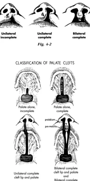
c. Submucous cleft (SMCP): occult cleft of the soft palate encompassing classic clinical triad (bifid uvula, notching of the hard palate, zona pellucida) 2. Classification a. Lip (Fig. 4-2) i. Unilateral (a) Complete (b) Incomplete ii. Bilateral (a) Complete (b) Incomplete iii. Median (a) Complete (b) Incomplete b. Palate (Fig. 4-3) 3. Prevalence
a. Cleft of lip with or without cleft palate (CL±CP) 1:750 in Caucasians, less in African-Americans (0.41 per 1000 live births), greater in Asians (1.41 per 1000 live births)
b. Cleft of palate alone (CP) 1:2500
33 32
4. Occurrence risk in offspring (Table 4-1) 5. Etiology
a. Multifactorial combination of heredity with or without environmental factors
b. Teratogenic agents — e.g. pheyntoin, alcohol c. Nutritional factors may contribute — folate
deficiency 6. Embryology
a. Cleft lip with palate forms at 4-6 weeks due to lack of mesenchymal penetration (merging) and fusion
b. Isolated cleft palate forms later, at 7-12 weeks, from lack of fusion
7. Pathophysiology and Functional Deficits a. Cleft lip
i. Inability to form fluid and air seal in eating or speech
ii. Malocclusion as a result of intrinsic deformities of alveolar process and teeth iii. Lack of continuity of skin, muscle and
mucous membrane of lip with associated nasal deformity and nasal obstruction iv. Deformity
b. Cleft palate
i. Inability to separate nasal from oral cavity so that air and sound escape through nose in attempted speech
ii. Feeding impaired by loss of sucking due to inability to create intra-oral negative pressure
iii. Loss of liquids and soft foods through nose due to common nasal-oral chamber iv. Middle ear disease in 100% of patients due
to Eustachian tube dysfunction, abnormal mucus
v. May be associated with Pierre-Robin sequence (cleft palate, micrognathia, glossoptosis). In these cases, airway obstruction and failure to thrive may be present. These cases may require ICU monitoring, prone positioning,
tracheostomy, and now mandibular distraction (moving the base of the tongue forward by mandibular advancement). Distraction has been used with some good effect in severe cases, avoiding
tracheostomy 8. Team concept
Because of multiple problems with speech, dentition, hearing, etc., management of the patient with a cleft should be by an interdisciplinary team, preferably in a cleft palate or craniofacial center. Team members include: plastic surgeon, orthodontist, dentist, geneticist, pediatrician, speech therapist, audiologist, social worker, and psychologist
9. Timing of Surgical Intervention
a. Cleft lip — most common 10 weeksof age. Once followed “rule of 10’s” (10 weeks of age, Hgb 10, 10 lbs.), but now this rule is more historical. Range of cleft lip repair varies from
0-3months of age in full-term, otherwise healthy, infant
b. Cleft palate — before purposeful sounds made (9 -12mos), depending upon health of infant, extent of cleft, but certainly before 18 months of age, if possible
c. Cleft nasal deformity — most centers perform primary correction at the time of lip repair, followed by secondary work at preschool age ( 4-5 years)
d. Alveolar cleft — most centers perform secondary bone grafting at the stage of mixed dentition (9-12years of age), just before eruption of the permanent canine, which is often affected by the cleft
e. Dentofacial skeletal abnormality — in most cleft patients, this manifests as maxillary
retrusion/hypoplasia. In 25% of cleft patients, orthognathic surgery (jaw-straightening procedure) has to be performed to correct a malocclusion (abnormal bite). Orthognathic surgery can only be performed in skeletally mature individuals (14-16years of age, women;
35 34
17-19years of age, men). With the advent of craniofacial distraction, surgical intervention can be performed earlier, but both patient and parents must be advised that the growing child may “outgrow” the correction, necessitating a repeat procedure
10. Principles of Primary Repair a. Cleft lip
i. Repair of skin, muscle and mucous
membrane to restore complete continuity of lip, symmetrical length and function ii. Simultaneous repair of both sides of a
bilateral cleft lip
iii. Preference for primary nasal reconstruction at time of lip repair
iv. In wide clefts (>10mm), presurgical orthodontics (palatal appliance, nasoalveolar molding) may be indicated, or a cleft lip adhesion (surgery to initially bring lip segments together, followed by definitive repair of lip 3 months later)
b. Cleft palate
i. One stage repair of both hard and soft palate
11. Secondary Repair a. Cleft lip
i. Revision of lip repair if needed ii. Revision of nose as required
iii. Repair of alveolar cleft (if present) with bone graft around 9 years of age (time of eruption of canine teeth)
b. Cleft palate
i. Correction of velopharyngeal inadequacy (nasal escape of sound and air due to remaining structural defect of palate): 4-6 years of age
ii. Repair of any palate fistula B. Other Congenital Anomalies
1. Craniosynostosis (343 out of 1,000,000 live births). a. Definition: Premature fusion of one or more
cranial vault sutures. Categorized into syndromic and nonsyndromic types
i. Nonsyndromic:
(a) Order of frequency according to suture type (ascending to descending): Sagittal, metopic, coronal, lambdoid, other)
(b) Characteristic head shape according to suture affected: Sagittal—
scaphocephaly (scapho, Gr., meaning boat-shaped); metopic—trigonocephaly (trigono, Gr., meaning triangular- or keel-shaped forehead); bicoronal – brachycephaly (brachy, Gr., meaning short in AP direction)
(c) Ongoing debate as to whether or not these patients have an increased incidence of developmental delay (d) Treatment: anterior vault reshaping
(fronto-orbital advancement/reshaping), total vault reshaping, or posterior vault reshaping, depending on location and severity of craniosynostosis. Usually performed within first year of life to take advantage of molding capacity of skull
ii. Syndromic:
(a) Major associated syndromes include Apert (craniosynostosis, exorbitism, midfacial retrusion with complex syndactyly of the 2-4 digits of the hands/feet), Crouzon (craniosynostosis, exorbitism, midfacial retrusion), and Pfeiffer (craniosynostosis, exorbitism, midfacial retrusion, broad thumbs and toes) syndromes
(b) Characteristic head shape involves turribrachycephaly (turri-, Gr., tower) (c) 50% of Apert syndrome patients have substantial mental delay; Crouzon and Pfeiffer syndrome patients usually develop normally
(d) Genetic defect identified in fibroblast growth factor receptor (FGFR) genes
(Apert, Crouzon---FGFR2, Pfeiffer— FGFR1)
(e) Goals of surgery: Release fused cranial sutures, correct profound exorbitism to prevent corneal exposure/blindness, improve craniofacial dysmorphism, correct malocclusions
(f) Surgical interventions:
Anterior/posterior/total vault reshaping (0-1 years), Monobloc (osteotomy and advance forehead and face
simultaneously with bone grafts/fixation) vs. Le Fort III (osteotomy and advance face) (4-6 years), with repeating procedures as necessary. Craniofacial distraction leads to greater advancement, less relapse than conventional procedures 2. Facial Dysostoses
a. Treacher-Collins Syndrome (Mandibulofacial Dysostosis)
i. Rare, autosomal dominant disorder ii. Affected gene on chromosome 5q iii. Variable penetrance
iv. Clinical manifestations: Lateral orbital wall deficiency/ midfacial retrusion due to hypoplasia/aplasia of the zygomatic bone; downward slanting palpebral fissures and colobomata; variable external ear malformations with deafness; mandibular hypoplasia with microretrognathia; underdeveloped lower jaw can lead to airway compromise, necessitating distraction or tracheostomy, or both v. Treatment: Skeletal and soft tissue
augmentation of deficient areas with autogenous bone (calvarium, rib, iliac crest) and autologous fat/tissue transfer,
respectively. Mandibular distraction may be necessary for achieving a stable airway
b. Hemifacial Microsomia
i. Third-most common congenital
malformation (following club foot and cleft lip and palate)
ii. 1:7000 live births affected
iii. No genetic defect ascribed; leading theory of cause is related to disruption of the stapedial artery during embryogenesis iv. Part of the oculoauriculovertebral (OAV)
spectrum
v. Usually associated with microtia vi. Manifestations include craniofacial or
hemifacial deficiency, both on skeletal and soft tissue level; microtia; mandibular hypoplasia; macrostomia; malocclusion from an abnormal cant (secondary to reduced vertical height of the ramus)
vii. Associated with Tessier #7 facial cleft and variable facial nerve palsy
viii. Pruzansky classification useful for mandibular discrepancy; OMENS
classification (orbit, mandible, ear, nerve, soft tissue) more comprehensive
ix. Treatment: Skeletal and soft tissue augmentation of deficient areas with autogenous bone (calvarium, rib, iliac crest) and autologous fat/tissue transfer,
respectively. Mandibular distraction may be necessary for achieving correction of malocclusion, versus conventional orthognathic procedures to correct jaw discrepancies in adolescence
c. Goldenhar Syndrome i. Variant of OAV spectrum
ii. Manifested by hemifacial microsomia, coloboma and epibulbar dermoids, vertebral spine abnormalities and renal abnormalities iii. Treatment as in ii.
d. Nager Syndrome 3. Embryologic Defects
a. Branchial cyst, sinus, or fistula
i. An epithelial-lined tract frequently in the lateral neck presenting along the anterior border of the sternocleidomastoid muscle. May present as a cyst or as a sinus connected with either the skin or oropharynx, or as a fistula between both skin and oropharynx openings
ii. Treatment — excision b. Thyroglossal duct cyst or sinus
i. Cyst in the mid-anterior neck over or just below the hyoid bone, with or without a sinus tract to the base of the tongue (foramen cecum)
ii. Treatment — excision c. Ear deformities
i. Types
(a) Complete absence (anotia) — very rare (b) Vestigial remnants or absence of part of
ear (microtia)
(c) Absence of part or all of external ear with mandibular deformity (hemifacial microsomia)
(d) Abnormalities of position (prominent ears)
ii. Treatment
(a) Anotia or microtia-construction from autogenous cartilage graft or synthetic implant, vascularized fascial flap, skin graft — usually requires more than one operation. (Traumatic loss of part or all of ear is treated similarly). Use of a prosthetic ear may be indicated in some patients
(b) Prominent ears — creation of an anthelical fold and/or re-positioning/ reduction of concha
II. TRAUMATIC
A. Facial soft tissue injuries
1. Evaluation of all systems by trauma team (ABCDE, primary survey)
2. Establishment of airway (may be obstructed by blood clots or damaged parts) by:
a. Finger (jaw thrust, e.g.) b. Suction
c. Endotracheal intubation
d. Cricothyroidotomy or tracheotomy
3. Control of active bleeding by pressure until control by hemostats and ligatures or cautery in operating room
4. Treatment of shock
5. Very conservative debridement of detached or nonviable tissue
6. Careful wound irrigation with physiologic solution 7. Remove all foreign materials
8. Palpate or explore all wounds for underlying bone injury; rule out injury to facial nerve, parotid duct, etc.
9. Radiologic evaluation
10. Repair as soon as patient’s general condition allows with meticulous reapproximation of anatomy a. Preferably less than 8 hours post-injury b. Primary closure may be delayed up to 24 hours
(dressing should be applied and antibiotics given while waiting)
11. Tetanus prophylaxis 12. Antibiotics if indicated B. Facial bone fractures
1. Classification
a. Mandible only — often bilateral (ring concept) i. Depending on anatomical region
(parasymphysis, body, angle, subcondyle) and overall function (malocclusion), open reduction and internal fixation (ORIF) may be indicated
ii. Panorex film and CT scan useful
iii. Key is displacement of bone segments and patient’s bite
iv. Approximately 10-13% of fractures in the mandible coincide with c-spine fracture; so, appropriate workup (x-rays) and c-spine stabilization must be performed prior to surgery
b. Zygomatic complex (Fig. 4-4)
i. Commonly associated with orbital floor fractures; therefore, must check extraocular movements and obtain opthalmology consultation if suspicious of globe injury ii. If severe displacement exists, must perform
ORIF with three-point fixation c. Maxillary — Le Fort I, II, III (Fig. 4-5) d. Naso-orbital-ethmoidal (NOE)
e. Isolated orbital floor fractures: blowout versus blow-in
i. Check for entrapment (failure to move eye in all directions)—if present, must decompress orbit within 48 hours
ii. Check for enopthalmos (position of globe in relation to unaffected globe in worm’s eye view). Must operate for enopthalmos 2mm or greater
f. Frontal sinus
g. Other isolated fractures — e.g. nasal h. Combination of above (panfacial fracture) i. Closed or open
j. Pediatric craniofacial fractures: Usually more conservative with operative repair in this patient population, due to growing facial skeleton and developing dentition
2. Diagnoses
a. Consider patient history
b. Physical examination for asymmetry, bone mobility, diplopia, extraocular muscle entrapment, sensory loss, malocclusion, local pain
c. Old (pre-injury) photographs often useful to assess baseline
d. X-rays
i. Skull (rare) and cervical spine ii. CT scan — axial and coronal — now
imaging modality of choice iii. Specialized views
(a) Waters view for facial bones (Fig. 4-6); good for orbital floor, now surpassed by CT