Introduction
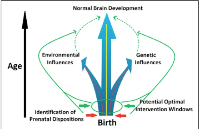
The explosive growth of brain structure and function in infancy is unparalleled by any other postnatal develop-mental period. This rapid expansion and organization is genetically determined but is also prone to epigenetic and environmental modifications. Therefore, the early brain develops with the highest level of plasticity, which facili-tates both adaptive changes, representing opportunity, and malformations, reflecting vulnerability. It is increasingly recognized that most neuropsychiatric disorders, mani-fested as complex combinations of cognitive, emotional, and behavioral deficits, have developmental origins that are, at least in part, rooted very early in the initial laying-out of the brain’s functional blue print (Beardslee and oth-ers 2011; Insel 2010). Therefore, a better undoth-erstanding of this most plastic period of human brain development is urgently needed to pave the way for early identification of risks and interventions that have the potential to alter the developmental trajectory at the earliest, most modifiable stage (Fig. 1). In the era with increasingly more interests in prevention strategies to reduce the burden of mental disorders, the importance of early identification and pre-ventive intervention cannot be overemphasized.
The large number of studies of pre- and postnatal struc-tural human brain growth have provided a relatively detailed understanding of the intricate processes of the early anatomical development. Briefly, the structural basis of later neural circuits, including neurogenesis, synapto-genesis, dendritic arborization, and formation/reformation of axons, starts and greatly expands in utero (Bystron and others 2008). After birth, the brain continues to grow at a remarkable pace with its total volume doubled in the first year, followed by another 15% increase during the second
1Biomedical Imaging Research Institute (BIRI), Department of
Biomedical Sciences and Imaging, Cedars-Sinai Medical Center, Los Angeles, CA, USA
2Department of Radiology and Biomedical Research Imaging Center,
University of North Carolina at Chapel Hill, NC, USA
3Departments of Psychiatry, Neurobiology, and Psychology, University
of North Carolina at Chapel Hill, NC, USA
4Department of Psychiatry, University of North Carolina at Chapel
Hill, NC, USA
Corresponding Author:
Wei Gao, Biomedical Imaging Research Institute (BIRI), Department of Biomedical Sciences and Imaging, Cedars-Sinai Medical Center, 116 N Robertson Blvd, PACT 800.7G, Los Angeles, CA 90048-0750, USA. Email: [email protected]
Abstract
Infancy is a critical and immensely important period in human brain development. Subtle changes during this stage may be greatly amplified with the unfolding of different developmental processes, exerting far-reaching consequences. Studies of the structure and behavioral manifestations of the infant brain are fruitful. However, the specific functional brain mechanisms that enable the execution of different behaviors remained elusive until the advent of functional connectivity fMRI (fcMRI), which provides an unprecedented opportunity to probe the infant functional brain development in vivo. Since its inception, a burgeoning field of infant brain functional connectivity study has emerged and thrived during the past decade. In this review, we describe (1) findings of normal development of functional connectivity networks and their relationships to behaviors and (2) disruptions of the normative functional connectivity development due to identifiable genetic and/or environmental risk factors during the first 2 years of human life. Technical considerations of infant fcMRI are also provided. It is our hope to consolidate previous findings so that the field can move forward with a clearer picture toward the ultimate goal of fcMRI-based objective methods for early diagnosis/identification of risks and evaluation of early interventions to optimize developing functional connectivity networks in this critical developmental window.
Keywords
(Knickmeyer and others 2008). Notably, this increase is largely accounted for by the growth of neural connections in gray matter (i.e., synapse and dendrites), long-range axons, and myelination, all of which are elements essen-tial for the organization of distributed functional networks (Tau and Peterson 2010). Although mostly progressive, regressive development including pruning of both syn-apses and axons are also evident during the infancy period, thus enabling reorganization of initially established func-tional circuits (Levitt 2003). Together, these early struc-tural elements establish the fundamental anatomical organization of the infant brain. However, directly linking the development of the brain’s structural elements and emergence of specific behaviors/functions, among which precursors of future problems may be hopefully identi-fied, is challenging given the unmatched specificity levels of the two domains.
In contrast, the characterization of neural circuits, typ-ically defined as networks of interconnected brain regions that integrate vast amounts of information and perform discrete sets of specific brain functions (Friston 2011), represents one of the most viable strategies to bridge between brain and behavior. Conceptually, in a region-specific manner, genes interact with a myriad of environ-mental factors to layout the determinants of neuronal birth, death, and cellular characteristics. Additionally, such gene-environmental interactions also dictate the for-mation and reforfor-mation of axons, dendrites, and syn-apses, critical elements for the building of different neural circuits with diverse configurations and functional attri-butes. Therefore, neural circuits represent more direct mediators of brain’s diverse functional capabilities. Their characterization during the critical infancy period may provide unprecedented insights into the brain basis of the infants’ fast-growing behavioral repertoire. Such
characterization would also prove invaluable for early identification of genetically and environmentally induced alterations that enhance risks for future onset of behav-ioral problems and/or mental disorders (Fig. 1).
Previously hindered by the lack of appropriate experi-mental fMRI protocols, little progress has been made on the delineation of the infants’ functional neural circuits prior to the new century. However, the field has seen a quantum jump since the advent and popularization of the functional connectivity MRI technique (fcMRI) (Biswal and others 1995; Raichle 2010). fcMRI does not require the performance of specific tasks in the scanner, thereby removing one of the most difficult obstacles in functional neuroimaging of the infant brain. Instead, fcMRI exam-ines the temporal correlation of spontaneous blood-oxy-gen level dependent (BOLD) signals in the absence of any external tasks. Based on the concept of “neurons fir-ing together wirfir-ing together,” fcMRI thus critically que-ries whether and how different brain areas are synchronized to form functionally coordinated networks during resting state—essentially providing a means to depict the brain’s functional organization. Based on this exciting new technique, there have been numerous arti-cles published on the functional development of infant (and fetal) brain in the past decade, resulting in an impres-sive body of work that has greatly improved our under-standing of this previously “dark period” of functional brain development. Therefore, a review and consolida-tion of these previous findings is warranted to capitalize on these exciting and tantalizing findings to help the field move forward. Although functional connectivity can also be inferred using other modalities (e.g., electroencepha-lography [EEG], magnetoencephaelectroencepha-lography, near-infrared spectroscopy, etc.), studies using fcMRI will be the pri-mary focus of this review. This review is not intended to be exhaustive but rather will focus on illustration of the poten-tial mechanisms underlying both normal and abnormal development based on some of the most relevant discover-ies. Specifically, findings describing the normative devel-opment of functional connectivity networks (FCNs) from the fetal period to the end of the second year will constitute the main body of this review. We will then describe devia-tions from the normative FCN development due to identifi-able genetic and/or environmental risk factors (e.g., maternal mental disorders, prenatal drug exposure, and premature birth). Following that, potential technical issues for infant fcMRI study will be discussed. Finally, we will present our conclusions and suggest several future direc-tions that deserve the field’s attention. Building on these previous findings, it is our hope that the field will move forward with a more systematic effort to tackle various risk factors that adversely affect normal early brain functional development and come up with tangible ways for early diagnosis/identification.
Normative Development of
Functional Connectivity and the
Behavior Associations during Infancy
Cortical Networks
The first article that used fcMRI to characterize infant brain FCNs was published by Fransson and colleagues in 2007 (Fransson and others 2007). This report was based on data from very preterm infants (gestational age [GA] <28 weeks) scanned at term-equivalent age (GA ~41 weeks). Premature birth represents a major risk factor for potentially abnormal functional connectivity develop-ment (Kwon and others 2015; Smyser and others 2010), so this study, together with others based on preterm infants, will be discussed later in the Premature Birth sec-tion. The first fcMRI studies in typically developing, full-term infants were published by Lin and colleagues (Lin and others 2008) and Liu and colleagues in 2008 (Liu and others 2008). Symmetric brain regions of primary senso-rimotor and visual networks were examined and shown to be functionally synchronized starting from birth. These findings were subsequently replicated using an indepen-dent sample by Gao and his colleagues (Gao and others 2015b). These results suggest that primary sensorimotor and visual networks are likely functioning at birth, which
first to be adult-like (which actually showed postnatal regressive growth in functional connectivity), followed by primary and secondary visual networks, then dorsal attention and DMN. In contrast, the frontoparietal execu-tive control networks are still in a premature form at the end of the first year. This series of studies, for the first time, delineated the developmental sequence of different functional brain networks during infancy and suggested a progressive maturation from primary to higher order net-works. One striking finding is that the DMN, thought to mainly govern a complex set of self-referential functions in addition to potentially other external ones (Elton and Gao 2015a; Gao and others 2013a), is one of the first higher order networks to show a well-distributed network structure by integrating distant medial frontal, medial/lat-eral parietal, and medial/latmedial/lat-eral temporal regions starting at 6 months of age (Fig. 2). This finding is consistent with the rapid emergence of self-awareness during the first year of life (Amsterdam 1972) and suggests that the development of theory-of-mind-related functions associ-ated with DMN likely serves as a foundation for other higher order functions to build on. This is consistent with the concept that social interaction lies at the core of infant cognitive and emotional development. Giving the rapid development of DMN during the first half of the first year, the report of its sensitivity to social and emotional environment is not surprising (Graham and others 2015). Actually, family social economic status (i.e., income) has been reported to show a trend of correlation with the functional connectivity pattern of DMN at 6 months of age (Gao and others 2015a), further underscoring the potential link between the social environment and DMN development. Therefore, the study of the DMN during the first year of life may serve as a window into the social/ emotional development, which likely influences subse-quent development of other higher order processes (Gao and others 2009).
Overall, functional connectivity development of dif-ferent networks during infancy generally follows a pri-mary-to-higher order sequence but different FCNs demonstrate unique timings and developmental trajecto-ries. These findings suggest that different networks likely possess different critical periods during development, and future studies are needed to more rigorously charac-terize such network-specific milestones and examine their behavioral associates so that clinical interventions aiming to rectify the growth of specific brain functions can be better informed.
Subcortical Networks
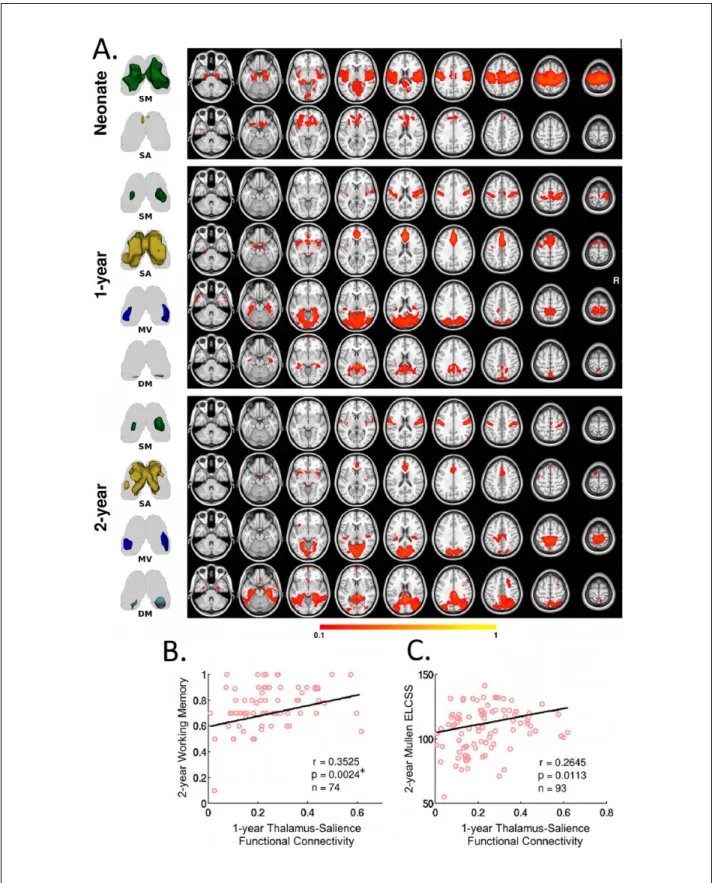
In addition to cortical networks, infant functional connec-tivity patterns of subcortical structures have also been characterized. The thalamus, especially, represents a
critical relay center and pacemaker of the whole brain system, and is of unique importance in early brain func-tional development (Jones 2000). In a longitudinal study of typically developing infants, Alcauter and colleagues delineated the thalamocortical functional connectivity pattern and correlated it with later behavioral outcomes during infancy (Fig. 3) (Alcauter and others 2014). They found that neonatal thalamic functional connectivity was dominated by connections to the sensorimotor/auditory/ visual networks, with a small medial anterior thalamic cluster projecting to insula and anterior cingulate cortex. The insula and anterior cingulate cortex constitute the main components of the brain’s salience network, which is thought to integrate internal and external information in order to assign salience to various events and produce a “sentient self” (Seeley and others 2007). With develop-ment, the nonspecific thalamo-primary functional net-work connectivity becomes more specialized, the thalamo-salience connections strengthen, and new pro-jections to the DMN emerge, underscoring the dynamic and network-specific growth of thalamocortical func-tional connections. Based on a different methodology and sample (only neonates), Toulmin and others has repli-cated the findings of dominant thalamo-sensorimotor connectivity and the connection between anterior medial thalamus and salience network-related regions in the newborns (Toulmin and others 2015), supporting the robustness of this set of results. However, given the repeatedly reported alterations of functional connectivity by premature birth (Kwon and others 2015; Smyser and others 2010), the study design of mixing data from both term and very preterm infants in Toulmin and others may have contributed to their results and cautions should be taken when comparing results from these two studies.
observed. This is characterized by synchronous activity of anterior insula with prefrontal regions, which progres-sively becomes more network-like with age (red color; Fig. 4) (Alcauter and others 2015a). Taken together with the observation that this network is the only one showing robust thalamic connectivity besides primary networks in neonates (Fig. 3A), these findings support the early func-tioning of the salience network. Given that one of the most critical prerequisites of infants’ learning experience is “paying attention,” which is guided by the salience detection function of the salience network, it is not sur-prising that thalamo-salience network connectivity, which likely relays critical information for proper evalu-ation of the salience weighting of different events, is essential for later cognitive performance. Taken together, the salience and DMN network might represent two of the unique higher order functional networks that may profoundly influence early brain functional brain devel-opment processes. Overall, these studies suggest that sub-cortical areas, including but not limited to the thalamus, play critical roles in normal brain development during infancy and warrant additional investigations.
Internetwork Connectivity
Given the critical importance of large-scale network-level interactions in both normal and abnormal adult brain functioning (Elton and Gao 2014, 2015a, 2015b; Gao and Lin 2012; Spreng and others 2010), when and how different FCNs begin to “talk” to each other during
early brain development is also an important question. As an initial attempt, Gao and others examined the early developmental course of the widely reported “anti-corre-lation” between the dorsal attention network and the DMN (Gao and others 2013b), thought to represent potential “competing” mechanisms between contrasting brain processes (Fox and others 2005; Gao and Lin 2012). They found that such anti-correlation is absent in neo-nates but appears at 1 year and strengthens during the sec-ond year of life (Fig. 5). Interestingly, the “growth” of such anti-correlations coincides with the changing sooth-ing practice in infants; startsooth-ing around 6 month of age, caregivers can sooth a crying baby (i.e., temporally stop-ping the “internal” distressed state) by drawing his/her attention to novel toys (i.e., external attention). Therefore, the observed developmental course of the anti-correlation between the dorsal attention network and DMN provides support for the “competition” hypothesis from a develop-mental perspective. However, it is important not to inter-pret this competition as an indicator of the “task-negative” nature of the DMN; although the anti-correlation between dorsal attention and DMN persists, there is an emerging body of work showing DMN’s increased connectivity with other task-related networks (e.g., salience and exec-utive control) during the performance of a wide variety of external tasks (Elton and Gao 2014, 2015a; Gao and Lin 2012; Gao and others 2013a; Spreng and others 2010). Actually, a separate study in adults by Gao and Lin sug-gests that salience and executive control networks likely act as “regulators” between the competing dorsal atten-tion and DMN networks and will flexibly coordinate with either one to facilitate corresponding task performances (Gao and Lin 2012). Notably, developmental changes of internetwork connections is not restricted to these higher order networks but represent a universal phenomenon associated with all identifiable FCNs (Gao and others 2015b). Going beyond the infancy period, the maturing architecture of such network-level interaction mecha-nisms has been further demonstrated in a study of 6-year-old children, providing critical support for the prolonged development of internetwork interactions (Emerson and others 2015). This is again expected given the need of extensive environment-based tuning for the establish-ment of effective communication strategies between pri-mary and higher order networks. Overall, investigations into network-level interactions during development deserve more attention and the associated behavioral sig-nificance needs to be better characterized.
The Integrated Whole Brain System
At the whole brain level, there is a growing interest in viewing the brain as an integrated system and character-izing its global properties based on graph theoretical Figure 4. Functional segregation of the insula and associated
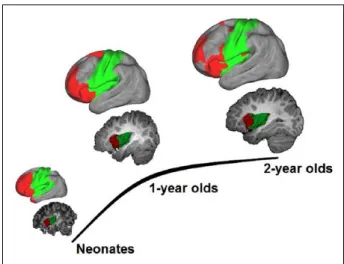
calculations (Rubinov and Sporns 2010). Although not a perfect model, the abstraction of the whole brain func-tional system to a “graph (i.e., brain regions as nodes and the functional connectivity between brain regions as edges)” is theoretically appealing in that one can obtain a series of summary metrics depicting differential but fundamental aspects of the information transferring property of the whole brain. For example, the concept of a “small-world” network concisely depicts a network that possesses both local (through dense short-range con-nections within local neighborhood) and global effi-ciency (through long-range short cuts between distant nodes) in information transferring. Using this approach, Gao and others showed that the neonatal brain already exhibits a “small-world” characteristic based on func-tional connectivity measures (Fig. 6) (Gao and others 2011). Fransson and others observed the same topology using a voxel-based approach (Fransson and others 2011). This indicates that the newborn brain is already equipped with a relatively optimized functional organi-zation that facilitates information transferring. However, it is also apparent that newborn brain lacks the “short-cut” long-range connections that are essential for global efficiency. These long-range connections only appear in 1-year-olds and strengthen in 2-year-olds, suggesting a developmental evolution of the brain’s functional topol-ogy toward a more efficient, globally optimized, system (Fig. 6) (Gao and others 2011).
Alterations of Normative Functional
Connectivity Development by
Identifiable Risks
Maternal Mental Illnesses
Individuals with mental disorders are at least as likely, if not more likely, than those without psychiatric illnesses to become parents. In the United States, an estimated 65% of women diagnosed with mental illness are mothers (Seeman 2002), resulting in a large population of off-spring at greater risks for later development of mental disorders/problems. Maternal mental health disorders represent a significant risk for disruption of normal brain development, due to known influences of maternal genetic factors on fetal brain growth (Satyanarayana and others 2011). However, intrauterine (e.g., disrupted hor-mone release) and postnatal environmental factors (e.g., poorer care giving and lower socioeconomic status) related to mental illness are also likely to contribute to the consequences, either independently or through interac-tions with genetic factors (Rice and Thapar 2010). Through imaging infants at the earliest stage of develop-ment, researchers can hopefully minimize some of the related postnatal environmental effects and discern new insights into the brain mechanisms underlying the genetic and intrauterine impact of maternal mental disorders.
amygdala (a brain region that is primarily involved in the brain’s emotional regulation) and various limbic and medial prefrontal regions in 6-month-old infants born to mothers with depression (Qiu and others 2015). This study suggests that neural correlates of the familial trans-mission of the phenotypes associated with maternal depression can be detected early in infancy. In our own work (unpublished data), amygdala and thalamus func-tional connectivity disruptions were detected in neonates whose mothers were diagnosed with either schizophrenia or bipolar disorder, advancing the timeline for detecting neural correlates of maternal mental disorders in off-spring. These findings are consistent with reports from adult patients with schizophrenia (Anticevic and others 2014) and/or adolescents at clinically high risk for devel-oping schizophrenia (Anticevic and others 2015; Gee and others 2012), suggesting that the amygdala, thalamus, and prefrontal areas might be among the most vulnerable areas for genetic risk of schizophrenia. However, future long-term longitudinal studies are needed to characterize how such early connectivity alterations enable prediction of later behavioral problems and/or onset of mental
illnesses. Moreover, the effect of medication associated with different maternal mental disorders may have con-tributed to the previous findings and need to be better modeled. Overall, the abnormal functional connectivity associated with genetic risks for mental disorders, par-ticularly those observed in affected infants, provides sup-port for future derivation of imaging-based biomarkers to identify risks and to develop very early interventions that can correct the final stages of circuit and behavioral development (Fig. 1).
Prenatal Drug Exposure
tiation, axonal elongation, and synaptogenesis, among others (Gaffuri and others 2012; Tortoriello and others 2014; Wu and others 2011), all of which are core building blocks of developing neural circuits. Therefore, prenatal drug exposure represents another significant threat to nor-mal functional brain development and has been linked to both short- and long-term developmental behavioral and cognitive consequences (Ackerman and others 2008; Bandstra and others 2010; Smeriglio and Wilcox 1999). Neuroimaging studies of the effects of prenatal exposure on brain structure and function are often done in later childhood or adolescence (Donald and others 2016; Li and others 2013; Roussotte and others 2012). Although infor-mative, these findings are more likely to reflect both ini-tial drug effects and the confounding postnatal influence of adverse environments associated with maternal drug abuse. Imaging performed in early infancy has the advan-tage of minimizing such confounds and provides a better depiction of the mechanisms by which functional connec-tions are affected by gestational exposure(s).
The first study of the prenatal drug effects on neonatal brain development was recently reported by Grewen and others (2014). This study focused on the effects of prena-tal cocaine exposure on neonaprena-tal brain structure. However, unlike animal models, in human studies women who use cocaine during pregnancy are also likely to use other drugs such as nicotine, alcohol, and marijuana. Therefore, researchers compared neonates exposed to cocaine and other drugs to a group of drug-naive infants and to a group exposed to a similar profile of other drugs including alco-hol, nicotine, marijuana, antidepressants, but without cocaine. This enabled detection of both drug-common and cocaine-specific effects. Prenatal cocaine was associated with reduced prefrontal gray matter volume compared with both non-cocaine drug-exposed and drug-free con-trol groups, after concon-trolling for covariates (birth weight, gestational age at scan, gender, total brain volume). Salzwedel and others then published the first study of pre-natal drug exposure effects on brain functional connectiv-ity in the same neonate sample (Salzwedel and others 2015). Results revealed hyperconnectivity between the
amygdala and a medial prefrontal cluster that was specific to prenatal cocaine exposure, after controlling for relevant covariates and other drug exposures (Fig. 7). Functional connectivity disruptions between the insula and medial prefrontal/sensorimotor areas were observed in both drug-exposed groups, suggesting a nonspecific vulnerability to multiple drugs for this connection. These modified func-tional connections may, at least partly, contribute to the long-lasting behavioral consequences previously reported in children with prenatal cocaine exposure. Indeed, in a follow-up study (unpublished work), more dramatic cocaine-specific functional connectivity disruptions were observed among thalamo-cortical connections. More importantly, greater alterations were related to lower over-all cognitive and fine motor scores measured at 3 months of age. These findings underscore the predictive power of early functional connectivity measures for later behavioral outcomes and support the exciting possibility of using early functional neuroimaging methods for the identifica-tion of risks for future behavioral problems.
In a more recent study of the effects of prenatal mari-juana exposure on neonatal functional connectivity, both marijuana-specific and drug-common effects were again observed (Grewen and others, 2015). Marijuana-specific connectivity disruptions were revealed for dorsal stria-tum and anterior insula seed regions, which all have high in utero Type 1 cannabinoid receptor (CB1R) expression. This early departure from typical network development may contribute to the deficits in motor and visual-spatial activity, integration and coordination (Willford and oth-ers 2010), attention (Goldschmidt and othoth-ers 2012), and social-emotional stability (Gray and others 2005) reported in children and adolescents prenatally exposed to mari-juana (Fried and Smith 2001, Fried and others 2003).
Taken together, these studies, for the first time, pro-vide compelling epro-vidence that prenatal exposure to
different drugs alters early brain structural growth and the orchestration of functional networks. These drug-related alterations likely arise from disrupted prenatal program-ming, since postnatal exposures were minimized is this sample of newborns. These results also indicate that indi-vidual drugs likely differentially affect discrete functional neural circuits. In the future, a longitudinal and more sys-tematic study with a larger sample size that is capable of discriminating the effects of various drugs during devel-opment is needed. Moreover, the potential drug-drug interaction effects deserve more attention, since polydrug use is the norm rather than the exception in most women who use and abuse substances during pregnancy.
Premature Birth
The exact causes for premature birth are still poorly understood but likely involve a complex set of genetic, biological, and environmental factors (Terao 1996). The rate of infants affected by premature birth is growing, partly due to the increased survival rate attributable to advances in perinatal and neonatal care. However, the last few weeks of gestation is a particularly critical period when most of the brain’s structural elements undergo an accelerated pace of development (Tau and Peterson 2010). Therefore, the transition from the womb to the outside environment during this critical period may adversely affect infants’ brain growth. Indeed, up to 40% of very premature infants (born at <30 GA) may develop motor deficits (Holsti and others 2002), and an estimated 30% to 60% experience long-term cognitive impairments and social/emotional difficulties (Anderson and others 2004; Taylor and others 2004). As mentioned above, Fransson and others published the first fcMRI study on very preterm infants scanned at term-equivalent age (Fransson and others 2007). They identified five FCNs including three primary (i.e., visual, sensorimotor, and auditory) and two higher order networks located at the anterior and posterior parts of the brain, respectively. However, since no term controls were included in this study, the effects of premature birth on infant brain development were beyond the scope of that study. A later study by Smyser and others examined the longitudinal development of FCNs in a cohort of very preterm infants and compared the data acquired at term-equivalent age with those from term controls (Smyser and others 2010). They reported functional connectivity developing from local to bilaterally symmetric connections even prior to term-equivalent age. Noticeably, premature infants dem-onstrate lower correlation values and limited distribu-tion, especially among long-range thalamocortical connections. Consistent with Smyer and others, another study (Kwon and others 2015) has shown disrupted lan-guage network lateralization based on functional
connectivity measures in very preterm infants compared with term controls. Notably, studies have extended find-ings of functional connectivity alterations related to pre-mature birth from term-equivalent age to childhood (Damaraju and others 2010) and adolescence (Mullen and others 2011; Schafer and others 2009), together with corroborating behavioral findings (Anderson and others 2004; Taylor and others 2004). These results underscore the far-reaching impact of this risk factor. Despite the seemingly converging findings pointing to abnormal functional connectivity development associated with premature birth, there are also reports showing negative findings (Doria and others 2011; Lee and others 2013). Therefore, future work is needed to more rigorously examine the effects of premature birth on the brain’s functional development. Importantly, the observed alter-ations likely reflect consequences of a combination of genetic/environmental factors leading to premature birth and/or the vastly different postnatal environments sur-rounding premature infants compared with the womb. Indeed, a study of effects of neonatal intensive care unit stay on the development of premature infants have shown that private room setup was associated with lower language scores and a trend for lower motor scores com-pared with open ward care (Pineda and others 2014). The authors suggest that their results underscore the impor-tance of sensory exposure during this sensitive period. On the other hand, Smith and others have shown that exposure to a greater number of stressors (e.g., vascular access/heel stick, radiology/diagnostic study, intubation, extubation, etc.) is associated with altered functional connectivity in the temporal lobe on top of structural alterations (Smith and others 2011). Therefore, further effort is warranted to more systematically examine the effect of environmental factors on the developing pre-mature brain to guide better postnatal care for this vul-nerable group. Overall, premature birth represents a significant threat to normal brain and behavioral devel-opment and viable objective ways (e.g., fcMRI-based) for the early detection of abnormality represents one of the highest priorities given the large and increasing num-ber of infants affected (Blencowe and others 2013).
Other Factors
The postnatal environment also represents a signifi-cant source of variation affecting normal functional brain development. The delicate process of infants’ social and emotional development, which lies in the core of early functional growth with critical influences on overall intellectual and cognitive development, requires a nurtur-ing and lovnurtur-ing environment to reach the greatest poten-tial. Consistent with this notion, early life stress, manifested as interparental conflict, has been shown to result in aberrant functional connectivity within the DMN, which subsequently is associated with greater neg-ative infant emotionality (Graham and others 2015). More generally, disrupted caregiving and parenting behaviors due to different causes (e.g., depression, sub-stance abuse, etc.) have been generally shown to affect normal functional connectivity development (Moses-Kolko and others 2014). Consistently, social economic status (e.g., maternal education and income) has been shown to directly correlate with the development of the motor and DMN networks during infancy (Gao and oth-ers 2015a).
Technical Considerations with fcMRI
in Infants
Technical Issues
Currently, the trend for imaging healthy normal infants is to perform the scan during natural sleep without sedation, which is safer and more acceptable to the infants/family and also practically less complicated. However, given a higher likelihood of subtle motion in naturally sleeping infants than cooperative awake adults, proper steps have to be taken to minimize motion artifacts beyond the stan-dard rigid-body motion correction and regression of motion parameters. Proposed by Power and others, data scrubbing represents an appealing way to remove the vol-umes contaminated by subtle motion (Power and others 2013). However, the by-product of disrupted temporal continuity introduced by scrubbing deserves further investigation. Regardless of which motion correction
bution of correlation values but does not appear to alter the relative differences across groups (Gao and others 2013a; Gao and others 2013b). Nonetheless, it is still rec-ommended that results obtained with GSR be compared with those without GSR and/or other normalization strat-egies (e.g., post hoc standardization; Yan and others 2013) to minimize the possibility of spurious results after GSR. Moreover, the negative correlation coefficients after GSR should be interpreted accordingly, given the known correlation distribution shift. Overall, similar to adult studies, the entire preprocessing pipeline for infant fcMRI data should be better optimized and standardized to facilitate comparisons across groups and replication of results, which is critical for the field to move forward. In this review, we specifically emphasized replicable find-ings where possible to highlight those potentially more robust results.
Sleeping Stage
infants immediately after they fall asleep and fix the sequence of scans across subjects so that the fcMRI scan will be acquired at approximately the same time after sleep for all subjects. If properly controlled, the relatively homogenous sleeping state across infant samples may actually provide a better context for the study of intrinsic functional networks considering the known intersubject variability of “resting-state” functional connectivity in awake adults due to potentially different cognitive/emo-tional states, metabolic rates, and so on (Laumann and others 2015).
Frequency
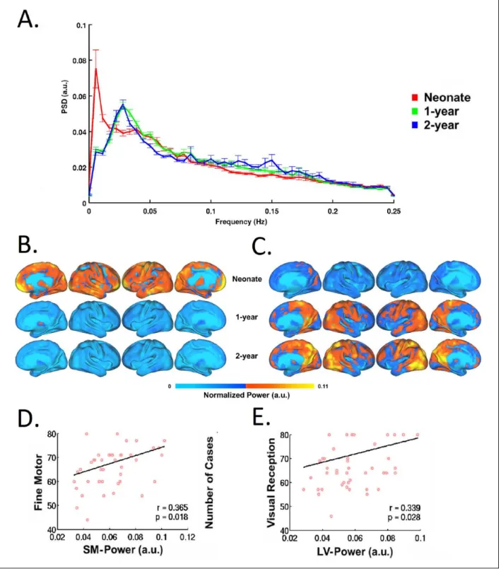
To date, most of infant fcMRI studies have focused on the frequency band of <0.1 Hz for the delineation of FCNs. This selection of frequency range is not driven physiolog-ically but more empirphysiolog-ically. Smith-Collins and others recently reported that a substantial proportion of signal power resides beyond this range and meaningful func-tional connectivity patterns can be observed using extended frequency bands in neonates (Smith-Collins and others 2015). In fact, the frequency distribution of BOLD signals and its changes across development likely reflect underlying maturation of either structural ele-ments (e.g., axons, myelination, etc.), functional pro-cesses (e.g., neural-vessel coupling), or both. The delineation of this process, by itself, is interesting. For example, Alcauter and others have shown that the peak frequency of BOLD signal shifts to a higher value during the first year of life (Fig. 8), and the power at peak-fre-quency for sensorimotor and visual networks in 1-year-olds correlates with motor and visual reception scores (Alcauter and others 2015b). This underscores the impor-tance of frequency-related properties of the BOLD signal in early brain development, which deserve more attention in future investigations.
Conclusions and Future Directions
In conclusion, numerous efforts from the past decade on functional connectivity study of the infant brain enabled a substantially improved understanding of the normative functional brain development process during infancy. For cortical networks, a maturation sequence from primary to higher order networks with the default-mode network highlighted as one of the earliest maturing higher order networks was observed. Subcortical areas are also estab-lishing cortical connectivity during infancy. Among them, the thalamo-salience network connectivity uniquely predicts later cognitive performances. Between networks, “cross-network” interactions are also evolving during infancy and highlight an emerging “competing”
mechanism between the default-mode network and the dorsal attention network. Finally, at the whole brain level, the neonatal brain already demonstrates an optimized topology qualifying as a “small-world” but global effi-ciency shows dramatic age-dependent improvement stressing the continued optimization of the brain’s func-tional topology for more efficient information transfer-ring. In addition to the delineation of the normative trajectories, infant fcMRI studies in different at-risk or diseased infant populations showed intriguing functional connectivity alterations associated with various risk and/ or pathological factors, underscoring the plastic and mod-ifiable nature of infant functional connectivity. These studies point to the exciting possibility of fcMRI-based objective ways for early diagnosis and/or identification of risks to facilitate the earliest possible intervention.
Declaration of Conflicting Interests
The author(s) declared no potential conflicts of interest with respect to the research, authorship, and/or publication of this article.
Funding
The author(s) disclosed receipt of the following financial sup-port for the research, authorship, and/or publication of this arti-cle: This work was supported by National Institutes of Health (R21NS088975 to WG; R03DA036645 to WG and KG; R01MH064065, R01HD05300 to JHG); Foundation of Hope for Research and Treatment of Mental Illness Award to WG; and Cedars-Sinai Institutional Support to WG.
References
Ackerman JP, Llorente AM, Black MM, Ackerman CS, Mayes LA, Nair P. 2008. The effect of prenatal drug exposure and caregiving context on children’s performance on a task of sustained visual attention. J Dev Behav Pediatr 29:467–74. Afif A, Bouvier R, Buenerd A, Trouillas J, Mertens P. 2007.
Development of the human fetal insular cortex: study of the gyration from 13 to 28 gestational weeks. Brain Struct Funct 212:335–46.
Alcauter S, Lin W, Smith JK, Gilmore JH, Gao W. 2015a. Consistent anterior-posterior segregation of the insula dur-ing the first 2 years of life. Cereb Cortex 25:1176–87. Alcauter S, Lin W, Smith JK, Goldman BD, Reznick JS,
Gilmore JH, and others. 2015b. Frequency of spontaneous BOLD signal shifts during infancy and correlates with cog-nitive performance. Dev Cogn Neurosci 12:40–50. Alcauter S, Lin W, Smith JK, Short SJ, Goldman BD, Reznick
JS, and others. 2014b. Development of thalamocortical connectivity during infancy and its cognitive correlations. J Neurosci 34:9067–75.
Amsterdam B. 1972. Mirror self-image reactions before age two. Dev Psychobiol 5:297–305.
Anderson PJ, Doyle LW, Victorian Infant Collaborative Study Group. 2004. Executive functioning in school-aged chil-dren who were born very preterm or with extremely low birth weight in the 1990s. Pediatrics 114:50–7.
Anticevic A, Cole MW, Repovs G, Murray JD, Brumbaugh MS, Winkler AM, and others. 2013. Characterizing thal-amo-cortical disturbances in schizophrenia and bipolar ill-ness. Cereb Cortex 24:3116–30.
Anticevic A, Haut K, Murray JD, Repovs G, Yang GJ, Diehl C, and others. 2015. Association of thalamic dysconnectivity and conversion to psychosis in youth and young adults at elevated clinical risk. JAMA Psychiatry 72:882–91. Ball G, Pazderova L, Chew A, Tusor N, Merchant N, Arichi T,
and others. 2015. Thalamocortical connectivity predicts cog-nition in children born preterm. Cereb Cortex 25:4310–8. Bandstra ES, Morrow CE, Mansoor E, Accornero VH. 2010.
Prenatal drug exposure: infant and toddler outcomes. J Addict Dis 29:245–58.
Beardslee WR, Chien PL, Bell CC. 2011. Prevention of men-tal disorders, substance abuse, and problem behaviors: a developmental perspective. Psychiatr Serv 62:247–54.
Behnke M, Smith VC; Committee on Substance Abuse; Committee on Fetus and Newborn. 2013. Prenatal sub-stance abuse: short- and long-term effects on the exposed fetus. Pediatrics 131:e1009–24.
Biswal B, Yetkin FZ, Haughton VM, Hyde JS. 1995. Functional connectivity in the motor cortex of resting human brain using echo-planar MRI. Magn Reson Med 34:537–41. Blencowe H, Cousens S, Chou D, Oestergaard M, Say L, Moller
AB, and others. 2013. Born too soon: the global epidemiology of 15 million preterm births. Reprod Health 10(Suppl 1):S2. Bystron I, Blakemore C, Rakic P. 2008. Development of the
human cerebral cortex: Boulder Committee revisited. Nat Rev Neurosci 9:110–22.
Chang C, Glover GH. 2009. Effects of model-based physiologi-cal noise correction on default mode network anti-correla-tions and correlaanti-correla-tions. Neuroimage 47:1448–59.
Damaraju E, Phillips JR, Lowe JR, Ohls R, Calhoun VD, Caprihan A. 2010. Resting-state functional connectivity differences in premature children. Front Syst Neurosci 4. doi:10.3389/fnsys.2010.00023.
Dinstein I, Pierce K, Eyler L, Solso S, Malach R, Behrmann M, and others. 2011. Disrupted neural synchronization in tod-dlers with autism. Neuron 70:1218–25.
Donald KA, Ipser JC, Howells FM, Roos A, Fouche JP, Riley EP, and others. 2016. Interhemispheric functional brain connectivity in neonates with prenatal alcohol exposure: preliminary findings. Alcohol Clin Exp Res 40:113–21. Doria V, Beckmann CF, Arichi T, Merchant N, Groppo M,
Turkheimer FE, and others. 2011. Emergence of resting state networks in the preterm human brain. Proc Natl Acad Sci U S A 107:20015–20.
Elton A, Alcauter S, Gao W. 2014. Network connectivity abnor-mality profile supports a categorical-dimensional hybrid model of ADHD. Hum Brain Mapp 35:4531–43.
Elton A, Di Martino A, Hazlett HC, Gao W. 2015. Neural connec-tivity evidence for a categorical-dimensional hybrid model of autism spectrum disorder. Biol Psychiatry. Epub Nov 2. Elton A, Gao W. 2014. Divergent task-dependent functional
connectivity of executive control and salience networks. Cortex 51:56–66.
Elton A, Gao W. 2015a. Task-positive functional connectiv-ity of the default mode network transcends task domain. J Cogn Neurosci 27:2369–81.
Elton A, Gao W. 2015b. Task-related modulation of functional connectivity variability and its behavioral correlations. Hum Brain Mapp 36:3260–72.
Emerson RW, Short SJ, Lin W, Gilmore JH, Gao W. 2015. Network-level connectivity dynamics of movie watching in 6-year-old children. Front Hum Neurosci 9:631. Fox MD, Snyder AZ, Vincent JL, Corbetta M, Van Essen DC,
Raichle ME. 2005. The human brain is intrinsically orga-nized into dynamic, anticorrelated functional networks. Proc Natl Acad Sci U S A 102:9673–8.
Fox MD, Zhang D, Snyder AZ, Raichle ME. 2009. The global signal and observed anticorrelated resting state brain net-works. J Neurophysiol 101:3270–83.
90:19–39.
Gao W, Alcauter S, Elton A, Hernandez-Castillo CR, Smith JK, Ramirez J, and others. 2015a. Functional network develop-ment during the first year: relative sequence and socioeco-nomic correlations. Cereb Cortex 25:2919–28.
Gao W, Alcauter S, Smith J, Gilmore J, Lin W. 2015b. Development of human brain cortical network architecture during infancy. Brain Struct Funct 220:1173–86.
Gao W, Gilmore JH, Alcauter S, Lin W. 2013a. The dynamic reorganization of the default-mode network during a visual classification task. Front Syst Neurosci 7:34.
Gao W, Gilmore JH, Giovanello KS, Smith JK, Shen D, Zhu H, and others. 2011. Temporal and spatial evolution of brain network topology during the first two years of life. PLoS One 6:e25278.
Gao W, Gilmore JH, Shen D, Smith JK, Zhu H, Lin W. 2013b. The synchronization within and interaction between the default and dorsal attention networks in early infancy. Cereb Cortex 23:594–603.
Gao W, Lin W. 2012. Frontal parietal control network regulates the anti-correlated default and dorsal attention networks. Hum Brain Mapp 33:192–202.
Gao W, Zhu H, Giovanello KS, Smith JK, Shen D, Gilmore JH, and others. 2009. Evidence on the emergence of the brain’s default network from 2-week-old to 2-year-old healthy pediatric subjects. Proc Natl Acad Sci U S A 106:6790–5. Gee DG, Karlsgodt KH, van Erp TG, Bearden CE, Lieberman
MD, Belger A, and others. 2012. Altered age-related tra-jectories of amygdala-prefrontal circuitry in adolescents at clinical high risk for psychosis: a preliminary study. Schizophr Res 134:1–9.
Goldschmidt L, Richardson GA, Willford JA, Severtson SG, Day, NL. 2012. School achievement in 14-year-old youths prenatally exposed to marijuana. Neurotoxicol Teratol 34:161–7. doi:10.1016/j.ntt.2011.08.009.
Graham AM, Pfeifer JH, Fisher PA, Carpenter S, Fair DA. 2015. Early life stress is associated with default system integrity and emotionality during infancy. J Child Psychol Psychiatry 56:1212–22.
Gray KA, Day NL, Leech S, Richardson GA. 2005. Prenatal marijuana exposure: effect on child depressive symp-toms at ten years of age. Neurotoxicol Teratol 27:439–48. doi:10.1016/j.ntt.2005.03.010.
Fukunaga M, and others. 2009. Decoupling of the brain’s default mode network during deep sleep. Proc Natl Acad Sci U S A 106:11376–81.
Horovitz SG, Fukunaga M, de Zwart JA, van Gelderen P, Fulton SC, Balkin TJ, and others. 2008. Low frequency BOLD fluctuations during resting wakefulness and light sleep: a simultaneous EEG-fMRI study. Hum Brain Mapp 29:671–82.
Imai M, Watanabe H, Yasui K, Kimura Y, Shitara Y, Tsuchida S, and others. 2014. Functional connectivity of the cortex of term and preterm infants and infants with Down’s syn-drome. Neuroimage 85(Pt 1):272–8.
Insel TR. 2010. Rethinking schizophrenia. Nature 468:187–93. Jones E. 2000. The thalamus. Cambridge, England: Cambridge
University Press.
Keehn B, Wagner JB, Tager-Flusberg H, Nelson CA. 2013. Functional connectivity in the first year of life in infants at-risk for autism: a preliminary near-infrared spectroscopy study. Front Hum Neurosci 7:444.
Knickmeyer RC, Gouttard S, Kang C, Evans D, Wilber K, Smith JK, and others. 2008. A structural MRI study of human brain development from birth to 2 years. J Neurosci 28:12176–82.
Kwon SH, Scheinost D, Lacadie C, Sze G, Schneider KC, Dai F, and others. 2015. Adaptive mechanisms of develop-ing brain: cerebral lateralization in the prematurely-born. Neuroimage 108:144–50.
Laumann TO, Gordon EM, Adeyemo B, Snyder AZ, Joo SJ, Chen MY, and others. 2015. Functional system and areal organization of a highly sampled individual human brain. Neuron 87:657–70.
Lee W, Morgan BR, Shroff MM, Sled JG, Taylor MJ. 2013. The development of regional functional connectivity in preterm infants into early childhood. Neuroradiology 55(Suppl 2):105–11.
Levitt P. 2003. Structural and functional maturation of the developing primate brain. J Pediatr 143:S35–45.
Li Z, Santhanam P, Coles CD, Ellen Lynch M, Hamann S, Peltier S, and others. 2013. Prenatal cocaine exposure alters functional activation in the ventral prefrontal cortex and its structural connectivity with the amygdala. Psychiatry Res 213:47–55.
functional connectivity in the developing brain. AJNR Am J Neuroradiol 29:1883–9.
Liu WC, Flax JF, Guise KG, Sukul V, Benasich AA. 2008. Functional connectivity of the sensorimotor area in natu-rally sleeping infants. Brain Res 1223:42–9.
Moses-Kolko EL, Horner MS, Phillips ML, Hipwell AE, Swain JE. 2014. In search of neural endophenotypes of postpar-tum psychopathology and disrupted maternal caregiving. J Neuroendocrinol 26:665–84.
Mullen KM, Vohr BR, Katz KH, Schneider KC, Lacadie C, Hampson M, and others. 2011. Preterm birth results in alter-ations in neural connectivity at age 16 years. Neuroimage 54:2563–70.
Murphy K, Birn RM, Handwerker DA, Jones TB, Bandettini PA. 2009. The impact of global signal regression on resting state correlations: are anti-correlated networks introduced? Neuroimage 44:893–905.
Pineda RG, Neil J, Dierker D, Smyser CD, Wallendorf M, Kidokoro H, and others. 2014. Alterations in brain struc-ture and neurodevelopmental outcome in preterm infants hospitalized in different neonatal intensive care unit envi-ronments. J Pediatr 164:52–60.e2.
Power JD, Barnes KA, Snyder AZ, Schlaggar BL, Petersen SE. 2013. Steps toward optimizing motion artifact removal in functional connectivity MRI; a reply to Carp. Neuroimage 76:439–41.
Qiu A, Anh TT, Li Y, Chen H, Rifkin-Graboi A, Broekman BF, and others. 2015. Prenatal maternal depression alters amygdala functional connectivity in 6-month-old infants. Transl Psychiatry 5:e508.
Raichle ME. 2010. Two views of brain function. Trends Cogn Sci 14:180–90.
Raichle ME, MacLeod AM, Snyder AZ, Powers WJ, Gusnard DA, Shulman GL, and others. 2001. A default mode of brain function. Proc Natl Acad Sci U S A 98:676–82. Rice F, Thapar A. 2010. Estimating the relative contributions of
maternal genetic, paternal genetic and intrauterine factors to offspring birth weight and head circumference. Early Hum Dev 86:425–32.
Ross EJ, Graham DL, Money KM, Stanwood GD. 2015. Developmental consequences of fetal exposure to drugs: what we know and what we still must learn. Neuropsychopharmacology 40:61–87.
Roussotte FF, Rudie JD, Smith L, O’Connor MJ, Bookheimer SY, Narr KL, and others. 2012. Frontostriatal connectivity in children during working memory and the effects of prena-tal methamphetamine, alcohol, and polydrug exposure. Dev Neurosci 34:43–57.
Rubinov M, Sporns O. 2010. Complex network measures of brain connectivity: uses and interpretations. Neuroimage 52:1059–69.
Salzwedel AP, Grewen KM, Vachet C, Gerig G, Lin W, Gao W. 2015. Prenatal drug exposure affects neonatal brain func-tional connectivity. J Neurosci 35:5860–9.
Satyanarayana VA, Lukose A, Srinivasan K. 2011. Maternal mental health in pregnancy and child behavior. Indian J Psychiatry 53:351–61.
Schafer RJ, Lacadie C, Vohr B, Kesler SR, Katz KH, Schneider KC, and others. 2009. Alterations in functional connectiv-ity for language in prematurely born adolescents. Brain 132:661–70.
Seeley WW, Menon V, Schatzberg AF, Keller J, Glover GH, Kenna H, and others. 2007. Dissociable intrinsic connectiv-ity networks for salience processing and executive control. J Neurosci 27:2349–56.
Seeman M. 2002. Women with schizophrenia as parents. Primary Psychiatry. http://primarypsychiatry.com/women-with-schizophrenia-as-parents/
Smeriglio VL, Wilcox HC. 1999. Prenatal drug exposure and child outcome. Past, present, future. Clin Perinatol 26:1–16.
Smith GC, Gutovich J, Smyser C, Pineda R, Newnham C, Tjoeng TH, and others. 2011. Neonatal intensive care unit stress is associated with brain development in preterm infants. Ann Neurol 70:541–9.
Smith-Collins AP, Luyt K, Heep A, Kauppinen RA. 2015. High frequency functional brain networks in neonates revealed by rapid acquisition resting state fMRI. Hum Brain Mapp 36:2483–94.
Smyser CD, Inder TE, Shimony JS, Hill JE, Degnan AJ, Snyder AZ. 2010. Longitudinal analysis of neural network devel-opment in preterm infants. Cereb Cortex 20:2852–62. Spreng RN, Stevens WD, Chamberlain JP, Gilmore AW,
Schacter DL. 2010. Default network activity, coupled with the frontoparietal control network, supports goal-directed cognition. Neuroimage 53:303–17.
Tau GZ, Peterson BS. 2010. Normal development of brain cir-cuits. Neuropsychopharmacology 35:147–68.
Taylor HG, Minich NM, Klein N, Hack M. 2004. Longitudinal outcomes of very low birth weight: neuropsychological findings. J Int Neuropsychol Soc 10:149–63.
Terao T. 1996. Causes of premature birth and its prevention. Nihon Sanka Fujinka Gakkai Zasshi 48:660–5.
Thomason ME, Dassanayake MT, Shen S, Katkuri Y, Alexis M, Anderson AL, and others. 2013. Cross-hemispheric func-tional connectivity in the human fetal brain. Sci Transl Med 5:173ra24.
Tortoriello G, Morris CV, Alpar A, Fuzik J, Shirran SL, Calvigioni D, and others. 2014. Miswiring the brain: Delta9-tetrahydrocannabinol disrupts cortical development by inducing an SCG10/stathmin-2 degradation pathway. EMBO J 33:668–85.
Toulmin H, Beckmann CF, O’Muircheartaigh J, Ball G, Nongena P, Makropoulos A, and others. 2015. Specialization and integration of functional thalamocortical connectivity in the human infant. Proc Natl Acad Sci U S A 112:6485–90. Willford JA, Chandler LS, Goldschmidt L, Day NL. 2010.
Effects of prenatal tobacco, alcohol and marijuana expo-sure on processing speed, visual-motor coordination, and interhemispheric transfer. Neurotoxicol Teratol 32:580–8. doi:10.1016/j.ntt.2010.06.012.
Wolff JJ, Gu H, Gerig G, Elison JT, Styner M, Gouttard S, and others. 2012. Differences in white matter fiber tract devel-opment present from 6 to 24 months in infants with autism. Am J Psychiatry 169:589–600.
Wu CS, Jew CP, Lu HC. 2011. Lasting impacts of prenatal can-nabis exposure and the role of endogenous cannabinoids in the developing brain. Future Neurol 6:459–480.