Article
Structure-Function Model for Kissing Loop
Interactions That Initiate Dimerization of Ty1 RNA
Eric R. Gamache1, Jung H. Doh1, Justin Ritz2, Alain Laederach2, Stanislav Bellaousov3, David H. Mathews3and M. Joan Curcio1,4,*
1 Laboratory of Molecular Genetics, Wadsworth Center, New York State Department of Health,
Albany, NY 12201, USA; [email protected] (E.R.G.); [email protected] (J.H.D.)
2 Department of Biology, University of North Carolina, Chapel Hill, NC 27599, USA;
[email protected] (J.R.); [email protected] (A.L.)
3 Department of Biochemistry and Biophysics and Center for RNA Biology,
University of Rochester Medical Center, Rochester, NY 14642, USA; [email protected] (S.B.); [email protected] (D.H.M.)
4 Department of Biomedical Sciences, University at Albany-SUNY, Albany, NY 12201, USA
* Correspondence: [email protected]; Tel.: +1-518-473-4213
Academic Editors: David J. Garfinkel and Katarzyna J. Purzycka
Received: 31 January 2017; Accepted: 21 April 2017; Published: 26 April 2017
Abstract:The genomic RNA of the retrotransposon Ty1 is packaged as a dimer into virus-like particles.
The 50terminus of Ty1 RNA harborscis-acting sequences required for translation initiation, packaging
and initiation of reverse transcription (TIPIRT). To identify RNA motifs involved in dimerization and packaging, a structural model of the TIPIRT domain in vitro was developed from single-nucleotide resolution RNA structural data. In general agreement with previous models, the first 326 nucleotides of Ty1 RNA form a pseudoknot with a 7-bp stem (S1), a 1-nucleotide interhelical loop and an 8-bp stem (S2) that delineate two long, structured loops. Nucleotide substitutions that disrupt either pseudoknot stem greatly reduced helper-Ty1-mediated retrotransposition of a mini-Ty1, but only mutations in
S2 destabilized mini-Ty1 RNA incisand helper-Ty1 RNA in trans. Nested in different loops of the
pseudoknot are two hairpins with complementary 7-nucleotide motifs at their apices. Nucleotide substitutions in either motif also reduced retrotransposition and destabilized mini- and helper-Ty1 RNA. Compensatory mutations that restore base-pairing in the S2 stem or between the hairpins rescued
retrotransposition and RNA stability incisand trans. These data inform a model whereby a Ty1 RNA
kissing complex with two intermolecular kissing-loop interactions initiates dimerization and packaging.
Keywords: long terminal repeat-retrotransposon; Ty1; Saccharomyces cerevisiae; RNA secondary structure; RNA packaging; RNA kissing complex; pseudoknot; kissing loop; SHAPE analysis
1. Introduction
Long terminal repeat (LTR)-retrotransposons and related families of endogenous retroviruses are mobile genetic elements that are widespread in eukaryotic genomes. These elements encode the enzymatic machinery to reverse transcribe RNA and integrate the resulting cDNA into the host genome. They mobilize their own RNA, that of non-autonomous mobile elements, and, more rarely, “hitchhiker” transcripts including coding and non-coding RNAs. The genomic incorporation of cDNA derived from cellular RNAs results in the duplication or replacement of cellular genes and the formation of novel chimeric genes and regulatory non-coding genes, insertional mutations and chromosomal rearrangements [1–4]. InSaccharomyces cerevisiae, for example, it has been argued that most protein coding genes have been replaced with cDNA copies lacking introns through the activity of retrotransposons [5–7]. In addition, chimeric cDNAs are incorporated at telomere ends in the absence of telomerase, leading to
Viruses2017,9, 93 2 of 23
gross chromosomal rearrangements [8]. Because of the mutagenic and regulatory potential of cDNAs
derived from cellular transcripts, the factors that govern the specificity of RNA selection for reverse transcription are of great interest, yet little is known about the principles that govern recognition of RNAs for packaging into virus-like particles (VLPs), the site of reverse transcription. This question is addressed here by investigating the determinants of Ty1 RNA packaging. Ty1 is the most active LTR-retrotransposon
family inS. cerevisiae[9]. The positive-strand genomic Ty1 RNA initiates in the 50LTR and terminates
in the 30LTR. Ty1 RNA is translated into p49-Gag and p199-Gag-Pol precursor proteins. These proteins
assemble into an immature VLP, with p49-Gag binding to Ty1 RNA as a dimer to encapsidate the RNA
genome [10]. Inside the VLP, p49-Gag is processed to form p45-Gag, resulting in VLP maturation, which
in turn results in stabilization of the Ty1 RNA dimer. The p199-Gag-Pol precursor is processed into p45-Gag, protease (PR), integrase (IN) and reverse transcriptase (RT). Ty1 RNA functions as a template for synthesis of cDNA that is transported to the nucleus and integrated into the genome.
A domain of Ty1 RNA consisting of the 53-nucleotide 50 UTR and 327 nucleotides of the
GAG coding region are required in cis for translation initiation, packaging and the initiation of
reverse transcription (TIPIRT domain; Figure1) [11]. Mutational analysis has identified several
RNA motifs within the TIPIRT domain that play a role in reverse transcription. These regions
include the primer-binding site (PBS; nucleotides 95–104), which is complementary to the 30 end
of tRNAiMet. The tRNAiMet is selectively packaged into Ty1 VLPs and serves as the primer for
initiation of reverse transcription [12,13]. Three adjacent 6- or 7-nucleotide regions of TIPIRT, known as Box 0 (nucleotides 110–116), Box 1 (nucleotides 144–149) and Box 2.1 (nucleotides 162–168) [14,15], are
complementary to sequences within the T or D hairpins of tRNAiMet. Analyses of mutations in both Ty1
RNA and tRNAiMethave established a role for an extended interaction between tRNAiMetand the PBS,
Box 0 and Box 1 regions of Ty1 RNA in the initiation of reverse transcription [15,16]. Overlapping Box
2.1 is a 14-nucleotide motif known as CYC5 (nucleotides 155–168), which is perfectly complementary
to a sequence in the 30UTR known as CYC3. CYC5:CYC3 complementarity promotes efficient reverse
transcription in vitro and retrotransposition in vivo [17,18]. In addition, intramolecular pairing of
nucleotides 1–7 to nucleotides 264–270 promotes efficient reverse transcription [19,20].
Viruses 2017, 9, 93 2 of 23
leading to gross chromosomal rearrangements [8]. Because of the mutagenic and regulatory potential of cDNAs derived from cellular transcripts, the factors that govern the specificity of RNA selection for reverse transcription are of great interest, yet little is known about the principles that govern recognition of RNAs for packaging into virus‐like particles (VLPs), the site of reverse transcription. This question is addressed here by investigating the determinants of Ty1 RNA packaging. Ty1 is the most active LTR‐retrotransposon family in S. cerevisiae [9]. The positive‐strand genomic Ty1 RNA initiates in the 5′ LTR and terminates in the 3′ LTR. Ty1 RNA is translated into p49‐Gag and p199‐Gag‐Pol precursor proteins. These proteins assemble into an immature VLP, with p49‐Gag binding to Ty1 RNA as a dimer to encapsidate the RNA genome [10]. Inside the VLP, p49‐Gag is processed to form p45‐Gag, resulting in VLP maturation, which in turn results in stabilization of the Ty1 RNA dimer. The p199‐Gag‐Pol precursor is processed into p45‐Gag, protease (PR), integrase (IN) and reverse transcriptase (RT). Ty1 RNA functions as a template for synthesis of cDNA that is transported to the nucleus and integrated into the genome.
A domain of Ty1 RNA consisting of the 53‐nucleotide 5′ UTR and 327 nucleotides of the GAG coding region are required in cis for translation initiation, packaging and the initiation of reverse transcription (TIPIRT domain; Figure 1) [11]. Mutational analysis has identified several RNA motifs within the TIPIRT domain that play a role in reverse transcription. These regions include the primer‐binding site (PBS; nucleotides 95–104), which is complementary to the 3′ end of tRNAiMet. The tRNAiMet is selectively packaged into Ty1 VLPs and serves as the primer for initiation of reverse transcription [12,13]. Three adjacent 6‐ or 7‐nucleotide regions of TIPIRT, known as Box 0 (nucleotides 110–116), Box 1 (nucleotides 144–149) and Box 2.1 (nucleotides 162–168) [14,15], are complementary to sequences within the T or D hairpins of tRNAiMet. Analyses of mutations in both Ty1 RNA and tRNAiMet have established a role for an extended interaction between tRNAiMet and the PBS, Box 0 and Box 1 regions of Ty1 RNA in the initiation of reverse transcription [15,16]. Overlapping Box 2.1 is a 14‐nucleotide motif known as CYC5 (nucleotides 155–168), which is perfectly complementary to a sequence in the 3′ UTR known as CYC3. CYC5:CYC3 complementarity promotes efficient reverse transcription in vitro and retrotransposition in vivo [17,18]. In addition, intramolecular pairing of nucleotides 1–7 to nucleotides 264–270 promotes efficient reverse transcription [19,20].
Figure 1. Schematic of the Ty1 element DNA, the Ty1 RNA 5′ TIPIRT domain and in vitro transcripts analyzed by SHAPE chemistry. Ty1 retrotransposon DNA consists of two 334 bp long terminal repeats (LTRs; represented by tripartite rectangles) composed of U3 (unique to the 3′ end of the RNA), R (repeated at the 5’ and 3′ ends of the RNA) and U5 (unique to the 5’ end of Ty1 RNA). LTRs flank a central coding region (black bar). The GAG and POL ORFs are denoted by rectangles above the element. Below the DNA, the 5′ leader of Ty1 RNA from nucleotide 1 (the beginning of “R”) to nucleotide 448 (in the GAG ORF), which includes the TIPIRT domain (nucleotides 1–380), is represented below the Ty1 element DNA. Vertical white rectangles denote sequences that are essential for initiation of reverse transcription (1/7 and 264/270 pseudoknot S1 stem; 95/104‐PBS; 110/116‐Box 0; 144/149‐Box 1; 155/168‐CYC5, including Box 2.1). The horizontal white rectangle spanning nucleotide 237–380 denotes a region required for Ty1 RNA packaging. The schematic at the bottom represents the in vitro transcript (nucleotides 1–513, plus an FTL tag indicated by the striped
A secondary structure model of the 50terminus of Ty1 RNA within VLPs was derived from SHAPE
(selective hydroxyl-acylation analyzed by primer extension) data [21]. In this model, nucleotides 1–325
form a long-range pseudoknot in virio. The pseudoknot core consists of two 7 bp stems with a 1-nucleotide interhelical connector, and long structured loops that bridge the stems. The model supports many aspects of earlier structural models that were based on secondary structure prediction and mutational analyses [16,19], including pairing of the tRNAiMetto the PBS, Box 0 and Box 1 regions
of TIPIRT and circularization of Ty1 RNA via the CYC5:CYC3 interaction. Moreover, the functionally defined pairing of nucleotides 1–7 to nucleotides 264–270 forms the S1 stem of the pseudoknot. All of the RNA motifs that are known to be required for reverse transcription are in S1 or its multibranched loop (L1), suggesting that this domain may be functionally as well as structurally distinct from S2 and its loop (L3). Using nucleotide substitutions and compensatory mutations, it was shown that the S2 stem is required for retrotransposition, but, an S2 stem-destabilizing mutation, U260C, had no effect on reverse transcription [20].
In contrast withcis-acting sequences required for reverse transcription, Ty1 RNA sequences
that are necessary for dimerization and packaging within VLPs have not been precisely defined [22].
An internally deleted mini-Ty1 RNA containing the 380-nucleotide TIPIRT domain and 357 nucleotides
of the 30 terminus of Ty1 RNA including the 30 polypurine tract and 30 LTR, was shown to be
sufficient for retrotransposition whenGAGandPOLproteins were expressed intransfrom a helper-Ty1
element [11]. Deletion of nucleotides 237–380 abolished retrotransposition and co-purification of
mini-Ty1 RNA with VLPs, suggesting thatcis-acting sequences required for Ty1 RNA packaging reside
in this domain. This region includes one strand of the S1 stem as well as the S2 stem and its structured
loop [21]. However, mutations that destabilize S1 pairing, or the U260C mutation in the S2 stem did
not diminish Ty1 RNA packaging [20].
RNA elements required for encapsidation of retroviral RNA within virions, known asψ(psi)
sites, are at least 100 nucleotides long, contain multiple stem-loop structures and are in the 50UTR,
sometimes extending intoGAG. RNA elements that facilitate dimerization are located near those
that promote RNA encapsidation, and dimerization and packaging are tightly coupled processes,
both facilitated by the nucleocapsid activity of Gag [23,24]. Prior to recruitment into assembling
virions, dimerization of retroviral genomes is initiated by an intermolecular “kissing loop” interaction between single-stranded loop sequences of stem-loops in the RNA. Subsequently the interaction
extends into palindromic sequences in the stems to form stable dimers. Purzycka et al. [21] identified
three palindromic sequences (PAL1–PAL3) in the 50terminus of Ty1 RNA that were less reactive in
virio than ex virio. Based on analogy with retroviral dimerization sites, the authors proposed that PAL sequences are sites where the nucleic acid chaperone activity of Gag could promote a transition from
intramolecular pairing to intermolecular pairing [25]. However, potential kissing loop sequences that
initiate dimerization of Ty1 RNA have not been identified.
In this work, we present a SHAPE-directed structural model of the 50 TIPIRT domain of Ty1
RNA in vitro. The model corroborates previously proposed models of the 50terminus of Ty1 RNA in
virio and in vitro [20,21]. We overlay nucleotide conservation ofSaccharomycesTy1 and Ty2 element
sequences onto the secondary structure model to identify conserved secondary structures with potential roles in packaging. The biological significance of structural elements was investigated by introducing mutations into mini-Ty1 RNA and measuring helper-Ty1-mediated retrotransposition
in vivo. We confirmed the requirement for both stems of the pseuodoknot in retrotransposition [20],
and found that separating the stems by four nucleotides has no effect on retrotransposition, raising the possibility that the stems play roles in non-overlapping functions. In addition, complementary 7-nucleotide motifs at the apices of two stem-loops, SL1a and SL3a, were shown to be required for
efficient retrotransposition. Unlike S1 stem mutations [20], nucleotides substitutions in the S2 stem
subject Ty1 RNA to rapid degradation incisand intrans. Also, SL1a and SL3a loop substitutions
result in slow and fast degradation, respectively, incisand intrans. Trans-complementation of the
Viruses2017,9, 93 4 of 23
motifs suggests that these motifs form intermolecular duplexes. Based on these data, we propose that intermolecular pairing between the apical motifs of these stem-loops, one of which has been implicated
in dimerization [21], and the other of which may be dependent on the S2 stem for its stability, forms a
Ty1 RNA kissing complex that initiates dimerization of Ty1 RNA for packaging into VLPs.
2. Materials and Methods
2.1. In Vitro Transcription and RNA Purification
The DNA template for in vitro transcription was generated by PCR with primers
PJ502 (50-CCTAATACGACTCACTATAGGGGAGGAGAACTTCTAGTATATTCTG-30) and PJ745
(50-ATGAGCTCCCAGATTCGTCAGAATTATCAGTAAATGTATTACCTGACTCAGG-30) and plasmid
pGTy1his3AI-[∆1] [26] as a template. The reaction yielded a DNA fragment with the T7 promoter,
513 bp corresponding to nucleotide 1–513 of Ty1-H3 RNA and 27 bp complementary to the Cy5-FTL primer. The PCR product was gel-purified using a Gel Extraction kit (Qiagen, Germantown, MD, USA).
In vitro transcription reactions were performed using ~150 ng of the purified DNA template in a 20µL
MEGAscript T7 transcription kit (Invitrogen, Carlsbad, CA, USA) reaction, which was incubated at
37◦C for 4 h. The RNA was purified using the MEGAclear kit (Invitrogen). RNA was stored at−80◦C.
2.2. Selective 20-Hydroxyl Acylation Analyzed by Primer Extension
A 10 picomole sample of the in vitro transcribed RNA was brought to a total volume of 12µL by
addition of 0.5×TE. The RNA was heated at 95◦C for 2 min and cooled on ice for 2 min. Following
addition of 6µL 3.3×RNA Folding Buffer (1×: 100 mM HEPES [pH 8.0], 20 mM MgCl2, 100 mM KCL),
the reaction was split into two samples of equal volume. RNA was renatured by incubation at 37◦C for
20 min. To one sample, 1µLN-methylisotoic anhydride (NMIA) in DMSO was added, and to the other,
1µL DMSO was added (control). Reactions were incubated at 37◦C for 45 min. The NMIA-modified
RNA and control RNA samples were ethanol-precipitated by adding 90µL H2O, NaCl to 44 mM,
glycogen to 44µg/µL and EDTA to 44µM. After adding 3.5× volumes of ethanol, the RNA was
precipitated at−80◦C for 30 min. The RNA was pelleted at 4◦C, and washed with 70% ethanol. Pellet
was dried in a Savant SpeedVac concentrator and resuspended in 10µL of 0.5×TE buffer (1×: 10 mM
Tris-HCl [pH 8], 1 mM EDTA). The Cy5-FTL primer (50-ATAATTCTGACGAATCTGGGAGCTCAT-30)
was annealed to the 30 end of each RNA at 65◦C for 15 min, and then 35◦C for 15 min. RNA was
reverse transcribed by first adding Superscript III First-Strand Synthesis buffer (Invitrogen), 5 mM
DTT, 40U of RNAseOUT (Invitrogen), and 500µM dNTPs to the reaction, followed by incubation
at 52◦C for 1 min. Superscript III Reverse Transcriptase (200 units; Invitrogen) was added, and the
reaction was incubated at 52◦C for 15 min. RNA was hydrolyzed by addition of NaOH to 180 mM,
followed by heating to 95◦C for 5 min. Reactions were neutralized by addition of an amount of HCl
equivalent to the NaOH added. The remaining nucleic acid was precipitated using sodium acetate
at a final concentration of 75 mM, MgCl2at 25 mM, and 3.3×volumes of ethanol followed by cold
centrifugation. The resulting pellet was washed with 70% ethanol and dried in a Savant SpeedVac
concentrator. The pellet was resuspended in 40µL Sample Loading Solution (Beckman Coulter,
Indianapolis, IN, USA), and 1.1µL of the 600-bp Beckman Coulter sequencing ladder was added.
Sequencing ladder reactions were performed in the same way as the control reaction above, with
addition of 2µL of one 5 mM dideoxyNTP (ddNTP). Two different ddNTP reactions were run for
each sample, using a different ddNTP for each. Primer extension products were resolved by capillary electrophoresis using a Beckman Coulter CEQ8000 Genetic Analysis System.
Experimental datasets from three technical replicates were individually corrected for signal variation, and peak intensities were integrated using MatLab (MathWorks, Inc., Natick, MA, USA)
and ShapeFinder [27]. Reactivities were normalized by dividing the peak intensities by the average
of the 10% most reactive peaks excluding outliers, which were determined by boxplot analysis as
(SD) of the normalized reactivities of each nucleotide position was calculated across datasets, and reactivities with SD > 0.7 were also excluded. Normalized reactivities of overlapping nucleotides from the three RNA species were averaged to obtain a composite dataset spanning nucleotides 1 to 615 of the Ty1 RNA. Composite reactivity data was used to determine a pseudo-free energy change
restraint added to the nearest-neighbor thermodynamic parameters [28,29]. Structure prediction was
performed using ShapeKnots [30]. Collapsed diagrams were generated using XRNA (http://RNA.
ucsc.edu/RNAcenter/xRNA/xRNA.html). Diagrams were edited using Adobe Illustrator. The raw
SHAPE data is available in SNRNASM format as supplemental data in this manuscript (Table S1) [31].
2.3. Conservation of Sequences in the Ty1 RNA 5' Terminus
Clustal X [32] was used to align sequences corresponding to nucleotides 1 to 615 of the transcript
of 31 genomic Ty1 elements and 15 Ty2 elements inS. cerevisiaestrain S288C (www.yeastgenome.org),
35 Ty1 and 17 Ty2 sequences from other strains ofS. cerevisiae (www.ncbi.nlm.nih.gov/genome?
term=txid4932[orgn]), and four Ty1-like elements from otherSaccharomycesspecies, including one
element fromSaccharomyces weihenstephen(accession number gb|ABPO01001678.1|); one element
from Saccharomyces mikatae (gb|AACH01000084.1|); one element from Saccharomyces paradoxus
(gb|AABY01000078.1|); and one element from Saccharomyces kluyveri (gb|AF492702.1|). Each
nucleotide position was assigned to one of three categories based on whether the nucleotide was conserved in all 102 Ty elements and if not, whether the nucleotide was conserved in the 66 Ty1 elements ofS. cerevisiae.
2.4. Plasmids
The helper-Ty1 plasmid, pEIB, was a kind gift of Leslie Derr and Jeffrey Strathern. It is a 2µ-based,
TRP1-marked plasmid harboring theGAL1promoter fused to nucleotides 241-5561 of Ty1-H3 DNA [33].
This region of Ty1-H3 includes the R and U5 regions of the 50 LTR andGAGandPOLORFs, but
lacks the plus-strand polypurine tract (PPT1) and the 30 LTR. The Ty1-H3 sequence in pEIB also
harbors mutations (T335C, T338A, A339T, G340C, C341A, C344A, T347C) that disrupt annealing of the
tRNAiMetprimer but preserve the amino-acid sequence ofGAG.
The mini-Ty1his3AIplasmid, pJC994, is a 2µ-based,URA3-marked plasmid that was constructed
by deleting the HpaI-SnaBI fragment of pGTy1his3AI-[∆1](nucleotides 818–5463 of Ty1-H3 DNA) [26].
Mutations were introduced into plasmid pJC994 using the QuikChange Lightning Site-Directed Mutagenesis kit (Agilent Technologies, Santa Clara, CA, USA) and the standard protocol. Plasmid DNA was purified and Ty1 sequences were confirmed by DNA sequencing. The sequence of primers used for site-directed mutagenesis is available upon request.
Plasmid pGAL1:GAGNT:GFPis a CEN-basedLEU2-marked plasmid consisting of vector pRS415
carrying an ApaI-EagI fragment containing theGAL1promoter, the 575 bp XhoI-HpaI fragment of
Ty1-H3 (nucleotides 241–815), a 7-nucleotide linker including aBamHI site, and theGFP(S65T)ORF
andADH1terminator from plasmid pFA6-GFP(S65T)-HIS3MX[34]. Mutations in Ty1-H3 sequence
were introduced into pGAL1:GAGNT:GFPby PCR-amplification of Ty1 sequences from derivatives of
pJC994 containing various mutations in mini-Ty1, digestion withXhoI-BamH1, and substitution of the
resulting fragment for the XhoI-BamHI fragment of pGAL1:GAGNT:GFP.
2.5. Quantitative Transposition Frequency Assay
Plasmids pEIB and pJC994 or its mutagenized derivatives were co-transformed into strain JC5839 (MATahis3∆1 ura3∆0 leu2∆0 met15∆0 trp1::hisG spt3∆::kanMX), a derivative of strain BY4741 [35].
Single colony isolates of each strain grown in SC-Ura-Trp 2% glucose broth at 30◦C for 2 days
were pelleted and resuspended in 5 volumes of SC-Ura-Trp 2% galactose, 2% raffinose broth. Each
culture was divided into seven 1-mL cultures, which were grown at 20◦C. After 48 h, 1 mL of YEPD
broth was added to each culture, which was incubated at 20◦C for 18 h. A 1µL aliquot of each
Viruses2017,9, 93 6 of 23
colony forming units. Aliquots of the remaining culture were plated onto SC-His 2% glucose agar.
The retrotransposition frequency for each culture is the number of His+prototrophs divided by the
total number of colony forming units in the same volume of culture. The median retrotransposition frequency among the seven biological replicates was determined, and the 95% confidence interval was calculated.
2.6. Northern Analysis
Single colony isolates of each strain were grown in SC-Ura-Trp 2% glucose broth at 30◦C. Cells
were pelleted, washed in water, and resuspended in SC-Ura-Trp 2% galactose 2% raffinose broth at an
OD600of 0.05 and grown for 20–24 h at 20◦C to an OD600of 0.4. Cells were pelleted and washed in
water, and cell pellets were frozen at−80◦C. RNA was extracted from cell pellets thawed on ice for
30 min using the MasterPure (Epicentre, Madison, WI, USA) kit. A 10µg sample of total RNA and an
equal volume of Ambion NorthernMax glyoxal loading buffer (Thermo Fisher Scientific, MA, USA)
was incubated for 30 min at 50◦C. Samples were fractionated on a 1% agarose gel in 10 mM NaPO4,
pH 6.5 at 100 V for 2.5 h. RNA was transferred to a Hybond-XL (GE Healthcare, Troy, NY, USA) membrane using alkaline transfer conditions for 3 h, and then crosslinked to the membrane using a Spectrolinker (Spectronics Corporation, Westbury, NY, USA) set to “optimal”. In vitro transcribed
RNA probes were synthesized using SP6 or T7 polymerase in conjunction with32P-rCTP. Antisense Ty1
RNA (nucleotides 815–2173) transcribed from plasmid pGEM-TyA1 [36] was used to specifically detect
helper-Ty1 RNA, sense-strandHIS3transcript from plasmid pGEM-HIS3 [36] detected mini-Ty1his3AI
RNA, and an antisense 18S rRNA transcript from plasmid pBDG512 [37] detected 18S rRNA. Probes
were incubated sequentially in NorthernMax hybridization buffer (Ambion). After washing, blots were exposed to phosphor screens and scanned using a Typhoon phosphorimager (GE Healthcare). Images were quantitated using ImageQuant software (GE Healthcare) by normalizing to the 18S rRNA
signal. Blots were stripped in boiling 0.1% SDS, rinsed and stored in 5×SSC before reprobing.
2.7. GFP Activity
Plasmid pGAL1:GAGNT:GFP, derivatives bearing nucleotide substitutions and vector pRS415
were transformed into thespt3∆::kanMXderivative of strain BY4741 [35]. Two transformants of each
plasmid were grown in SC-Leu 2% glucose overnight at 30◦C. Cells were spun down, and pellets
resuspended in an equal volume of SC-Leu 2% raffinose 2% sucrose. A 1:20 dilution in SC-Leu 2%
raffinose 2% sucrose broth was grown overnight at 20◦C. Cultures were diluted to an OD600of 0.2 and
grown for 3 h at 20◦C. Galactose (2% final) was added and cultures were incubated for 2.5 h at 20◦C.
A 1 mL aliquot of each culture was spun down at 1000×gfor 10 min. The medium was aspirated
and cell pellets were resuspended in 500µL sterile water. The geometric mean of the GFP activity in
10,000 cells was quantified by flow cytometry using a FACSCalibur (Becton, Dickinson and Company, Franklin Lakes, NJ, USA), and the average of the geometric mean of GFP activity in the two biological replicates of each strain was determined.
3. Results
3.1. Secondary Structure Model of Ty1 RNA TIPIRT Domain
The goal of this study was to identify RNA secondary structures and motifs within the Ty1 TIPIRT domain that are involved in RNA dimerization and packaging into VLPs. To begin, a secondary structure model of the Ty1 RNA leader sequences was developed using average SHAPE reactivities and
the ShapeKnots algorithm [30]. SHAPE analysis involves treating a folded RNA with an electrophilic
agent that forms 20-O-ester adducts with reactive nucleotides in RNA. The SHAPE reactivity of each
nucleotide is inversely correlated to the contribution of that nucleotide to base-pairing or tertiary interactions. Adduct formation on each nucleotide is measured as the degree of impediment to primer
algorithm with one that reconciles experimental SHAPE reactivities with traditional free energy rules to obtain a structure that is maximally compatible with the experimental data.
An in vitro transcript corresponding to nucleotides 1–513 of Ty1-H3 RNA, which encompasses the
Ty1 TIPIRT domain, plus a 27-nucleotide tag was subject to SHAPE analysis (Figure1). The transcript
was folded in 100 mM KCl and 6.7 mM MgCl2and then treated withN-methylisotoic anhydride
(NMIA), which forms 20-O-ester adducts with reactive nucleotides. The reaction was performed
under conditions that promote the formation of a single adduct per RNA molecule. The reactivity of individual nucleotides was determined by reverse transcriptase-mediated primer extension analysis of the transcript that was treated with NMIA or, as a control, untreated. Extension reactions were
performed using a fluorescently labeled primer hybridized to the 27-nucleotide tag at the 30end of
the transcript. The products of primer extension reactions were resolved by capillary electrophoresis.
Nucleotides modified by 20-O-adducts were detected as stops to primer extension, resulting in a peak.
The reactivity of each nucleotide was determined by integrating individual peaks from NMIA-treated samples. Three independent repetitions were performed and the average SHAPE reactivity at each nucleotide was determined. The average SHAPE reactivities were used to restrain computational predictions of secondary structure models by the ShapeKnots algorithm.
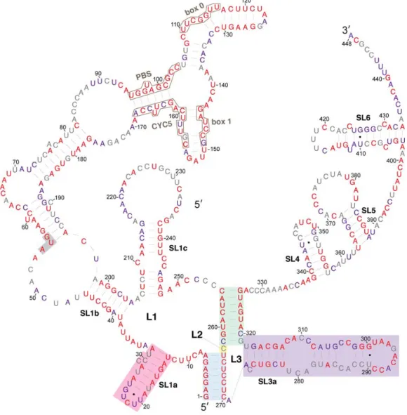
A model of the secondary structure of the Ty1 RNA TIPIRT domain annotated by the average
SHAPE reactivity of each nucleotide position is shown in Figure2. A prominent feature of the model
is a pseudoknot formed by long-range interactions of sequences spanning the first 326 nucleotides of Ty1 RNA, which is within the functionally defined 380-nucleotide TIPIRT domain. This pseudoknot is
similar to those predicted previously in the 50terminus of in vitro transcribed Ty1 RNA and in Ty1
RNA isolated from VLPs, although earlier modeling did not make use of a pseudoknot discovery algorithm [20,21]. The pseudoknot core consists of the 7-bp S1 pairing (Figure2, blue shading) and the
8-bp S2 pairing (Figure2, green shading) connected by a 1-nucleotide interhelical loop (L2) (Figure2,
yellow shading). The S1 stem of the pseudoknot, formed by pairing of the seven 5'-terminal nucleotides
of Ty1 RNA to nucleotides 264–270, has an established function during retrotransposition [19,20].
Nucleotides 255–262 interact with nucleotides 319–326 of Ty1 RNA to form the S2 pairing of the
pseudoknot (Figure2, green shading). The S2 stem contains an additional base-pair (C255–G326) that
was not predicted in earlier models [20,21].
All but one of the nucleotides within the pseudoknot core had low reactivity with NMIA, including the unpaired L2 nucleotide, suggesting that the pseudoknot is a thermodynamically stable tertiary interaction within the Ty1 RNA. This conclusion is supported by the fact that other RNA structure
prediction algorithms that do not employ SHAPE data, such as pknotsRG and IPknot [38,39], also
predict a pseudoknot with identical S1 and S2 stems and L2 nucleotide in the 50leader of Ty1 RNA
(Figures S1 and S2).
The multibranched L1 loop (8/254) of the pseudoknot, formed by stem S1, contains three nested stem-loops (SL1a-SL1c). The first stem-loop (13/32; SL1a) has a single bulged nucleotide and short loop
(Figure2, pink shading). PAL1 and PAL2 sequences, which were proposed to interact intermolecularly
in the dimeric RNA of VLPs [25], are contained in the SL1a hairpin. The second stem-loop (39/204;
SL1b) is an extended domain containing two nested stem-loops. SL1b contains the sequences that pair
with tRNAiMetand with 30terminal sequences of Ty1 RNA (Figure2, black outlines) in the model of
Ty1 gRNA in virio [21]. The third (206/248; SL1c) is a stem-loop with a bulge loop and an internal
loop. L1 sequences include the entire 50UTR (1/53) of Ty1 RNA and the AUG codon ofGAG(Figure2,
highlighted in grey).
The L3 loop of the pseudoknot (271/318) is formed by the S2 pairing and composed almost entirely
of the low reactivity SL3a stem-loop (272/318) (Figure2, purple shading), which has two small internal
loops. S2 and L3 are within a region of Ty1 RNA that is necessary for packaging into VLPs (238/380) [11].
Beyond the pseudoknot, the 30terminal region of the Ty1 in vitro transcript harbors three stem-loops,
Viruses2017,9, 93 8 of 23
Viruses 2017, 9, 93 8 of 23
Figure 2. SHAPE reactivities and secondary structure model of the 5′ leader of Ty1 RNA. Nucleotides are colored according to their SHAPE reactivities, which are indicated on the color bar at the bottom left. Regions of low reactivity have a high probability of being constrained within secondary or tertiary structure. The AUG nucleotides shaded in grey comprise the start codon of GAG. The pseudoknot core contains stem S1 (blue shading), loop L2 (nucleotide 263, yellow shading), and stem S2 (green shading). Pseudoknot loops L1 (nucleotides 9–254) and L3 (nucleotides 271–318) are not shaded. The SL1a hairpin (pink shading) and SL3a hairpin (purple shading) are indicated.
Regions of the structural model that differ from previous SHAPE analysis‐derived structural
models of Ty1 RNA in virio [21] and the 5′ terminus of Ty1 RNA in vitro [20] include: (a) the presence
of the 255C‐326G base‐pair in the S2 pseudoknot stem, as noted above; (b) extension of the SL1a stem
by two base‐pairs by inclusion of a 1‐nucleotide bulge in our model; (c) the presence of a large loop at
the apex of stem‐loop SL1c in our model, compared to a bulge‐stem‐loop structure at the apex of SL1c
in previous models; (d) extension of the SL3a stem‐loop by two base‐pairs by inclusion of a
1‐nucleotide bulge in our model; and (e) the presence of SL4, which is not present in previous models.
As expected, no evidence of interactions seen in virio between motifs in SL1b and tRNAiMet or
between CYC5 and CYC3 was observed because neither tRNAiMet nor CYC3 are present in our system.
The location of hairpin SL3a within an essential packaging domain prompted us to look for features
that could function in the formation of a Ty1 RNA kissing complex. We noticed that the ACAGAAU
(293/299) sequence in the SL3a loop is perfectly complementary to an AUUCUGU (19/25) motif in the Figure 2.SHAPE reactivities and secondary structure model of the 50leader of Ty1 RNA. Nucleotides are colored according to their SHAPE reactivities, which are indicated on the color bar at the bottom left. Regions of low reactivity have a high probability of being constrained within secondary or tertiary structure. The AUG nucleotides shaded in grey comprise the start codon ofGAG. The pseudoknot core contains stem S1 (blue shading), loop L2 (nucleotide 263, yellow shading), and stem S2 (green shading). Pseudoknot loops L1 (nucleotides 9–254) and L3 (nucleotides 271–318) are not shaded. The SL1a hairpin (pink shading) and SL3a hairpin (purple shading) are indicated.
Regions of the structural model that differ from previous SHAPE analysis-derived structural models of Ty1 RNA in virio [21] and the 50terminus of Ty1 RNA in vitro [20] include: (a) the presence of the 255C-326G base-pair in the S2 pseudoknot stem, as noted above; (b) extension of the SL1a stem by two base-pairs by inclusion of a 1-nucleotide bulge in our model; (c) the presence of a large loop at the apex of stem-loop SL1c in our model, compared to a bulge-stem-loop structure at the apex of SL1c in previous models; (d) extension of the SL3a stem-loop by two base-pairs by inclusion of a 1-nucleotide bulge in our model; and (e) the presence of SL4, which is not present in previous models.
As expected, no evidence of interactions seen in virio between motifs in SL1b and tRNAiMetor between
The location of hairpin SL3a within an essential packaging domain prompted us to look for features that could function in the formation of a Ty1 RNA kissing complex. We noticed that the ACAGAAU (293/299) sequence in the SL3a loop is perfectly complementary to an AUUCUGU (19/25) motif in the loop and two apical base-pairs of the SL1a stem (G-U and U-A). The tertiary structure of the pseudoknot might allow these complementary motifs to pair intramolecularly. However, 4 of the 7 nucleotides (296/299) in the SL3a loop are highly reactive in SHAPE analysis of RNA in vitro (Figure2) [20]; therefore, it is unlikely that the SL1a and SL3a motifs are base-paired in vitro. The loop of SL3a is also highly reactive in virio [21], suggesting that the SL1a and SL3a motifs are also not base-paired in VLPs. Another intriguing possibility is that the complementary apical motifs of SL1a and SL3a base-pair intermolecularly to form
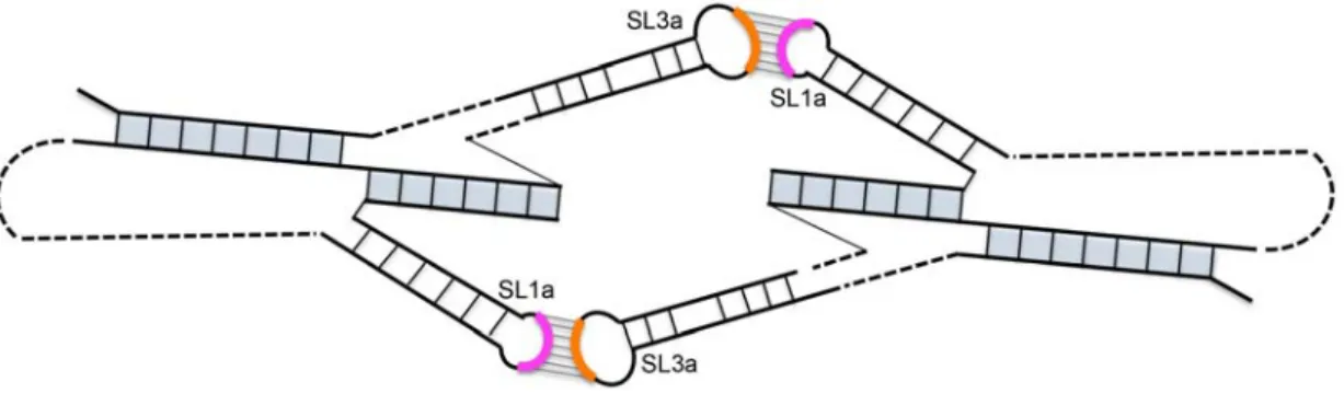
a symmetrical Ty1 RNA kissing complex with two kissing loops (Figure3). In vitro, where the TIPIRT
domain RNA is monomeric in the absence of Gag [17,40], and in VLPs, where the Ty1 RNA is a mature
dimer [10,21], the motif in SL3a is mostly reactive, arguing against base-pairing of the complementary
SL1a-SL3a motifs in these RNA forms. Nonetheless, pairing between the SL1a and SL3a apical motifs on different Ty1 RNA molecules could form a transient symmetrical kissing complex that initiates packaging of Ty1 RNA into VLPs, and then is converted to a stable dimer linkage within the mature VLP.
Viruses 2017, 9, 93 9 of 23
loop and two apical base‐pairs of the SL1a stem (G‐U and U‐A). The tertiary structure of the pseudoknot
might allow these complementary motifs to pair intramolecularly. However, 4 of the 7 nucleotides
(296/299) in the SL3a loop are highly reactive in SHAPE analysis of RNA in vitro (Figure 2) [20];
therefore, it is unlikely that the SL1a and SL3a motifs are base‐paired in vitro. The loop of SL3a is also
highly reactive in virio [21], suggesting that the SL1a and SL3a motifs are also not base‐paired in VLPs.
Another intriguing possibility is that the complementary apical motifs of SL1a and SL3a base‐pair
intermolecularly to form a symmetrical Ty1 RNA kissing complex with two kissing loops (Figure 3). In
vitro, where the TIPIRT domain RNA is monomeric in the absence of Gag [17,40], and in VLPs, where
the Ty1 RNA is a mature dimer [10,21], the motif in SL3a is mostly reactive, arguing against base‐pairing
of the complementary SL1a‐SL3a motifs in these RNA forms. Nonetheless, pairing between the SL1a
and SL3a apical motifs on different Ty1 RNA molecules could form a transient symmetrical kissing
complex that initiates packaging of Ty1 RNA into VLPs, and then is converted to a stable dimer
linkage within the mature VLP.
Figure 3. Model of a symmetrical Ty1 RNA kissing complex containing two Ty1 RNA pseudoknots interacting via two 7‐base‐pair intermolecular RNA duplexes formed between apical motifs in stem‐ loop SL1a (pink arc) and SL3a (orange arc). The pseudoknot stems are shaded in blue.
3.2. Conservation of Ty1 RNA TIPIRT Domain
We compared the conservation of nucleotides within the Ty1 RNA 5′ terminus to the secondary
structure model to ascertain whether there are conserved structural features that could function in cis
in retrotransposition. Because most S. cerevisiae Ty1 elements are mobile or recently mobile [41], and
therefore have a high degree of sequence identity [42,43], we also compared their sequences to that of
Ty2 elements, a closely related family of LTR‐retrotransposons in S. cerevisiae. The 5′ terminal sequence
of 66 Ty1 elements and 32 Ty2 elements from a variety of laboratory, industrial and natural S. cerevisiae
strain genomes [44], as well as four Ty1 elements from other Saccharomyces species were aligned. Each
nucleotide position was assigned to one of three categories based on the degree of conservation at
that position: (1) conserved in all 102 Saccharomyces Ty1 and Ty2 elements (Figure 4, red coloring);
(2) conserved in all 66 S. cerevisiae Ty1 elements (Figure 4, purple coloring); or (3) variable among the
S. cerevisiae Ty1 elements analyzed (Figure 4, grey coloring).
The alignment indicates that nucleotides in the pseudoknot core are very highly conserved. S1
nucleotides are invariant in all Saccharomyces Ty1 and Ty2 elements. S2 nucleotides, including C255 and
G326, whose pairing is predicted uniquely in the structural model presented here, are invariant, with
the exception of three nucleotides at the base of S2. Two of these nucleotides (C262 and C320) are
substituted in a few Ty2 elements, while the third nucleotide, G319, is a U nucleotide in four of the 66
S. cerevisiae Ty1 elements, but is otherwise conserved. Similarly, the L2 nucleotide C263 is substituted
by an A nucleotide in three S. cerevisiae Ty1 elements. Thus, every residue of the pseudoknot core is
invariant or has limited variation, in agreement with the conclusion of Huang et al. [20].
The entire 326‐nucleotide pseudoknot domain has a high degree of conservation overall. Sequences
that are very highly conserved among S. cerevisiae Ty1 elements include those that bind tRNAiMet
(PBS, Box 0 and Box 1; Figure 4, black outlines) and those within sequence regions that are predicted
to be base‐paired, including the SL1a stem, regions of the SL1b stem such as the pairing between
nucleotides 39–45 and 198–204 and the SL1c stem. While most regions that are predicted to be single Figure 3.Model of a symmetrical Ty1 RNA kissing complex containing two Ty1 RNA pseudoknots interacting via two 7-base-pair intermolecular RNA duplexes formed between apical motifs in stem-loop SL1a (pink arc) and SL3a (orange arc). The pseudoknot stems are shaded in blue.
3.2. Conservation of Ty1 RNA TIPIRT Domain
We compared the conservation of nucleotides within the Ty1 RNA 50terminus to the secondary
structure model to ascertain whether there are conserved structural features that could function incis
in retrotransposition. Because mostS. cerevisiaeTy1 elements are mobile or recently mobile [41], and
therefore have a high degree of sequence identity [42,43], we also compared their sequences to that of
Ty2 elements, a closely related family of LTR-retrotransposons inS. cerevisiae. The 50terminal sequence
of 66 Ty1 elements and 32 Ty2 elements from a variety of laboratory, industrial and naturalS. cerevisiae
strain genomes [44], as well as four Ty1 elements from otherSaccharomycesspecies were aligned. Each
nucleotide position was assigned to one of three categories based on the degree of conservation at
that position: (1) conserved in all 102SaccharomycesTy1 and Ty2 elements (Figure4, red coloring);
(2) conserved in all 66S. cerevisiaeTy1 elements (Figure4, purple coloring); or (3) variable among the
S. cerevisiaeTy1 elements analyzed (Figure4, grey coloring).
The alignment indicates that nucleotides in the pseudoknot core are very highly conserved.
S1 nucleotides are invariant in allSaccharomycesTy1 and Ty2 elements. S2 nucleotides, including C255
and G326, whose pairing is predicted uniquely in the structural model presented here, are invariant, with the exception of three nucleotides at the base of S2. Two of these nucleotides (C262 and C320) are substituted in a few Ty2 elements, while the third nucleotide, G319, is a U nucleotide in four of the 66 emphS. cerevisiae Ty1 elements, but is otherwise conserved. Similarly, the L2 nucleotide C263 is
substituted by an A nucleotide in threeS. cerevisiaeTy1 elements. Thus, every residue of the pseudoknot
Viruses2017,9, 93 10 of 23
The entire 326-nucleotide pseudoknot domain has a high degree of conservation overall. Sequences
that are very highly conserved amongS. cerevisiae Ty1 elements include those that bind tRNAiMet
(PBS, Box 0 and Box 1; Figure4, black outlines) and those within sequence regions that are predicted to be base-paired, including the SL1a stem, regions of the SL1b stem such as the pairing between nucleotides 39–45 and 198–204 and the SL1c stem. While most regions that are predicted to be single stranded have low nucleotide conservation, nucleotides 8–12, nucleotides 34–38, nucleotides 63–69, and the SL3a loop
are conserved. The SL1a loop is conserved inS. cerevisiaeTy1 elements but not in Ty2 elements. Within
the 53-nucleotide 50UTR, 34 nucleotides (64%) are invariant amongst all 102SaccharomycesTy1 and Ty2
elements analyzed, while 44 nucleotides (83%) are conserved among 66S. cerevisiaeTy1 elements.
Viruses 2017, 9, 93 10 of 23
stranded have low nucleotide conservation, nucleotides 8–12, nucleotides 34–38, nucleotides 63–69, and the SL3a loop are conserved. The SL1a loop is conserved in S. cerevisiae Ty1 elements but not in Ty2 elements. Within the 53‐nucleotide 5′ UTR, 34 nucleotides (64%) are invariant amongst all 102
Saccharomyces Ty1 and Ty2 elements analyzed, while 44 nucleotides (83%) are conserved among 66
S. cerevisiae Ty1 elements.
Figure 4. Relative evolutionary conservation of each nucleotide overlayed on the secondary structure model of the 5′ leader of Ty1 RNA. The color of each RNA base indicates its degree among conservation among 102 Ty1 and Ty2 elements from the genus Saccharomyces. Categories of conservation are as follows: red, 100% conserved among 102 Ty1 and Ty2 elements in the genus Saccharomyces; purple, 100% conserved in 66 Saccharomyces cerevisiae Ty1 elements; grey, not 100% conserved in either set.
3.3. Requirement for Pseudoknot Stems S1 and S2 in Retrotransposition
To identify the role of Ty1 RNA secondary structures in retrotransposition, we used an established helper‐Ty1/mini‐Ty1 assay in which two defective but complementing Ty1 elements are co‐expressed, each from a plasmid‐based GAL1 promoter (Figure 5) [11]. The helper‐Ty1 element encodes functional Gag and Gag‐Pol proteins, and its RNA is packaged in VLPs but cannot be used in reverse transcription because it harbors silent substitutions in the PBS and lacks the 3′ polypurine tract and LTR [11]. The mini‐Ty1his3AI element has an internal deletion of most of the GAG ORF and the entire POL ORF;
Figure 4.Relative evolutionary conservation of each nucleotide overlayed on the secondary structure model of the 50leader of Ty1 RNA. The color of each RNA base indicates its degree among conservation among 102 Ty1 and Ty2 elements from the genusSaccharomyces. Categories of conservation are as follows: red, 100% conserved among 102 Ty1 and Ty2 elements in the genusSaccharomyces; purple, 100% conserved in 66Saccharomyces cerevisiaeTy1 elements; grey, not 100% conserved in either set.
3.3. Requirement for Pseudoknot Stems S1 and S2 in Retrotransposition
To identify the role of Ty1 RNA secondary structures in retrotransposition, we used an established helper-Ty1/mini-Ty1 assay in which two defective but complementing Ty1 elements are co-expressed,
Gag and Gag-Pol proteins, and its RNA is packaged in VLPs but cannot be used in reverse transcription
because it harbors silent substitutions in the PBS and lacks the 30 polypurine tract and LTR [11].
The mini-Ty1his3AIelement has an internal deletion of most of theGAGORF and the entirePOLORF;
nonetheless, 50 leader sequences corresponding to nucleotides 1–575 of Ty1 RNA as well as the last
357 nucleotides of Ty1, including the 30polypurine tract and LTR, are retained. Together, these regions
are sufficient for mini-Ty1 RNA to be used as a template for retrotransposition when Ty1 proteins are
supplied in trans. Mini-Ty1his3AIalso carries thehis3AIretrotransposition indicator gene, which allows
cells harboring transposed reverse transcripts to be detected as His+prototrophs [45]. The plasmids
were expressed in an spt3∆ strain, which lacks expression of endogenous Ty1 RNA. The median
retrotransposition frequency in the strain co-expressing the mini-Ty1his3AIwith wild-type sequences
and the helper-Ty1 was 1.82×10⁻⁶. The frequency of His+prototrophs in the absence of helper-Ty1
was 1.8% of that in its presence. This background of His+prototrophs may be due to a low frequency of
recombination events that introduces full-length genomic Ty1 sequences into the mini-Ty1his3AIplasmid.
Viruses 2017, 9, 93 11 of 23
nonetheless, 5′ leader sequences corresponding to nucleotides 1–575 of Ty1 RNA as well as the last 357 nucleotides of Ty1, including the 3′ polypurine tract and LTR, are retained. Together, these regions are sufficient for mini‐Ty1 RNA to be used as a template for retrotransposition when Ty1 proteins are supplied in trans. Mini‐Ty1his3AI also carries the his3AI retrotransposition indicator gene, which allows cells harboring transposed reverse transcripts to be detected as His+ prototrophs [45]. The plasmids were expressed in an spt3∆ strain, which lacks expression of endogenous Ty1 RNA. The median retrotransposition frequency in the strain co‐expressing the mini‐Ty1his3AI with wild‐type sequences and the helper‐Ty1 was 1.82 × 10⁻⁶. The frequency of His+ prototrophs in the absence of helper‐Ty1 was 1.8% of that in its presence. This background of His+ prototrophs may be due to a low frequency of recombination events that introduces full‐length genomic Ty1 sequences into the mini‐Ty1his3AI plasmid.
Figure 5. Assay for helper‐mediated retrotransposition of mini‐Ty1his3AI. A complete Ty1 element is shown at the top for reference. The mini‐Ty1his3AI element and helper‐Ty1 element are each expressed from the GAL1 promoter (labeled rectangle), which is fused to the transcription start site of Ty1‐H3 at the first nucleotide of the R domain in the 5′ LTR. GAL1:mini‐Ty1his3AI is carried on a URA3‐based plasmid and GAL1:helper‐Ty1 is contained on a TRP1‐based plasmid (not illustrated). The elements are co‐expressed in an spt3∆ strain lacking endogenous Ty1 element transcription. The internally deleted mini‐Ty1his3AI element contains 5′ sequences corresponding to nucleotides 1–575 of Ty1 RNA, as well as the last 357 nucleotides of Ty1, including the 3′ polypurine tract (not illustrated) and 3′ LTR. The his3AI retrotransposition indicator gene, consisting of the HIS3 marker gene interrupted by an antisense intron (boxed arrowhead), is inserted in the mini‐Ty1 between the 5′ leader and 3′ LTR. The direction of mini‐Ty1his3AI transcription from the GAL1 promoter (denoted by the arrow atop the GAL1 rectangle) is opposite to the direction of his3AI transcription (denoted by an arrow atop the HIS3 rectangle), so the intron is only be spliced from the Ty1his3AI transcript. The helper‐Ty1 element carries functional GAG and POL ORFs, but the polypurine tract and 3′ LTR are deleted. In addition, silent nucleotide substitutions in the PBS (denoted by a white rectangle marked with an “X”) block the binding of tRNAiMet. Splicing is illustrated by removal of the boxed arrowhead representing the intron from the rectangle that denotes the HIS3 gene. Gag and Gag‐Pol proteins translated from the helper‐Ty1 RNA form VLPs that package the spliced mini‐Ty1HIS3 RNA, which is reverse transcribed to form Ty1HIS3 cDNA. Integration of the cDNA into the host genome allows the cell to be detected as a His+ prototroph.
Mutations were introduced into structural elements of the TIPIRT domain of the mini‐Ty1his3AI plasmid. All mutations and compensatory mutations introduced into GAG maintained an open reading
Figure 5.Assay for helper-mediated retrotransposition of mini-Ty1his3AI. A complete Ty1 element is shown at the top for reference. The mini-Ty1his3AIelement and helper-Ty1 element are each expressed from theGAL1promoter (labeled rectangle), which is fused to the transcription start site of Ty1-H3 at the first nucleotide of the R domain in the 50LTR.GAL1:mini-Ty1his3AIis carried on aURA3-based plasmid andGAL1:helper-Ty1 is contained on aTRP1-based plasmid (not illustrated). The elements are co-expressed in anspt3∆strain lacking endogenous Ty1 element transcription. The internally deleted mini-Ty1his3AIelement contains 50sequences corresponding to nucleotides 1–575 of Ty1 RNA, as well as the last 357 nucleotides of Ty1, including the 30polypurine tract (not illustrated) and 30LTR. Thehis3AI
retrotransposition indicator gene, consisting of theHIS3marker gene interrupted by an antisense intron (boxed arrowhead), is inserted in the mini-Ty1 between the 50 leader and 30 LTR. The direction of mini-Ty1his3AItranscription from theGAL1promoter (denoted by the arrow atop theGAL1rectangle) is opposite to the direction ofhis3AItranscription (denoted by an arrow atop theHIS3rectangle), so the intron is only be spliced from the Ty1his3AItranscript. The helper-Ty1 element carries functionalGAGand
POLORFs, but the polypurine tract and 30LTR are deleted. In addition, silent nucleotide substitutions in the PBS (denoted by a white rectangle marked with an “X”) block the binding of tRNAiMet. Splicing is
Viruses2017,9, 93 12 of 23
Mutations were introduced into structural elements of the TIPIRT domain of the mini-Ty1his3AI
plasmid. All mutations and compensatory mutations introduced into GAG maintained an open
reading frame but not necessarily the amino acid sequence of the truncated Gag product. An UC264AG
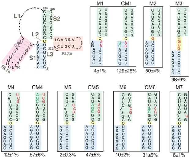
substitution that disrupts S1 complementarity in mini-Ty1his3AIRNA reduced helper-Ty1 mediated
retrotransposition to 4% of that of the mini-Ty1his3AI with wild-type sequence (Figure 6, M1).
A compensatory mutation that reestablishes S1 complementarity restored retrotransposition to
levels equivalent to the wild-type mini-Ty1his3AI (Figure6, CM1). Similar results were obtained
with the identical substitutions in a previous study [20]; therefore, these findings validate the
helper-Ty1/mini-Ty1 assay and confirm the role of the S1 pairing in retrotransposition [11,19].
Viruses 2017, 9, 93 12 of 23
frame but not necessarily the amino acid sequence of the truncated Gag product. An UC264AG
substitution that disrupts S1 complementarity in mini‐Ty1his3AI RNA reduced helper‐Ty1 mediated
retrotransposition to 4% of that of the mini‐Ty1his3AI with wild‐type sequence (Figure 6, M1). A
compensatory mutation that reestablishes S1 complementarity restored retrotransposition to levels
equivalent to the wild‐type mini‐Ty1his3AI (Figure 6, CM1). Similar results were obtained with the
identical substitutions in a previous study [20]; therefore, these findings validate the helper‐Ty1/mini‐Ty1
assay and confirm the role of the S1 pairing in retrotransposition [11,19].
Figure 6. Retrotransposition of mini‐Ty1his3AI elements with mutations in the Ty1 pseudoknot core. The schematic (top left) shows the secondary structure of the Ty1 pseudoknot core and portions of the L1 and L3 loops. Blue shading, stem S1; yellow shading, loop L2; green shading, stem S2; pink shading, SL1a hairpin, a segment of the L1 loop; orange shading, SL3a hairpin, a portion of the L3 loop. Dotted lines represent bases in loops L1 and L3 that are not shown. Labeled, boxed schematics show the nucleotide substitutions or additions in each mutant mini‐Ty1his3AI element analyzed. Black letters represent wild‐ type nucleotides; red letters represent nucleotide substitutions or additions; and green letters represent compensatory substitutions that restore base‐pairing with nucleotide substitutions. The percentage below each box is the median frequency of helper‐mediated retrotransposition of the mini‐Ty1his3AI bearing the indicated mutation divided by the median helper‐mediated retrotransposition frequency of the mini‐Ty1his3AI element with wild‐type Ty1‐H3 sequence, +/− the 95% confidence interval.
We analyzed the requirement for pseudoknot stem S2 by introducing double and triple nucleotide
substitutions that disrupt S2 complementarity. These mutations reduced retrotransposition to
2%–12% of that of the wild‐type mini‐Ty1his3AI (Figure 6, M4, M5 and M6). Even the single C320U
substitution, which is predicted to change a G‐C base‐pair to a G‐U base‐pair, reduced retrotransposition
to 6% of wild‐type activity (Figure 6, M7). Reestablishing S2 complementarity in the mutants harboring
double and triple nucleotide substitutions by introduction of compensatory mutations restored
retrotransposition up to 31%–57% of the wild‐type mini‐Ty1his3AI (Figure 6, CM4, CM5 and CM6). Figure 6.Retrotransposition of mini-Ty1his3AIelements with mutations in the Ty1 pseudoknot core. The schematic (top left) shows the secondary structure of the Ty1 pseudoknot core and portions of the L1 and L3 loops. Blue shading, stem S1; yellow shading, loop L2; green shading, stem S2; pink shading, SL1a hairpin, a segment of the L1 loop; orange shading, SL3a hairpin, a portion of the L3 loop. Dotted lines represent bases in loops L1 and L3 that are not shown. Labeled, boxed schematics show the nucleotide substitutions or additions in each mutant mini-Ty1his3AIelement analyzed. Black letters represent wild-type nucleotides; red letters represent nucleotide substitutions or additions; and green letters represent compensatory substitutions that restore base-pairing with nucleotide substitutions. The percentage below each box is the median frequency of helper-mediated retrotransposition of the mini-Ty1his3AIbearing the indicated mutation divided by the median helper-mediated retrotransposition frequency of the mini-Ty1his3AI
element with wild-type Ty1-H3 sequence, +/−the 95% confidence interval.
We analyzed the requirement for pseudoknot stem S2 by introducing double and triple nucleotide substitutions that disrupt S2 complementarity. These mutations reduced retrotransposition to 2–12%
which is predicted to change a G-C base-pair to a G-U base-pair, reduced retrotransposition to 6% of
wild-type activity (Figure 6, M7). Reestablishing S2 complementarity in the mutants harboring
double and triple nucleotide substitutions by introduction of compensatory mutations restored
retrotransposition up to 31–57% of the wild-type mini-Ty1his3AI(Figure6, CM4, CM5 and CM6).
Compensatory mutations may not fully reconstitute the activity of the wild-type mini-Ty1his3AI
because the base composition of S2 or ensemble folding of mini-Ty1his3AIRNA is altered. Together,
these data suggest that the S2 stem of the pseudoknot is as critical for retrotransposition as the S1 stem. Many pseudoknots have 0 to 1-nucleotide interhelical loops that promote a stable pseudoknot
conformation in which individual stems stack coaxially [46]. It has been suggested that S1 and S2 of
the TIPIRT domain pseudoknot stack coaxially [20,21], even though the unreactive L2 nucleotide can be
substituted without major effects on pseudoknot structure or function [20]. To determine the consequences of disrupting the potential for coaxial stacking of the pseudoknot stems, we increased the length of L2
from one to four nucleotides by addition of a GCG triplet (Figure6, M3). This mutation had no effect on
retrotransposition of mini-Ty1his3AI. We also confirmed that the C236G substitution of the L2 nucleotide
reduced retrotransposition only modestly (50%) (Figure6, M2). In summary, our data demonstrate that
neither the length nor composition of L2 is a major determinant of pseudoknot conformation; therefore, coaxial stacking of S1 and S2 is not likely to be necessary for pseudoknot function.
3.4. Requirement for Complementary Motifs in SL1a and SL3a Hairpins in Retrotransposition
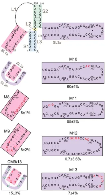
The SL3a hairpin (272/318) is in a region of the TIPIRT domain that contains essential Ty1 RNA
packaging sequences [11]. The ACAGAAU (293/299) motif in the loop of SL3a is complementary to the
AUUCUGU motif (19/25) encompassing the 3-nucleotide loop and first two base-pairs of the SL1a stem (Figure7). Except for 1 nucleotide (G296) in SL3a, both sequences are invariant inS. cerevisiaeTy1 elements. Therefore, we hypothesized that intermolecular “kissing loop” interactions between the complementary
sequences in SL1a and SL3a (Figure3) could initiate dimerization of Ty1 RNA. To determine whether
these complementary motifs are individually required for retrotransposition, we substituted UCUCUAA
for ACAGAAU (293/299) in the SL3a loop, which reduced helper-Ty1-mediated mini-Ty1his3AI
retrotransposition to 7% of wild-type activity (Figure7, M13). Substitution of UUAGAGA for AUUCUGU
(19/25) in SL1a reduced retrotransposition to 8% (Figure7, M9). Both the AUUCUGU19UUAGAGA
mutant and wild-type RNA have an A-U and G-U base-pair at the apex of the SL1a stem; thus, the retrotransposition defect of the AUUCUGU19UUAGAGA mutant is probably not due to disruption of the SL1a stem. Instead our findings indicate that complementary motifs in SL1a and SL3a are required incisin Ty1 retrotransposition.
To determine whether reestablishing complementarity between apical sequences of the SL1a and SL3a hairpins restores retrotransposition, both AUUCUGU19UUAGAGA and ACAGAAU293UCUCUAA
were introduced into a single mini-Ty1his3AIelement. This double mutant transposed at 15% of the
Viruses2017,9, 93 14 of 23
Viruses 2017, 9, 93 14 of 23
Figure 7. Retrotransposition of mini‐Ty1his3AI elements with mutations in stem‐loops SL1a and SL3a. The schematic (top) shows the secondary structure of the Ty1 pseudoknot core and loops L1, L2 and L3, with the SL1a hairpin (pink shading) and SL3a hairpin (purple shading) highlighted. A second schematic (second from top, left) shows the proposed kissing loop interaction between the seven apical nucleotides of hairpin SL1a (pink shading) and seven apical sequences of the SL3a hairpin. Labeled, boxed schematics show the nucleotide substitutions or additions in each mutant mini‐Ty1his3AI element analyzed. Black letters indicate wild‐type sequence; red letters indicated nucleotide substitutions or additions; and green letters indicate compensatory substitutions that are predicted to restore base‐pairing with nucleotide substitutions. The percentage below each box is the median frequency of helper‐mediated retrotransposition of each mini‐Ty1his3AI bearing the indicated mutation divided by the median helper‐mediated retrotransposition frequency of the mini‐Ty1his3AI element with wild‐ type Ty1‐H3 sequence, +/− the 95% confidence interval.
Figure 7. Retrotransposition of mini-Ty1his3AI elements with mutations in stem-loops SL1a and SL3a. The schematic (top) shows the secondary structure of the Ty1 pseudoknot core and loops L1, L2 and L3, with the SL1a hairpin (pink shading) and SL3a hairpin (purple shading) highlighted. A second schematic (second from top, left) shows the proposed kissing loop interaction between the seven apical nucleotides of hairpin SL1a (pink shading) and seven apical sequences of the SL3a hairpin. Labeled, boxed schematics show the nucleotide substitutions or additions in each mutant mini-Ty1his3AIelement analyzed. Black letters indicate wild-type sequence; red letters indicated nucleotide substitutions or additions; and green letters indicate compensatory substitutions that are predicted to restore base-pairing with nucleotide substitutions. The percentage below each box is the median frequency of helper-mediated retrotransposition of each mini-Ty1his3AIbearing the indicated mutation divided by the median helper-mediated retrotransposition frequency of the mini-Ty1his3AI
element with wild-type Ty1-H3 sequence, +/−the 95% confidence interval.
To examine the role of the SL3a bulged stem in retrotransposition, we introduced double mutations near the base and the loop of the SL3a stem. Nucleotides C324 and A325, and the bases with which they are predicted to pair (275/276) are invariant among Ty1 and Ty2 elements; however, disruption of this
substitution of CA for GG (301/302) near the SL3a loop also resulted in a minor retrotransposition
defect (Figure7, M10). In contrast, substitution of six nucleotides within the bulged stem of SL3a
strongly decreased retrotransposition (Figure7, M12).
Sequences that comprise the SL1a stem-loop are mostly conserved, particularly inS. cerevisiaeTy1
elements, despite the fact that this region is non-coding. A 7-nucleotide substitution that completely
disrupts pairing in the S1 stem strongly reduced retrotransposition (Figure7, M8). Mini-Ty1his3AIRNA
with a two-nucleotide substitution in the SL1a stem could not be co-transformed with helper-Ty1 into the same yeast strain, even though several transformation strategies were attempted. In summary, major nucleotide substitutions in the stems of SL1a and SL3a hairpins strongly decreased retrotransposition, but it remains to be determined whether the secondary structure of the stems is the critical feature required.
3.5. Role for the S2 Stem and SL1a-SL3a Kissing Loops in Ty1 RNA Stability
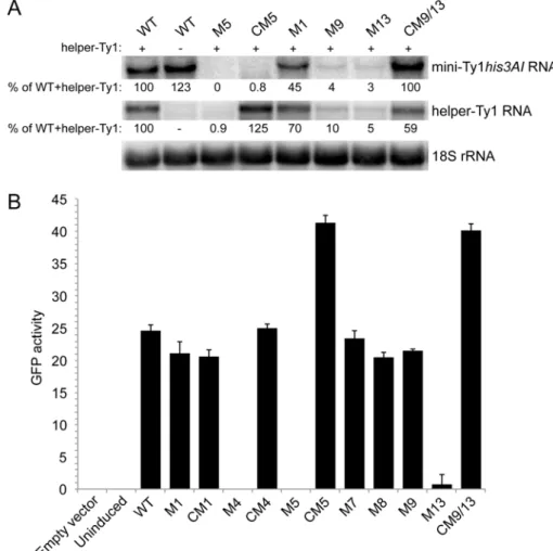
Because the S2 stem and SL3a hairpin overlap with a region required for Ty1 RNA packaging, mutations in the S2 stem and SL3a loop, as well as apical mutations in the SL1a hairpin hypothesized to interact with SL3a, might inhibit retrotransposition by blocking packaging of Ty1 RNA. To explore this possibility, we first determined whether mutations in stem S2 and hairpins SL1a and SL3a affect
RNA stability. The level of transcript from wild-type and mutant pGAL1:mini-Ty1his3AIelements was
monitored by northern analysis using a probe specific tohis3AI. Helper-Ty1 RNA was also quantitated
using a probe in the Ty1POLregion; a discrete band of ~5.5 kb was detected despite the absence of
the termination signal in the 30LTR. Strains were induced by growth in galactose for 24 h at 20◦C to
mimic the conditions used in the retrotransposition assay. Levels of mini-Ty1his3AIRNA in the presence
and absence of helper-Ty1 RNA were equivalent (Figure8A, compare WT lanes plus (+) and minus (−)
helper-Ty1), demonstrating that packaging of mini-Ty1his3AIRNA is not required for stability. The level
of mini-Ty1his3AIRNA with a UC264AG mutation in pseudoknot stem S1 was decreased about 2-fold
(Figure8A, M1). This result is consistent with previous analyses of this and other stem S1 mutations
in a full-length pGAL1:Ty1his3AIelement in the absence of helper-Ty1 [20]. Thus, disruption of stem
S1 minimally affects Ty1 RNA stability. In contrast, mini-Ty1his3AIRNA bearing the AUG321GCU
mutation in stem S2 was undetectable (Figure8A, M5). Surprisingly, helper-Ty1 RNA was also absent,
indicating that expressing mini-Ty1his3AIRNA with the AUG321GCU mutation destabilizes helper-Ty1
RNA intrans. Mini-Ty1his3AIRNA with double compensatory mutations AUG321GCU/CAU258AGC
was also present at very low levels, but the level of helper-Ty1 RNA in this strain was completely restored
(Figure8A, CM5). These findings support the idea that base-pairing of stem S2 is necessary for mini-Ty1
RNA and helper-Ty1 RNA stability. Instability of the AUG321GCU/CAU258AGC mini-Ty1 RNA was unexpected, because this compensatory mutant transposes at 47% of the frequency of the wild-type
mini-Ty1his3AI. A possible explanation for this inconsistency is that two temporally or structurally
distinct pools of the AUG321GCU/CAU258AGC mutant exist, one that is successfully packaged into VLPs and is used in retrotransposition, and another that is degraded.
To explore this possibility, we used a second, more sensitive approach to measure mini-Ty1 RNA
levels, this time in the absence of helper-Ty1. The Ty1 sequences from each pGAL1:mini-Ty1his3AI
plasmid was subcloned into an expression plasmid, creating an in-frame fusion of the 50UTR and first
522 nucleotides ofGAGto theGFPORF (GagNT:GFP). The pGAL1:mini-Ty1(GagNT:GFP) plasmids
were introduced into thespt3∆strain, and expression was induced for 2.5 h in galactose at 20◦C.
The mean GFP activity in 10,000 cells bearing a plasmid with wild-type or mutant Ty1 sequences was measured by flow cytometry to monitor the presence of Ty1 RNA after a brief galactose-induction
(Figure8B). The GFP activities in isolates with plasmid pGAL1:mini-Ty1(GagNT:GFP) containing the
UC264AG mutation or the UC264AG/GA6UC compensatory mutation in stem S1 were comparable
to that of the plasmid with wild-type Ty1 sequence (Figure 8B, compare M1 and CM1 to WT),
supporting the idea that mutations in pseudoknot stem S1 minimally destabilize Ty1 RNA [20].